Method for detecting protein-protein interactions in cells

a protein-protein interaction and living cell technology, applied in the field of living cell protein-protein interaction detection, can solve the problems of inability to apply methods for bacterial cells, inability to understand protein-protein interactions (ppi), and limitations of in-vitro analysis of dynamic protein-protein interactions in-vivo

Active Publication Date: 2013-02-28
KOREA RES INST OF BIOSCI & BIOTECH
View PDF8 Cites 18 Cited by
  • Summary
  • Abstract
  • Description
  • Claims
  • Application Information

AI Technical Summary

Problems solved by technology

Thus, understanding of protein-protein interactions (PPI) in cells is an important issue in current biological research.
However, there are limitations to the in-vitro approach for the analysis of dynamic protein-protein interactions in-vivo.
However, a Y2H system is disadvantageous in that it must be performed in yeast, nuclear translocation is required, and it relies on the indirect readout of secondary induced transcriptional activation and effects, not in direct contact, resulting in false-positives.
Even though the two methods are useful for the analysis of protein interactions in animal cells using a high speed sorter flow cytometry, the methods are difficult to apply for bacterial cells.
For example, the detection level of FRET is too low to detect weak signals from bacterial cells considerably smaller than eukaryotic cells.
Fluorescence complementation assay may show non-uniform protein expression in cells of a large size, and is influenced by other factors in bacterial cells, leading to false-positive results.
Fluorescence co-localization is used by localization in particular organelles of eukaryotic cells, but cannot be applied to a bacterial system that has no cell organelles.

Method used


Image

  • Method for detecting protein-protein interactions in cells
  • Method for detecting protein-protein interactions in cells
  • Method for detecting protein-protein interactions in cells

Examples


example 1

Gene and Enzyme Acquisition

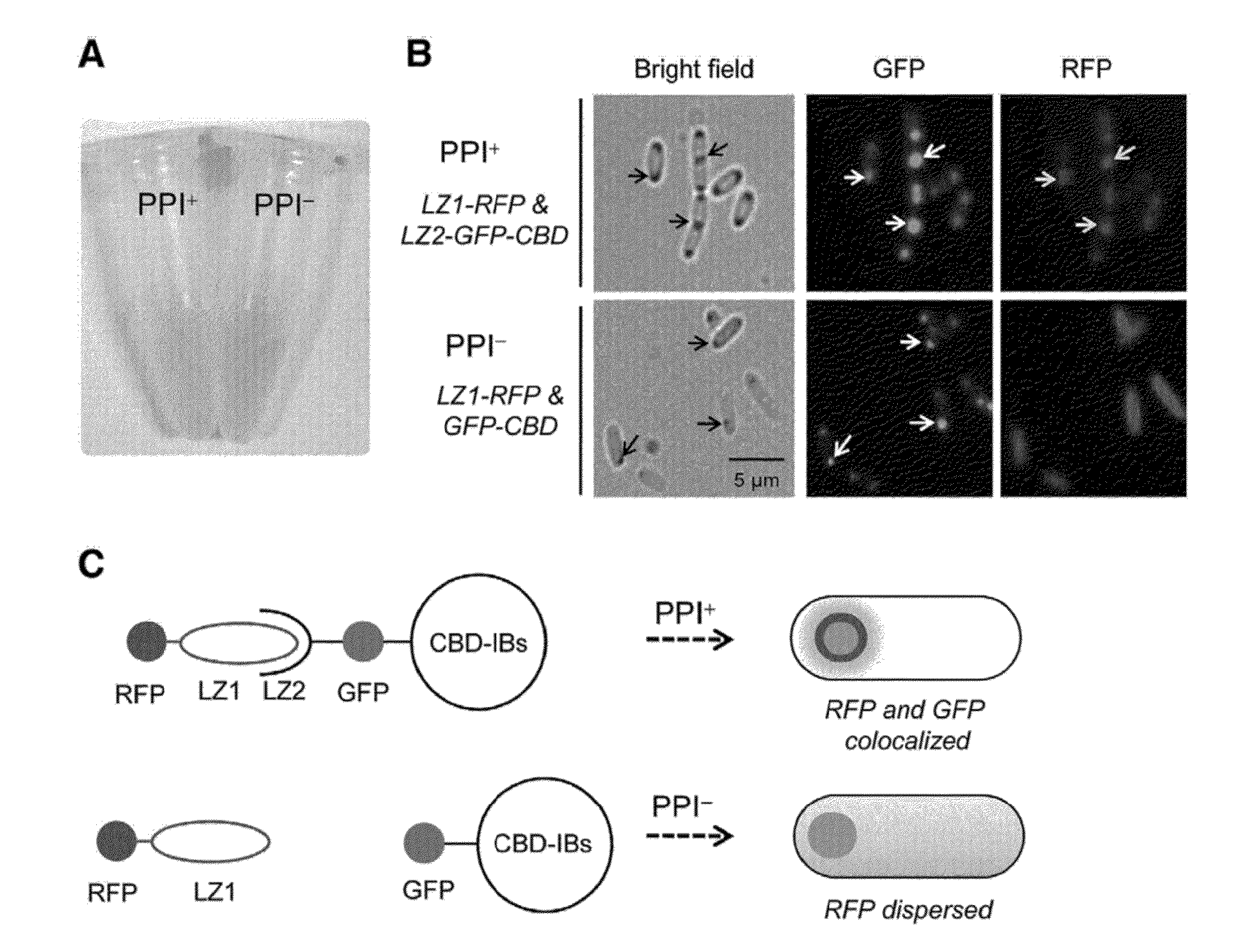
[0060]Family II CBD (cellulose-binding domain) gene was cloned from the exoglucanase gene of Cellulomonas fimi KCTC 9143 strain deposited at the Korean Collection of Type Cultures (KCTC), at the Korea Research Institute of Bioscience and Biotechnology. The improved GFP (green fluorescent protein) gene was obtained from a commercial plasmid pEGFP (Clontech, CA). A plasmid pRFP having RFP (monomeric red fluorescent protein 1) gene was provided by KAIST (Daejeon, Korea). Two antiparallel leucine zipper genes, LZ1 (EQLEKKLQALEKKLAQLEWKNQALEKKLAQ; charged residues are underlined, SEQ ID NO. 1) and LZ2 (ALKKELQANKKELAQLKWELQALKKELAQ; charged residues are underlined, SEQ ID NO. 2) were obtained from pMRBAD-Z-CGFP and pETlla-Z-NGFP which were provided by Dr. Regan at Yale university (Magliery, T. J., et al., 2005. Detecting protein-protein interactions with a green fluorescent protein fragment reassembly trap: scope and mechanism. J. Am. Chem. Soc. 127:146-57.). A...

example 2

DNA Manipulation

[0061]All primers were synthesized by Bioneer in Daejeon, Korea. Primers used in the present invention are shown in the following Table 1. NdeI and XhoI restriction sites are represented in bold.

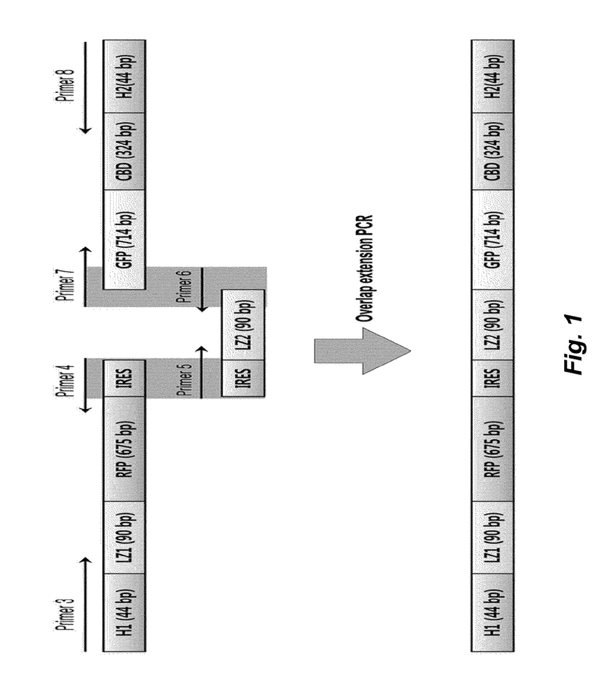
TABLE 1 SEQPrimerIDnameSequence (5′→3′)NO.Primer 1GATATACATATGGTGAGCAAGGGCGAG3Primer 2GGTGCTCGAGTTACTTGTACAGCTTGTCCATGCC4Primer 3CCTCTAGAAATAATTTTGTTTAACTTTAAGAAGGAGATATACATATGGCAAGCGAGCAGCTGGAA5Primer 4CAATTCTTTTTTGAGGGCCATATGATAATCTCCTTCTTAAAGTTAAACAAAATTATTTTAGGCGCCGG6TGGAGTGGCGGCCPrimer 5GGCCGCCACTCCACCGGCGCCTAAAATAATTTTGTTTAACTTTAAGAAGGAGATTATCATATGGCCCT7CAAAAAAGAATTGPrimer 6CAGCTCCTCGCCCTTGCTCACCTGCGCCAGTTCCTTTTTCAG8Primer 7CTGAAAAAGGAACTGGCGCAGGTGAGCAAGGGCGAGGAGCTG9Primer 8GCAGCCAACTCAGCTTCCTTTCGGGCTTTGTTAGCAGCCGGATCTCAGCCGACCGTGCAGGGCGTGCC10

[0062]The gfp gene was amplified from pEGFP using primer 1 (SEQ ID NO. 3) and primer 2 (SEQ ID NO. 4), cleaved using NdeI and XhoI, and cloned into a pET21a vector (Invitrogen, CA) to prepare pGFP. The gfp and cbd genes were fused ...

example 3

Protein Expression

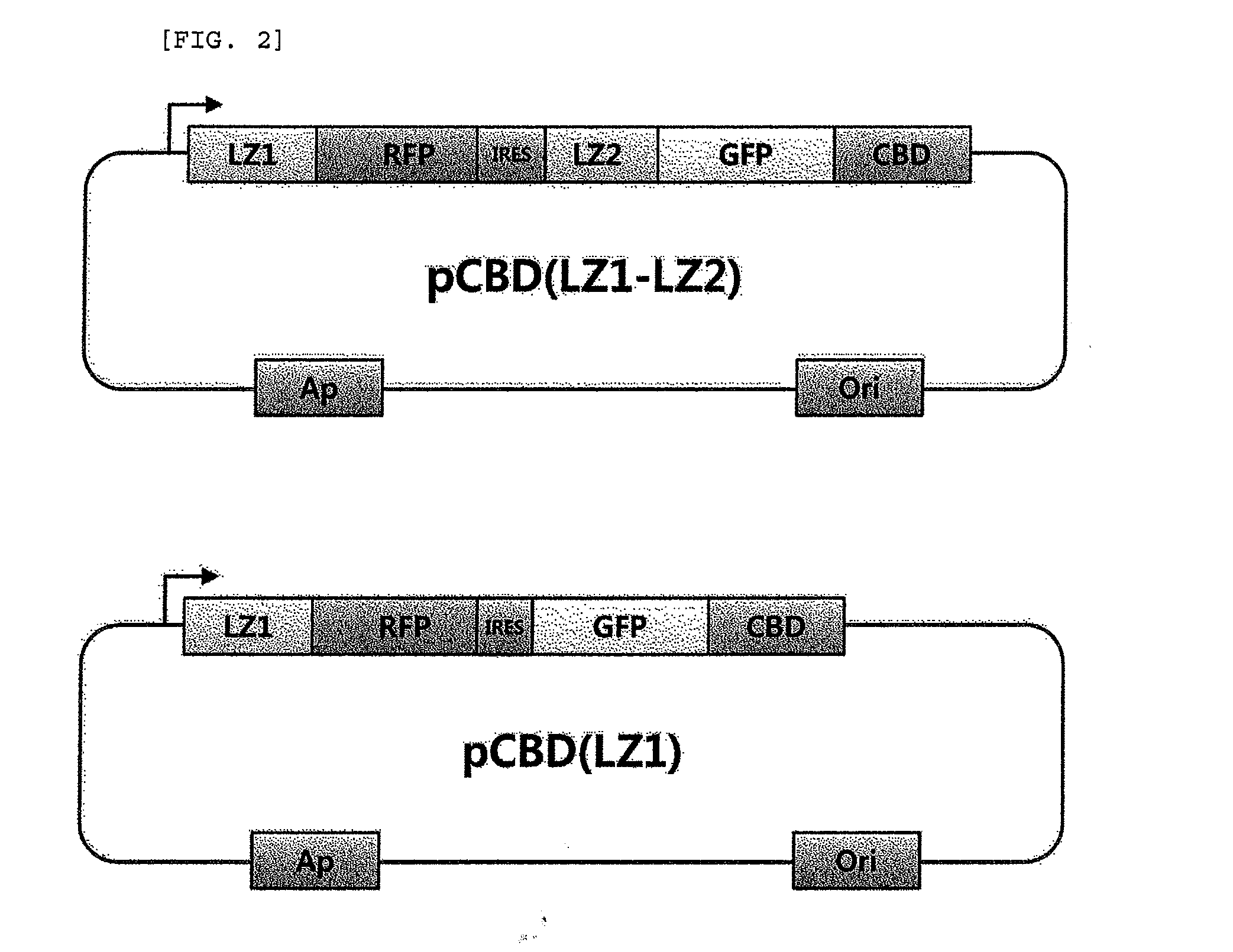
[0064]E. coli BL21 (DE3) was used as an expression host cell. The cells were transformed with pCBD(LZ1-LZ2) or pCBD(LZ1) prepared in Example 2, and cultured in LB media containing ampicillin (50 μg / ml). When OD at 600 nm reached 0.5, IPTG (isopropyl-1-thio-β-D-galactopyranoside, 1 mM) was added for the induction of CBD-induced inclusion bodies (CBD-IB), and the cells were incubated at 37° C. for 6 hours. Formation of inclusion bodies in the cultured cells was observed under a microscope. E. coli cells were disrupted by sonication on ice, and expression of the fusion protein was examined by SDS-PAGE and Western blotting. Non-aqueous fraction or CBD-IB were separated and obtained by centrifugation at 16,300×g for 10 minutes, and resuspended in PBS buffer (pH 7.4) containing Triton X-100 (0.50) to remove the membrane-bound precipitate.


PUM

PropertyMeasurementUnit
ODaaaaaaaaaa
pHaaaaaaaaaa
pHaaaaaaaaaa

Abstract

The present invention relates to a method for detecting protein-protein interactions in living cells, and more particularly, to a method for providing cells comprising a first construct and a second construct, wherein the first construct comprises a polynucleotide encoding a first fusion protein which comprises a bait protein, a first fluorescent protein and a CBD (cellulose-binding domain) protein, and wherein the second construct comprises a polynucleotide encoding a second fusion protein which comprises a prey protein and a second fluorescent protein so as to allow formation of inclusion bodies, and detecting interactions between the bait protein and the prey protein that are displayed by inclusion bodies, a method for isolating the prey protein bound to the bait protein using the cells comprising the constructs, the cells, and a kit for detecting protein-protein interactions, comprising the constructs.

Description

CROSS-REFERENCE TO RELATED APPLICATIONS[0001]This application claims the benefit of Korean Patent Application Nos. 10-2011-0086057, filed Aug. 26, 2011, and 10-2012-0063804, filed Jun. 14, 2012.STATEMENT REGARDING SEQUENCE LISTING[0002]The Sequence Listing associated with this application is provided in text format in lieu of a paper copy, and is hereby incorporated by reference into the specification. The name of the text file containing the Sequence Listing is 550077—402_SEQUENCE_LISTING_.txt. The text file is 9 KB, was created on Jun. 15, 2012, and is being submitted electronically via EFS-Web.BACKGROUND OF THE INVENTION[0003]1. Field of the Invention[0004]The present invention relates to a method for detecting protein-protein interactions in living cells, and more particularly, to a method for providing cells comprising a first construct and a second construct, wherein the first construct comprises a polynucleotide encoding a first fusion protein which comprises a bait protein, ...

Claims


Application Information

Patent Timeline
Patent Type & Authority Applications(United States)
IPC IPC(8): C12P21/02C12N1/21C12N5/10G01N21/64
CPCG01N21/6428G01N33/542G01N33/5008
Inventor LEE, SEUNG GOOCHOI, SU-LIMGAM, JONG SIKSONG, JAE JUNLEE, SANG JUN
Owner KOREA RES INST OF BIOSCI & BIOTECH
Why Eureka
Try Eureka