Arterial spin-labeled (ASL) multiplexed echo planar imaging (m-epi)

a multiplexed echo and planar imaging technology, applied in the field of magnetic resonance imaging, can solve the problems of limited slice size, epi has not been able to satisfactorily image perfusion in the entire brain, and asl perfusion, so as to reduce the time to scan the brain, reduce the time to sequential echo trains, and improve the interdependence of slice saturation effects on arterial input functions and recorded mr signals.

Inactive Publication Date: 2013-04-04
FEINBERG DAVID
View PDF28 Cites 16 Cited by
  • Summary
  • Abstract
  • Description
  • Claims
  • Application Information

AI Technical Summary

Benefits of technology

[0005]The replacement of EPI with M-EPI in ASL imaging has several advantages. For example, the complex interdependence of slice saturation effects on arterial input functions and recorded MR signal is greatly improved as far fewer time sequential echo trains are utilized. The time sequential echo trains can be reduced by a factor of N (the number of simultaneously images slices) in M-EPI compared with conventional EPI. With ASL using M-EPI combined with SIR, the entire brain can be scanned in, e.g., in 400 ms or less time with 30 to 60 images, as determined by N images=SIR×MB factors. If SIR=1, then N=MB alone, and this is one example included in the scope of the new process described in this patent specification. Ultimately, all slices can be acquired in a single echo train if the SIR and/or MB factors are large enough for the product to create enough slices to cover the entire brain or at least the portion of interest. It can be very important to cover the entire brain or organ, or at least the portion of interest, with slices to record the entire state of perfusion. This has not been possible for the entire brain with ASL based on conventional EPI due to the limited time window for measuring the hemodynamic perfusion effects because it was not

Problems solved by technology

Another limitation is that tissue blood is moving through different vascular compartments including arterial, capillary and venous compartments, so the time window of imaging can be constrained to a few hundred milliseconds for the capillary compartment.
Echo planar imaging (EPI) has been used as a readout image for the ASL perfusion technique but is limited in the number of slices that can be imaged due to the normal physiological time course of labeled blood moving through the different vascular compartments.
ASL with conventional multi-slice 2D EPI has not been able to satisfactorily image perfusion in the entire brain because of timing limitations.
This has not been possible for the entire

Method used


Image

  • Arterial spin-labeled (ASL) multiplexed echo planar imaging (m-epi)
  • Arterial spin-labeled (ASL) multiplexed echo planar imaging (m-epi)
  • Arterial spin-labeled (ASL) multiplexed echo planar imaging (m-epi)

Examples


Embodiment Construction

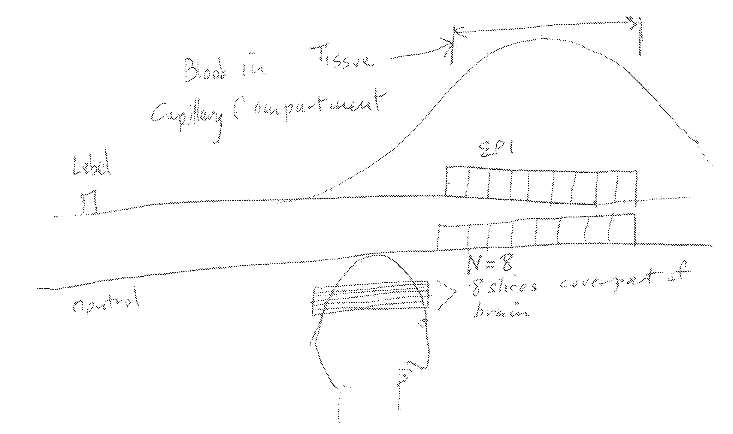
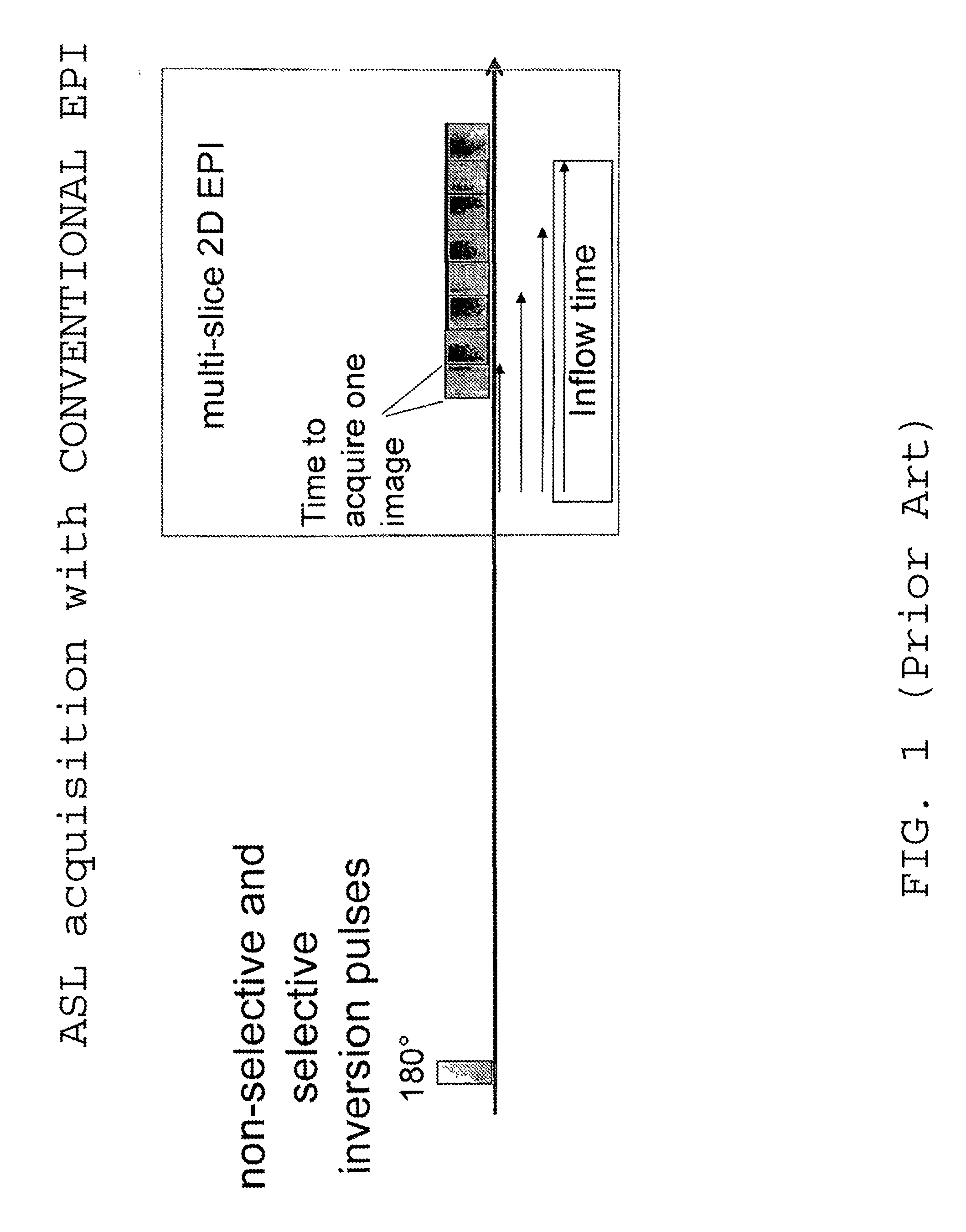
[0014]FIG. 1 illustrates conventional multi-slice 2D EPI in which several slice images are acquired one after the other in time. The different blood inflow times in consecutively acquired slice images limit the accuracy of blood flow quantization. Different vascular compartments are filled at different times, resulting in undesirable coupling to spatial slice position.

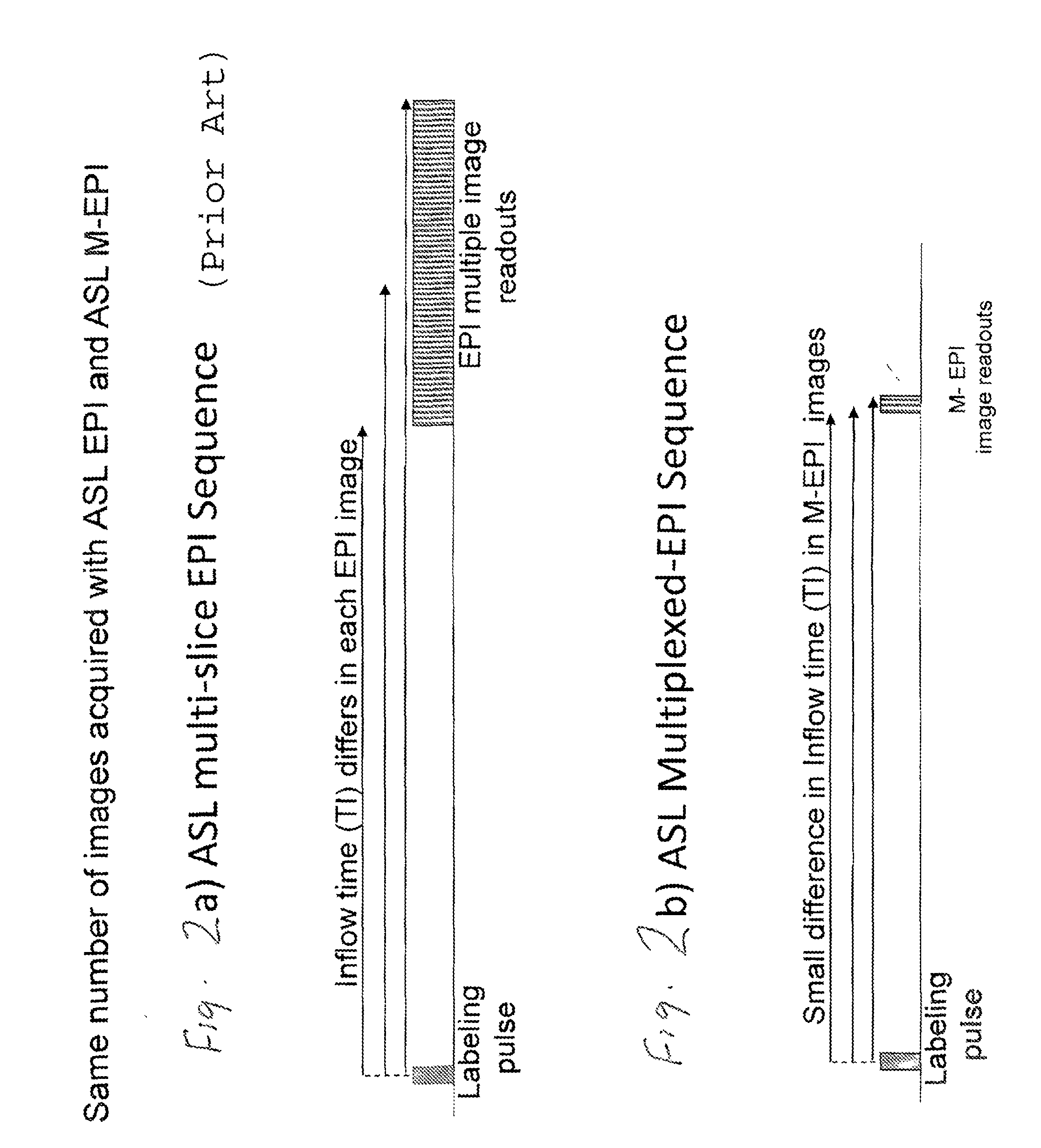
[0015]FIGS. 2a and 2b illustrate important differences between ASL multi-slice EPI imaging and ASL multiplexed EPI imaging. The example of FIG. 2a illustrates that MR signals for multiple 2D slice images are obtained in time sequence over a time span that is much longer than that seen in FIG. 2b, where MR signals for the same number of multiple 2D slice images are obtained using M-EPI essentially simultaneously.

[0016]FIG. 3 illustrates some important consequences of differences between ASL using conventional EPI and the new approach of using ASL with M-EPI. With the old approach, the acquisition time of MR signals for ...


PUM


Abstract

An MRI system and method for imaging perfusion in an arterial spin labeled (ASL) process in which multiplexed echo-planar imaging (M-EPI) is used rather than conventional EPI, to thereby speed up imaging and generate sets of images that show different phases of perfusion and provide additional benefits. A single multiband RF excitation pulse can be used to excite multiple slices for imaging, or a time sequence of multiband pulses can be used to further increase the number of slices.

Description

CROSS-REFERENCE TO RELATED APPLICATIONS AND INCORPORATION BY REFERENCE[0001]This patent application is a continuation-in-part of (a) PCT International Application No. PCT / US11 / 57161, filed Oct. 20, 2011, which claims the benefit of U.S. Provisional Application No. 61 / 394,929, filed Oct. 20, 2010, and (b) U.S. patent application Ser. No. 13 / 632,941, filed Oct. 1, 2012, which is a continuation of U.S. patent application Ser. No. 13 / 397,634, filed Feb. 15, 2012, which claims the benefit of U.S. Provisional Application Nos. 61 / 443,215, filed Feb. 15, 2011, 61 / 444,031, filed Feb. 17, 2011, and 61 / 444,039, filed Feb. 17, 2011. This patent specification incorporates by reference the entire contents of each of these applications, including their drawings and the appendices attached thereto.FIELD[0002]This patent specification is in the field of magnetic resonance imaging (MRI). More specifically it pertains to imaging tissue perfusion with MRI.BACKGROUND AND SUMMARY OF THE DISCLOSURE[0003]A...

Claims


Application Information

Patent Timeline
IPC IPC(8): A61B5/026
CPCA61B5/0263G01R33/543G01R33/56366G01R33/4835G01R33/5616A61B5/0042
Inventor FEINBERG, DAVID
Owner FEINBERG DAVID
Why Eureka
Try Eureka