Method of using near infrared fluorescent dyes for imaging and targeting cancers

a near infrared fluorescent dye and cancer technology, applied in the field of imaging methods for detecting tumor cells, can solve the problems of notoriety for heterogeneity, and achieve the effect of increasing the delivery of molecules

Inactive Publication Date: 2013-04-25
GEORGIA STATE UNIV RES FOUND INC
View PDF4 Cites 19 Cited by
  • Summary
  • Abstract
  • Description
  • Claims
  • Application Information

AI Technical Summary

Benefits of technology

[0030]Various embodiments of the present invention provide a method, comprising: providing a composition comprising a near-infrared (NIR) organic carbocyanine dye conjugated or complexed to a molecule; administering the composition comprising the NIR organic carbocyanine dye-molecule conjugate or complex to a subject; and (i) determining the pharmacokinetics and / or pharmacodynamics of the molecule in a cancer cell or tumor cell in the subject, (ii) imaging the subject to follow the movement of the molecule in the subject, or (iii) increasing the delivery of the molecule to a cancer cell or tumor cell in the subject.

Problems solved by technology

One limitation of these approaches is that the previous NIR moieties only detect specific cancer cell types with well-characterized surface properties, whereas tumors are notorious for their heterogeneity (14, 15).

Method used


Image

  • Method of using near infrared fluorescent dyes for imaging and targeting cancers
  • Method of using near infrared fluorescent dyes for imaging and targeting cancers
  • Method of using near infrared fluorescent dyes for imaging and targeting cancers

Examples


example 1

The Utility of IR-780 and IR-783 for In Vitro Imaging of Human and Mouse Cells: Molecular Mechanisms of Dye Uptake and Retention

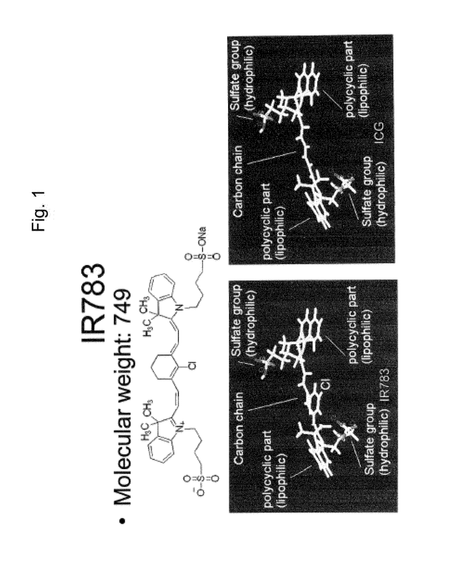
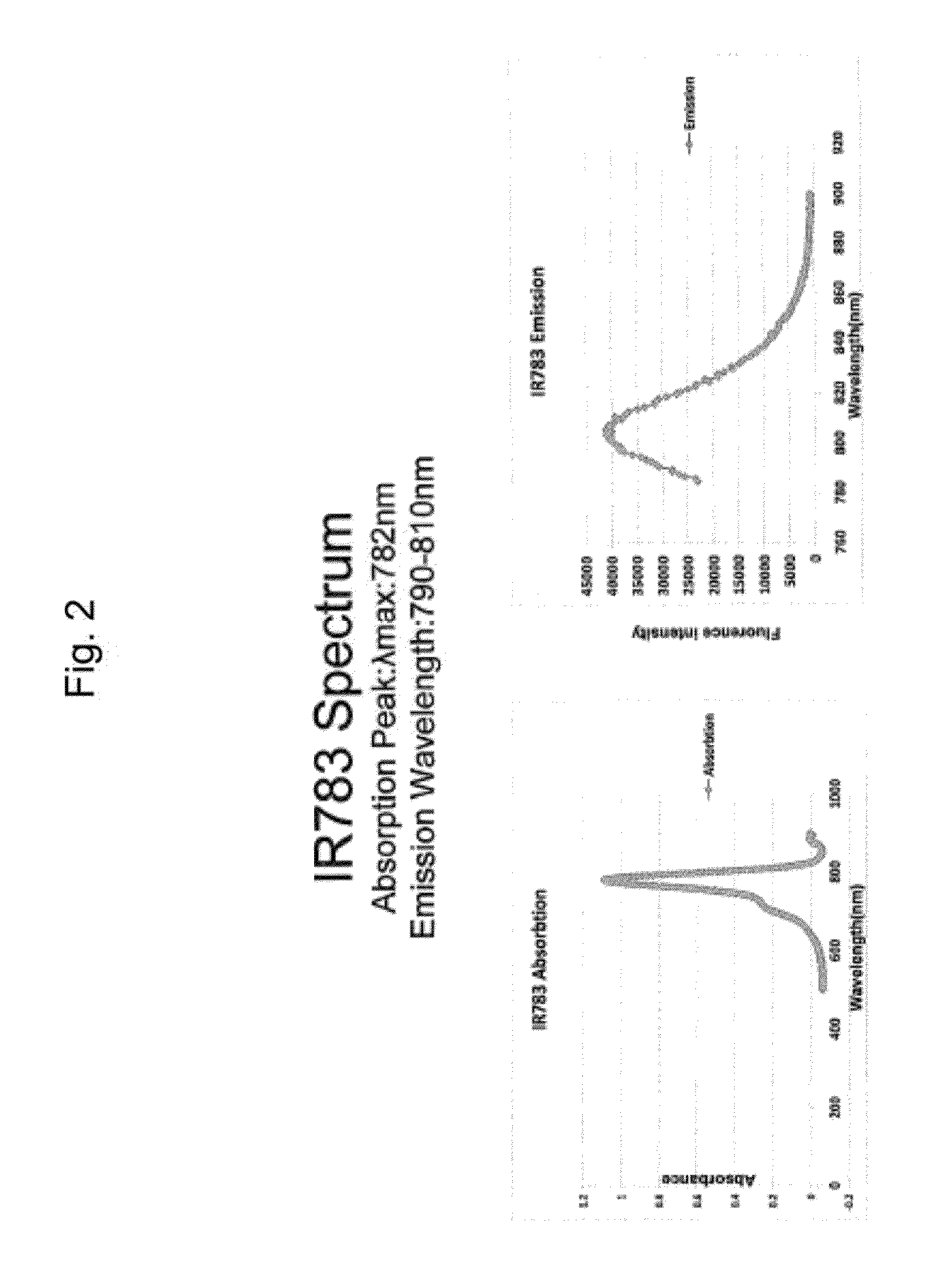
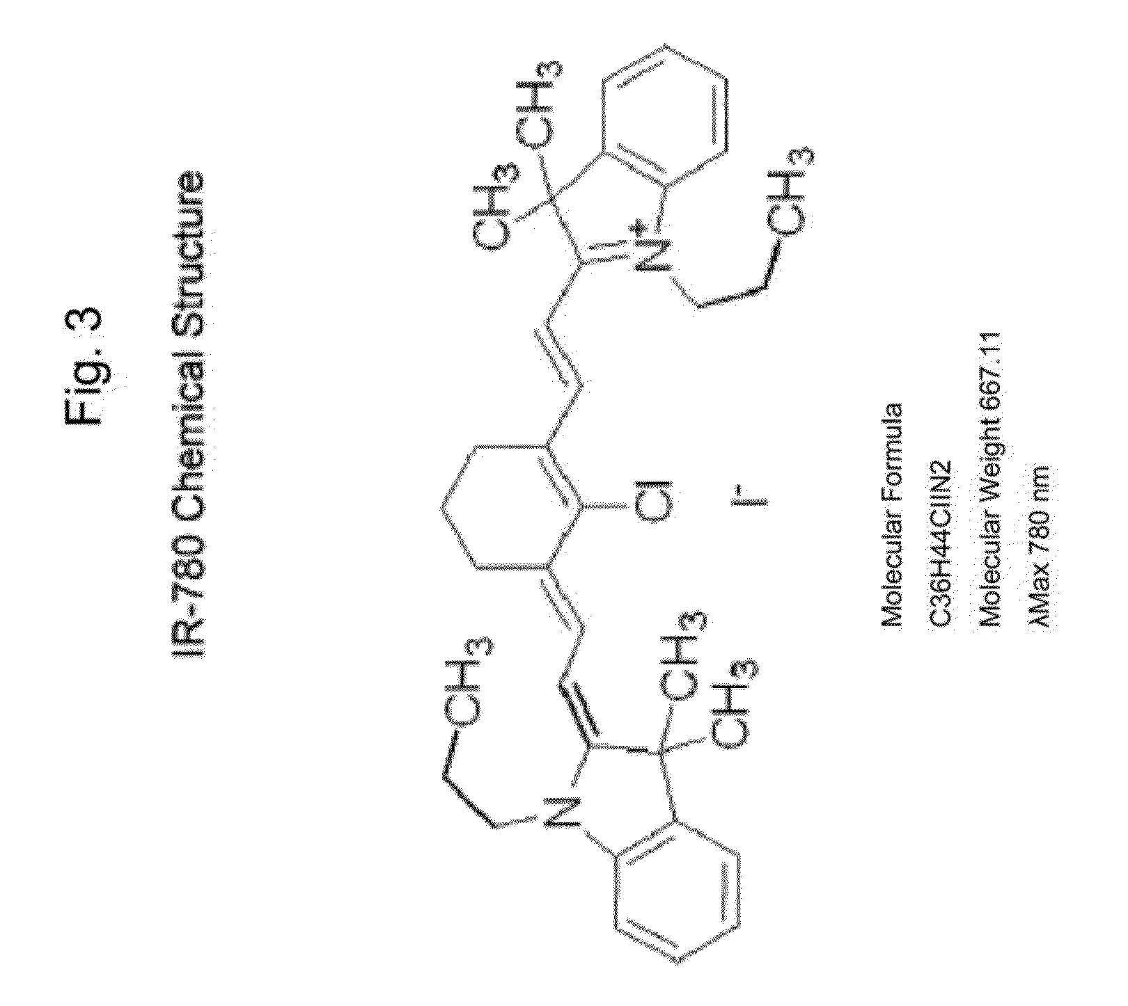
[0178]The inventors have evaluated extensively the NIR organic carbocyanine dyes, IR-780 and IR-783 in vitro in a wide range of cultured human and mouse cancer and normal cells. The chemical structures of these dyes and their light absorption and emission are shown FIGS. 1-3.

[0179]Cancer cells were cultured in T-Medium with 5% FBS at 37° C. incubator. When cells reached confluence of 80-90%, they were trypsinized and plated into 4-well chamber slides with the concentration of 5,000-8000 cells each. The cells were incubated overnight and ready to be used for dye uptake study. The NIR organic dye of interest was dissolved by DMSO at the concentration of 10 mM as the stock solution. The stock solutions were diluted with fresh T-Medium (5% FBS) to the concentration of 50 μM, cells were washed with PBS for 1 min, 2 times at room temperature, and then 800 μL of t...

example

The Utility of IR-780 and IR-783 for In Vivo Studies

[0181]To determine the dye uptake into normal mouse tissues or into tumor xenografts or tumors developed in situ in transgenic mice, NIR dye of interest was injected at a dose of 100 μL at a concentration of 100 μM through tail veins. After variable time of the dye injection, the mice were anesthetized and imaged by using the Kodak animal in vivo imaging system, which can obtain both the NIR signal and the X-ray picture and these images were merged. It was observed that there was no dye uptake into normal mouse tissues (FIG. 14). The uptake of either IR-780 or IR-783 was found in a broad spectrum of human xenograft implants in mice, either grown subcutaneously, orthotopically or intraosseously (FIGS. 15-22). IR-783 was also found to accumulate in prostate tumors and their metastases in a transgenic TRAMP mouse, a transgenic mouse strain overexpressed large and small T-antigen in the prostate gland under the control of a prostate-sp...

example 3

The Comparative Aspect of the Toxicities of IR-780 and IR-783 in Inbred Strain of Mice

[0182]The toxicities of IR-780 and IR-783 in mice was determined by the daily intraperitoneal injection of these dyes at 100× excess of the imaging concentration of these dyes in inbred strain of Balb / c mice. In control mice, the same volume of PBS was injected. The body weight and lethality of these mice were closely followed for 28 days. FIG. 29 showed that while IR-780 appears to be toxic to mice at this elevated dose, IR-783 was found to be completely safe when used at the 100× imaging dose with no lethality in treated mice and all animals gained weight during the period of monitoring with no statistical differences between IR-783-treated and PBS-treated mice. In contrast, IR-780 was found to be highly toxic and killed all mice at day 2 after the injection of this dye at 100× excess of the imaging dose of this dye; no toxicity was detected by injecting mice with indocyanine green (ICG) as evide...


PUM


Abstract

The present invention describes methods of identifying, detecting, imaging, isolating and locating cancer cells in a subject. The method invokes the use of near-infrared (NIR) organic carbocyanine dyes, particularly, near infrared heptamethine cyanine dyes and the detection of the fluorescence of these NIR dyes. The uptake of these dyes by cancer cells and not by normal cells, as well as their high intensity, among other things, allow for the detection of cancerous cells in a subject and facilitate their subsequent isolation. Further, detection of many tumor types and tumor cell populations under cell culture and in vivo conditions are described.

Description

FIELD OF INVENTION[0001]This invention relates to imaging methods for detecting tumor cells and imaging cells of interest.BACKGROUND[0002]All publications herein are incorporated by reference to the same extent as if each individual publication or patent application was specifically and individually indicated to be incorporated by reference. The following description includes information that may be useful in understanding the present invention. It is not an admission that any of the information provided herein is prior art or relevant to the presently claimed invention, or that any publication specifically or implicitly referenced is prior art.[0003]The future of personalized oncology and medicine relies on the improvement of imaging methods for detecting tumor cells and metastatic deposits earlier in patients, by providing a sensitive imaging probe for real-time support for surgeons, oncologists, pathologists and laboratory medicine personnel. They also require the development of ...

Claims


Application Information

Patent Timeline
Patent Type & Authority Applications(United States)
IPC IPC(8): C12Q1/04A61K49/00
CPCA61B5/0059A61B5/417A61B2503/40C12Q1/04G01N33/5091G01N33/574A61K49/006G01N21/6428C09B23/0016C09B23/0033C09B23/0066C09B23/086
Inventor YANG, XIAOJIANZHAU, HAIYEN E.WANG, RUOXIANGCHUNG, LELAND W.K.
Owner GEORGIA STATE UNIV RES FOUND INC
Why Eureka
Try Eureka