Hypermethylation Biomarkers for Detection of Cervical Cancer

a biomarker and hypermethylation technology, applied in the field of cervical cancer, can solve the problems of low single-test sensitivity, poor reproducibility of equivocal and minor abnormalities, and limited application prospects

Active Publication Date: 2013-05-02
THE JOHN HOPKINS UNIV SCHOOL OF MEDICINE
View PDF1 Cites 2 Cited by
  • Summary
  • Abstract
  • Description
  • Claims
  • Application Information

AI Technical Summary

Problems solved by technology

Although oncogenic HPV is necessary for the transformation of cervical-epithelial cells, it is not a sufficient cause, and a variety of cofactors and molecular events influence whether cervical cancer will develop[13].
While cytological screening and HPV infection tests has substantially reduced cervical cancer incidence and mortality where it has been successfully implemented, it is limited by low single-test sensitivity and poor reproducibility for equivocal and minor abnormalities [14, 15].

Method used


Image

  • Hypermethylation Biomarkers for Detection of Cervical Cancer
  • Hypermethylation Biomarkers for Detection of Cervical Cancer
  • Hypermethylation Biomarkers for Detection of Cervical Cancer

Examples


example 1

Methods

Materials and Methods

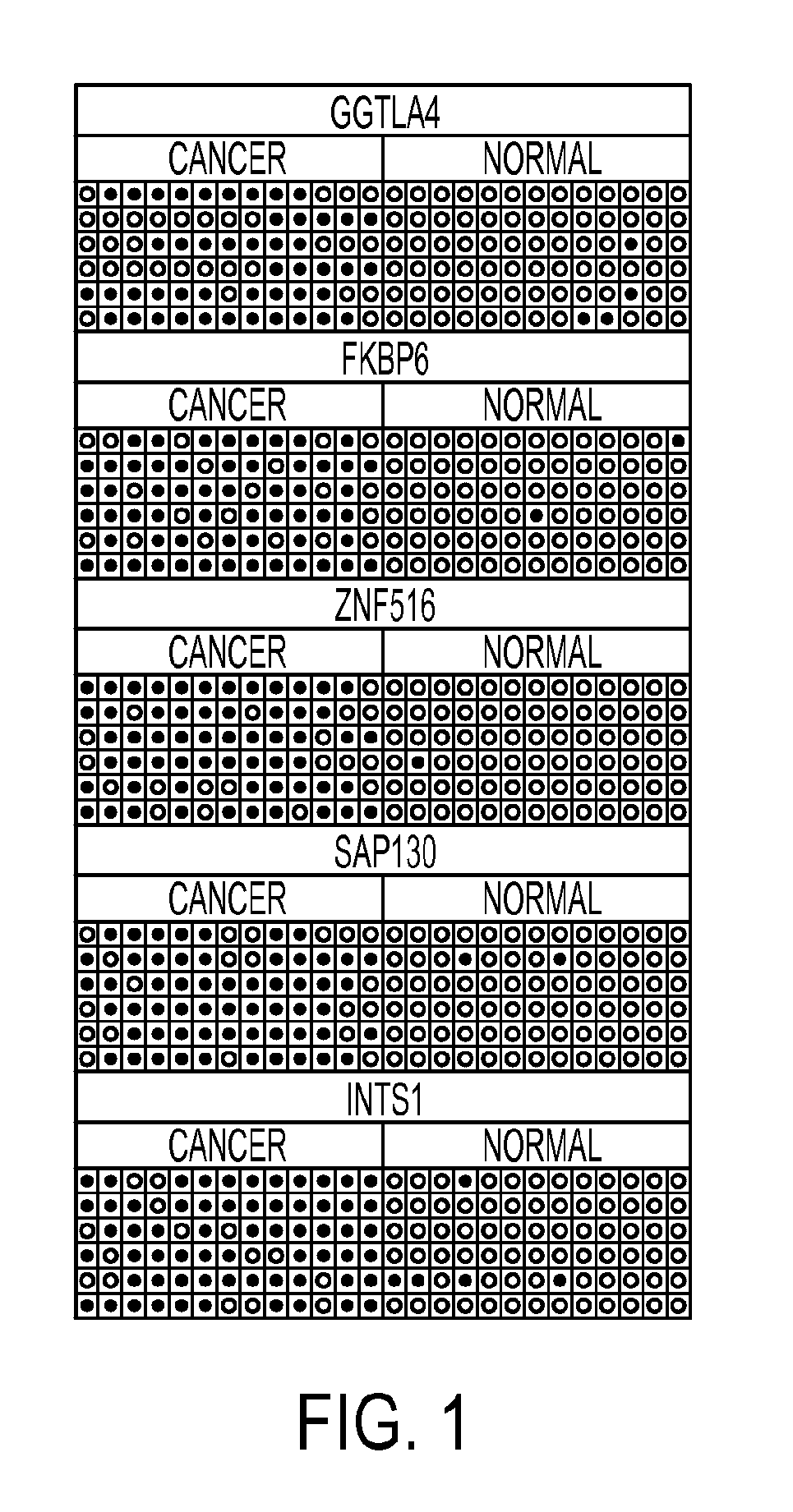
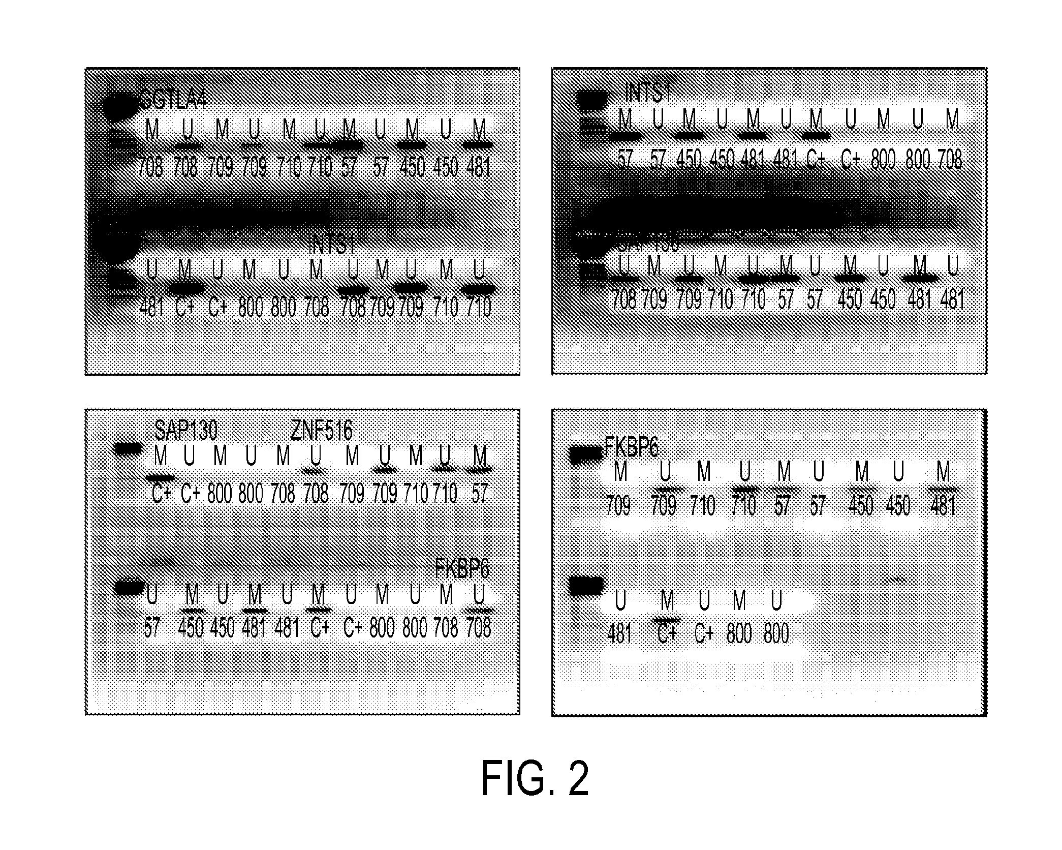
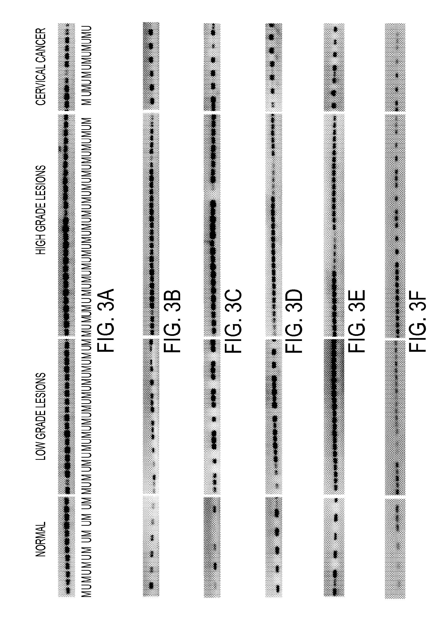
[0059]Clinical samples: The samples were collected at Doctor Hernán Henríquez Aravena (HHHA) tertiary care regional hospital in Temuco, Chile, between 2004-2008. The diagnosis was confirmed by histological examination (biopsy) during which a team of three pathologists from HHHA read the results and achieved a consensus diagnosis. A random set of pathology slides from the study samples was sent for diagnostic and immunohistochemistry confirmatory review by a pathologist at Johns Hopkins School of Medicine. The protocol for this study was approved by the Institutional Review Boards (IRB) of the Hernan Henriquez Aravena Hospital and the Johns Hopkins School of Medicine. To determinate the methylation status of promoter regions in the genome, 12 normal samples and 7 cervical cancers were used to perform the hybridization to an oligonucleotide tiling array (385K CpG Islands plus Promoter arrays, Nimblegen, Wis.). MSP validation was performed on 221 HPV genotyp...

example 2

Characteristics of the Patients

[0075]Two groups of patients were used in this study: One for the MSP validation (A) and other one for the IHQ analysis (B). Clinical and demographic variables were different in cases patients (LSIL, HSIL and CC) and controls. The age of the patients into three groups:50 years (A:22.6%, B:50.0%). Both groups of samples the lesion status was associated with age as expected. In the group A the most of low grade lesions (63.6%) were seen in patients 50 years old. In the group B normal patients were mostly seen (40.7%) among 31-50 year old patients, and 61.9% of invasive carcinoma patients were >50 years old. The ethnic descent of the patients was divided between native Chilean people (Mapuche) (A:22.2%, B: 28.7%) and Hispanic European (A:77.8%, B: 71.3%). The majority of the samples came from patients that were not Mapuches. The patients are all public assistance patients who receive different amounts of health care subsidy according to their income level...

example 3

HPV Genotyping.

[0076]PCR and Reverse line blot analyses revealed that 176 (79.6%) samples were HPV positive and 45 (20.4%) were HPV negative. Among normal samples 12% were HPV positive, most for single infection of HPV 16. Among LSIL 82% were HPV positive, most with single infections with HPV16 (41%) or HPV 11 (9%). Among the HPV positive LSIL lesions, 14% were multiple infections, most with HPV 16 and 18. Among the HSIL 95% were HPV positive, most of which (78%) were single infections with HPV 16 (50%) and HPV 18 (13%). Among tumor samples 99% were HPV positive, most of which (85%) were single infections with HPV16 (63%) and HPV 18 (10%).

[0077]Most of the patients (79.6%) were HPV positive. The largest number of HPV positive patients were in the 30-50 years old age category (51.7%), followed by >50 yrs old (24.4%) and <30 yrs old (23.9%). Most of the HPV positive patients were Non-Mapuches (80.1%) and revealed a socio-economic gradient similar to the study population: A Indigent (5...


PUM

PropertyMeasurementUnit
areaaaaaaaaaaa
methylation densityaaaaaaaaaa
heterogeneityaaaaaaaaaa

Abstract

Pap smears and HPV infection tests do not distinguish between lesions that will progress to an invasive carcinoma and those that will not. We aimed to identify epigenetic biomarkers for diagnosis and progression monitoring of premalignant lesions in cervical cancer. Hypermethylated genes were identified as potential biomarkers after validation by MSP, including GGTLA4 and ZNF516. The methylation frequency for these two genes was higher in tumor: GGTLA4 (100%) and ZNF516 (96%); than in normal samples: GGTLA4 (12%) and ZNF516 (16%). The methylation status of GGTLA4 showed a progression in methylation frequency from normal samples to invasive carcinoma. The immunohistochemical expression was lower in tumor for both: GGTLA4 (50.8%) and ZNF516 (66.2%); than in normal samples: GGTLA4 (71.2%) and ZNF516 (88.1%) (p<0.05). In conclusion, we identified methylation biomarkers for the molecular screening and characterization of cervical cancer.

Description

[0001]This invention was made using funds from the U.S. government. Under the terms of grant no. U01 CA84986 from the National Cancer Institute, the U.S. government retains certain rights in the invention.TECHNICAL FIELD OF THE INVENTION[0002]This invention is related to the area of cervical cancer. In particular, it relates to detection and characterization of cervical cancer.BACKGROUND OF THE INVENTION[0003]Cervical cancer (CC), the second most prevalent cancer in women and the fifth cause of death by cancer among women worldwide is a considerable public health problem[1]. Around 470,000 new cases of cervical cancer are detected annually, mostly in developing nations, of which approximately half will die [2].[0004]Cervical cancer is a cellular alteration that originates in the epithelium of the cervix and is initially apparent through slow and progressively evolving precursor lesions that can occur in various stages of cervical intraepithelial neoplasia (CIN), grouped into low and...

Claims


Application Information

Patent Timeline
Patent Type & Authority Applications(United States)
IPC IPC(8): C12Q1/68
CPCC12Q1/6869C12Q1/6886C12Q2600/154C12Q2600/156C12Q2600/106C12Q2523/125
Inventor GUERRERO-PRESTON, RAFAEL ENRIQUESIDRANSKY, DAVIDBREBI-MIEVILLE, PRISCILLA
Owner THE JOHN HOPKINS UNIV SCHOOL OF MEDICINE
Why Eureka
Try Eureka