

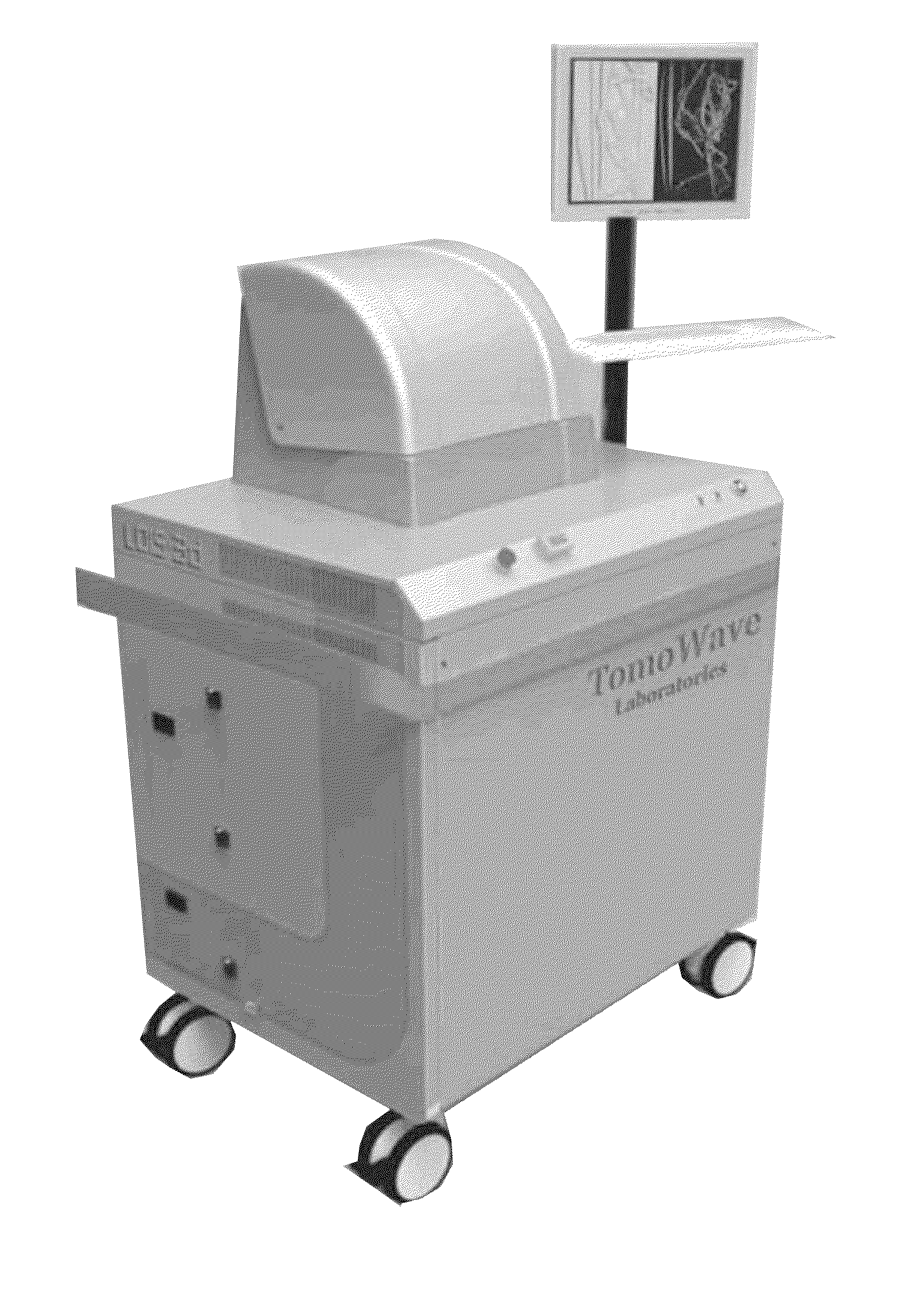
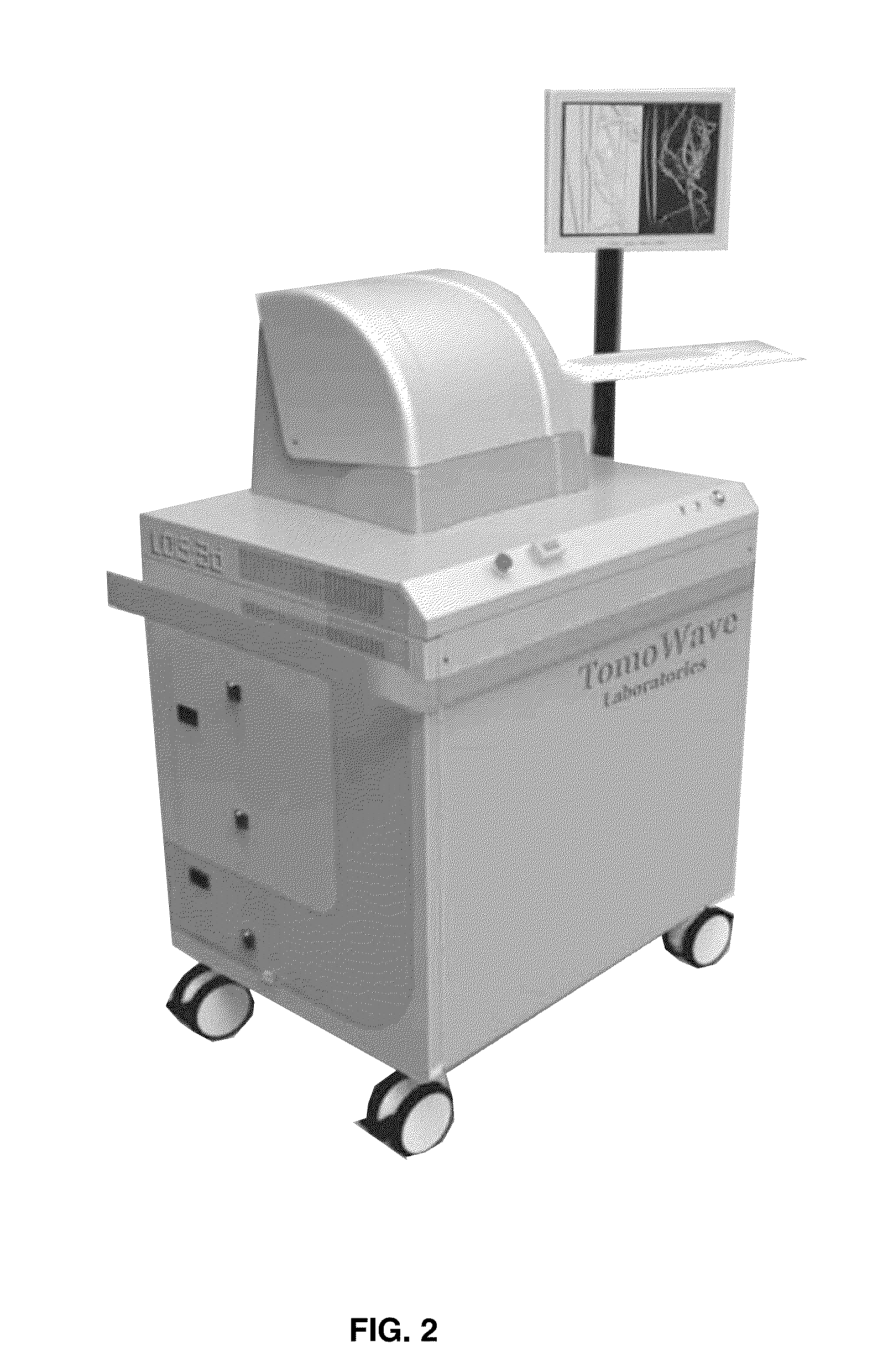
Laser Optoacoustic Ultrasonic Imaging System (LOUIS) and Methods of Use
an optoacoustic and ultrasonic imaging technology, applied in the field of biomedical imaging, can solve the problems of not being able to use biomedical imaging design, not three-dimensional tomography systems, and insufficient tissue comprehensive information in the prior art, so as to improve contrast, resolution and accuracy of quantitative information.
- Summary
- Abstract
- Description
- Claims
- Application Information
AI Technical Summary
Benefits of technology
Problems solved by technology
Method used
Image

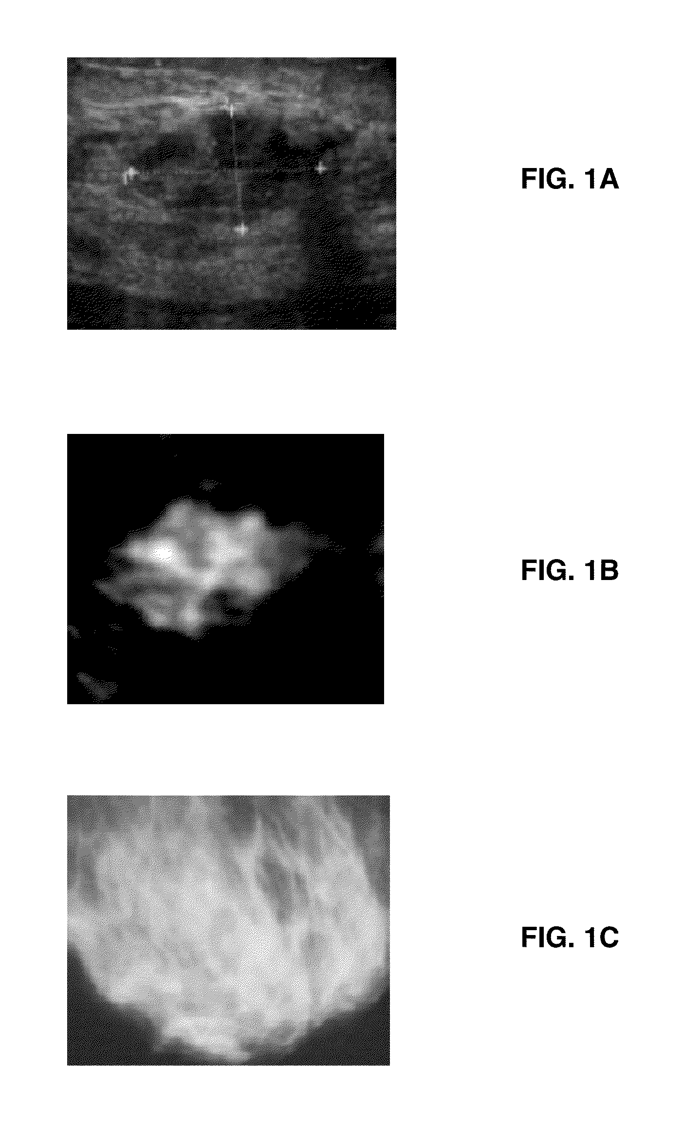
Examples
Embodiment Construction
[0041]As used herein in the specification, “a” or “an” may mean one or more. As used herein in the claim(s), when used in conjunction with the word “comprising”, the words “a” or “an” may mean one or more than one.
[0042]As used herein “another” or “other” may mean at least a second or more of the same or different claim element or components thereof. Similarly, the word “or” is intended to include “and” unless the context clearly indicates otherwise. “Comprise” means “include.”
[0043]As used herein, the term “about” refers to a numeric value, including, for example, whole numbers, fractions, and percentages, whether or not explicitly indicated. The term “about” generally refers to a range of numerical values (e.g., + / −5-10% of the recited value) that one of ordinary skill in the art would consider equivalent to the recited value (e.g., having the same function or result). In some instances, the term “about” may include numerical values that are rounded to the nearest significant figu...
PUM
 Abstract
Description
Claims
Application Information