Scanning confocal endoscope system

Inactive Publication Date: 2013-09-19
HOYA CORP
View PDF7 Cites 59 Cited by
  • Summary
  • Abstract
  • Description
  • Claims
  • Application Information

AI Technical Summary

Benefits of technology

The patent is about a technology that controls the distribution of light across a scanning area. It uses a point source control to ensure an even distribution of excitation light and a duty ratio control to ensure the light is evenly distributed during scanning. The technology also uses image signal detection to control the gain of the light. This results in a more uniform and accurate scan of the area, improving the quality and speed of the process.

Problems solved by technology

If fluorescence is discolored and thereby a shot image becomes blurred, detection of a diseased portion or an accurate judgment on a diseased portion by a doctor may be affected, which is undesirable.

Method used


Image

  • Scanning confocal endoscope system
  • Scanning confocal endoscope system
  • Scanning confocal endoscope system

Examples


Embodiment Construction

[0023]In the following, a scanning confocal endoscope system according to an embodiment of the invention is explained with reference to the accompanying drawings.

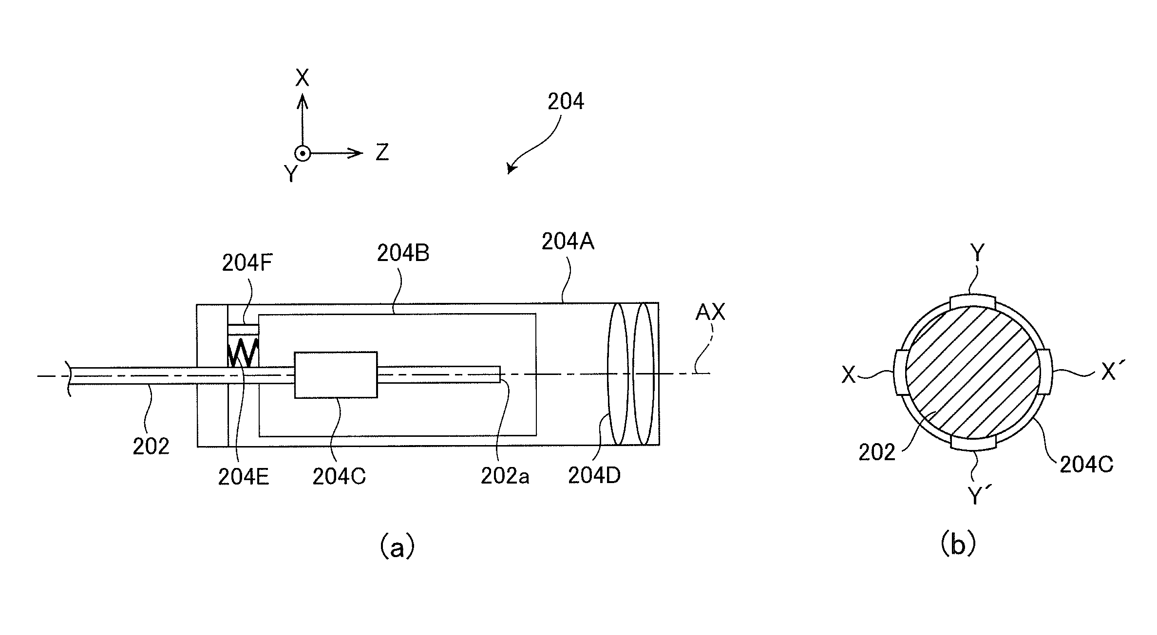
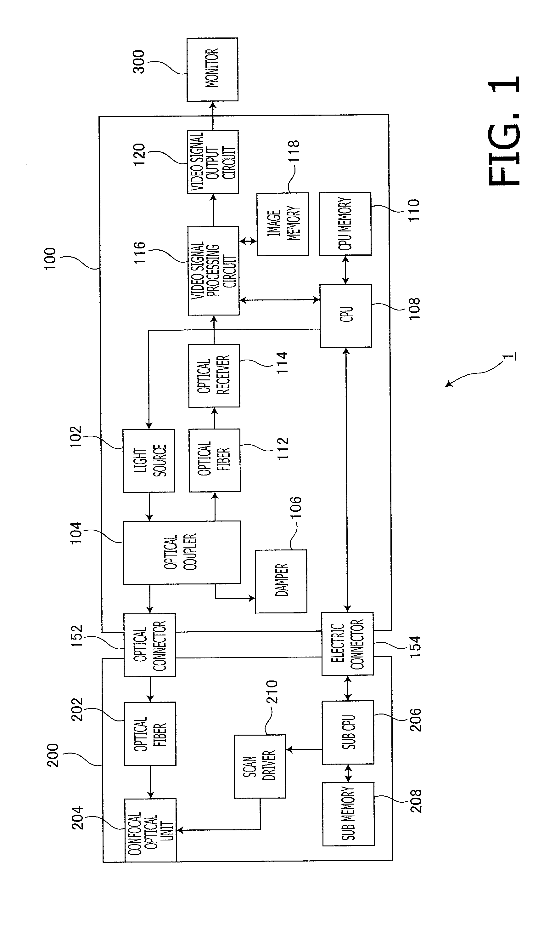
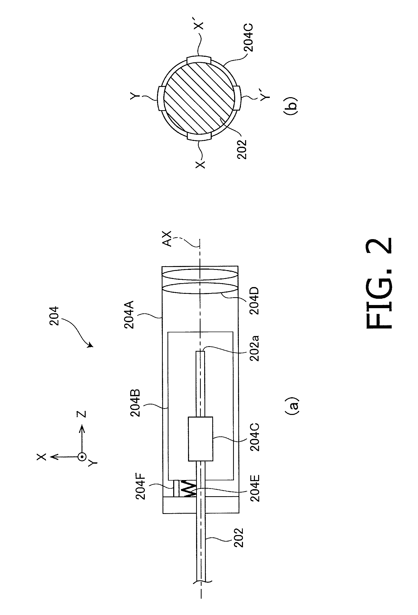
[0024]FIG. 1 is a block diagram illustrating a configuration of a scanning confocal endoscope system 1 according to the embodiment of the invention. The scanning confocal endoscope system 1 according to the embodiment of the invention is a system designed by making use of a fundamental principle of a confocal microscope, and is configured suitable for observing a subject at a high magnification and a high resolution. As shown in FIG. 1, the scanning confocal endoscope system 1 includes a system main body 100, a confocal probe 200 and a monitor 300. Confocal observation using the scanning confocal endoscope system 1 is performed in a state where a tip face of the tube-like confocal probe 200 having flexibility is operated to contact a subject.

[0025]The system main body 100 includes a light source 102, an optical coupler 104,...


PUM


Abstract

A scanning confocal endoscope system is configured by: a point source that scans on a subject with excitation light by periodically moving in a two-dimensional plane; a point source control means that controls the point source so that irradiation density of the excitation light becomes smaller than or equal to predetermined density over a whole scanning area; a confocal pinhole arranged at a position conjugate with a converging point of the excitation light; an image signal detection means that detects an image signal by receiving fluorescence emitted from the subject being excited by the excitation light via the confocal pinhole; and an image generation means that generates a confocal image using the detected image signal.

Description

TECHNICAL FIELD[0001]The present invention relates to a scanning confocal endoscope system which detects and images only light, obtained through a pinhole arranged at a position conjugate with a focal point of a confocal optical system, of fluorescence emitted from a subject excited by excitation light.BACKGROUND ART[0002]A scanning confocal endoscope system for observing a living tissue in a body cavity is known. The scanning confocal endoscope system scans, with excitation light, on a subject into which medicine is given. The scanning confocal endoscope system detects, with a photodetector, only a component, obtained through a pinhole arranged at a position conjugate with a focal point of a confocal optical system, of light emitted from a subject being scanned. The scanning confocal endoscope system generates an image with a high magnification and a high resolution relative to an image observed through a normal electronic scope or a normal fiber scope, based on a signal generated ...

Claims


Application Information

Patent Timeline
IPC IPC(8): A61B1/00
CPCA61B1/00009A61B1/00172A61B1/043G02B23/2476A61B5/0084G02B21/0024A61B5/0068
Inventor KOBAYASHI, SHOTARO
Owner HOYA CORP
Why Eureka
Try Eureka