Gelc-ms using stain free technology

a gelcms and stain-free technology, applied in the field of proteomics, can solve the problems of complex mass spectra, insufficient purity of biological samples containing diverse populations of proteins, and inability to submit directly to mass spectroscopy

Active Publication Date: 2013-11-21
BIO RAD LAB INC
View PDF1 Cites 4 Cited by
  • Summary
  • Abstract
  • Description
  • Claims
  • Application Information

AI Technical Summary

Benefits of technology

[0023]Further provided herein is a method of identifying one or more protein in a sample. The method includes performing GeLC-MS using a stain-free gel, wherein one or more amino acid in the protein is modified by the stain-free gel. In some embodiments, the method includes: electrophoretically separating the sample into a lane of the stain-free gel; isolating the lane, or portion thereof, from the gel; digesting the protein in the lane or portion thereof; and performing mass spectrometry (MS) on the digested protein. In one such embodiment, the performing comprises correlating MS data of the digested protein to a database of protein mass and charges.

Problems solved by technology

Crude biological samples that contain diverse populations of proteins are often not pure enough to submit directly to mass spectroscopy.
Furthermore, proteins cannot be easily identified by mass spectroscopy if too many different proteins are passed through the mass spectrometer at once.
This can lead to highly complex mass spectra where individual peaks, and in turn, individual protein sequences cannot be easily resolved.
Protein stains allow robust and sensitive detection, but hinder the rapid processing of biological samples for proteomic mass spectroscopy.
The process of applying the stain to the gel (staining) prior to detection can take hours.
Similarly time consuming is the removal of the stain after detection (destaining), which can be necessary in order to obtain accurate masses of proteins and protein fragments.
During these processes, some proteins (particularly low-molecular-weight and hydrophilic proteins) can diffuse out of the gel and into the buffer, thereby becoming lost to subsequent analysis.

Method used


Image

  • Gelc-ms using stain free technology
  • Gelc-ms using stain free technology
  • Gelc-ms using stain free technology

Examples


example

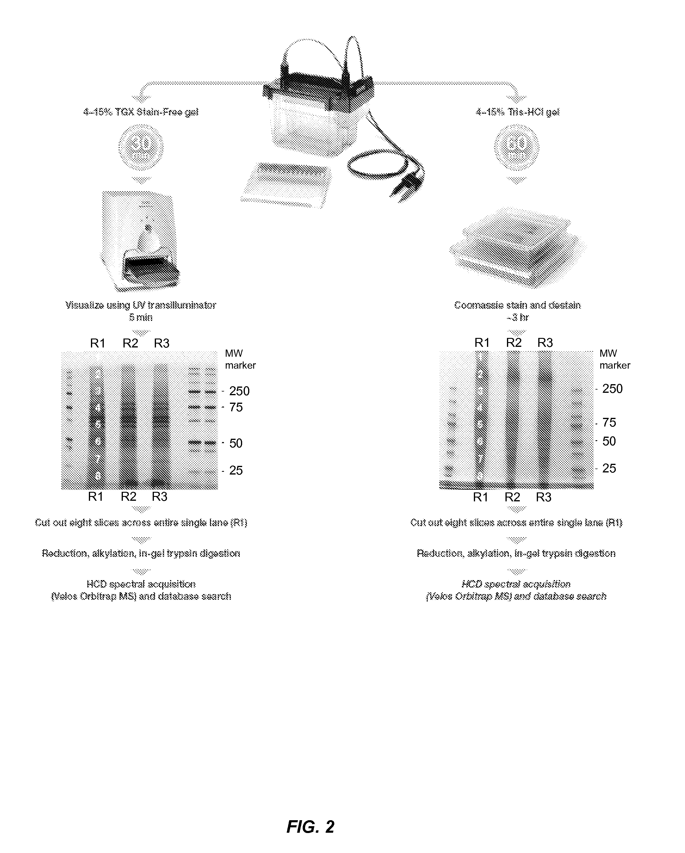
[0068]Protein Electrophoresis and Collection of Individual Gel Slices.

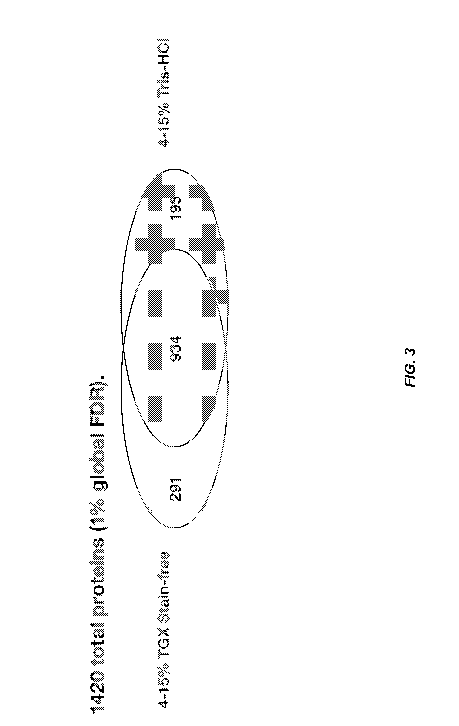
[0069]50 μg protein from 3T3 whole cell lysates was electrophoresed on 4-15% Criterion Tris-HCl or Criterion TGX Stain-Free gels in triplicate. After electrophoresis, Tris-HCl gels were visualized by Coomassie staining and destaining (−3 hr). After destaining was completed, eight slices covering one single entire lane were cut and processed for in-gel trypsin digestion and post-digestion sample processing. In the case of samples run on TGX Stain-Free gels, gels were visualized using a Gel Doc EZ imager with 5 min UV activation and eight slices covering an entire single lane cut out identically as above for in-gel digestion and post-digestion peptide purification. Each gel slice was assumed to contain ˜6 μg protein. See FIG. 2.

[0070]In-Gel Digestion (Adapted from Shevchenko et al. 1996).

[0071]Gel slices were initially washed with water, twice with 1:1 100 mM NH4HCO3 / acetonitrile for 15 min each, and then washed onc...


PUM

PropertyMeasurementUnit
wavelengthaaaaaaaaaa
temperatureaaaaaaaaaa
wavelengthsaaaaaaaaaa

Abstract

Disclosed herein is a method of preparing a protein sample for mass spectroscopy. The method includes separating proteins of the sample on an electrophoresis gel; contacting the proteins with a halo-substituted organic compound; exposing the gel to UV light; detecting fluorescence emitted from the electrophoresis gel; excising at least one portion of the electrophoresis gel based upon the detected fluorescence, wherein said at least one portion contains proteins of the protein sample; and subjecting proteins from the at least one portion to mass spectroscopy. Using this method, more proteins can be identified by GeLC-MS than when the electrophoresis gel is treated with a protein stain or subjected to the gel handling steps accompanying such treatment.

Description

CROSS-REFERENCES TO RELATED APPLICATIONS[0001]The present application claims priority from U.S. Provisional Application No. 61 / 648,875, entitled “GeLC-MS Using Stain-Free Technology” and filed May 18, 2012, the entire contents of which are herein incorporated by reference for all purposes.BACKGROUND OF THE INVENTION[0002]The growing field of proteomics is concerned with identifying large numbers of proteins in living organisms, understanding the functions and interactions of these proteins, and characterizing how the repertoire of proteins in an organism is modulated by factors such as developmental stage, disease state, and environment. Proteomics also aims to address how the set of proteins needed to sustain life varies among individual cells, cell types, tissues, individual organisms, groups of organisms, species, and groups of species. An important tool in experimental proteomics is mass spectroscopy, which allows identification of many proteins from complex biological samples. ...

Claims


Application Information

Patent Timeline
Patent Type & Authority Applications(United States)
IPC IPC(8): G01N27/447G01N30/06H01J49/00
CPCG01N27/44721H01J49/0036G01N30/06G01N33/6848H01J49/26
Inventor BELISLE, CHRISTOPHER
Owner BIO RAD LAB INC
Why Eureka
Try Eureka