Plant derived cell culture material

a cell culture material and plant technology, applied in the field of plant derived cell culture and cell delivery compositions, can solve the problems that the 3d cell culture biomaterials do not allow the transfer of hydrogel matrix with a needle without serious damage to the cultured cells

Active Publication Date: 2013-12-26
UPM-KYMMENE OYJ
View PDF4 Cites 17 Cited by
  • Summary
  • Abstract
  • Description
  • Claims
  • Application Information

AI Technical Summary

Benefits of technology

[0016]A significant advantage of the present invention is that cells can be maintained (and proliferated) on or in the biomaterial media without animal or human based chemicals originating outside the cells. The cells are evenly dispersed on or in the media / matrix containing cellulose nanofibers or a derivative thereof. Cells divide on or in the media, start to proliferate and the cell clusters start to grow spontaneously without the accumulation of cells on the bottom of the cell culture platform. The homogenous dividing of the cells in the cellulose nanofibers or a derivative thereof is a prerequisite for the biomaterial to function as 3D cell culture media.
[0017]Further advantages of the present invention include: cellulose nanofibers and / or derivatives thereof are inert and give no fluorescent background. The media comprising cellulose nanofibers or a derivative thereof can be injected. Injectability is explained by the rheological properties. The injection can be performed so that the cells stay stable inside matrix and they are homogeneously distributed in the matrix after injection.

Problems solved by technology

Existing 3D cell culture biomaterials do not allow transferring the hydrogel matrix with a needle without seriously damaging the cultured cells.

Method used


Image

  • Plant derived cell culture material
  • Plant derived cell culture material
  • Plant derived cell culture material

Examples


example 1

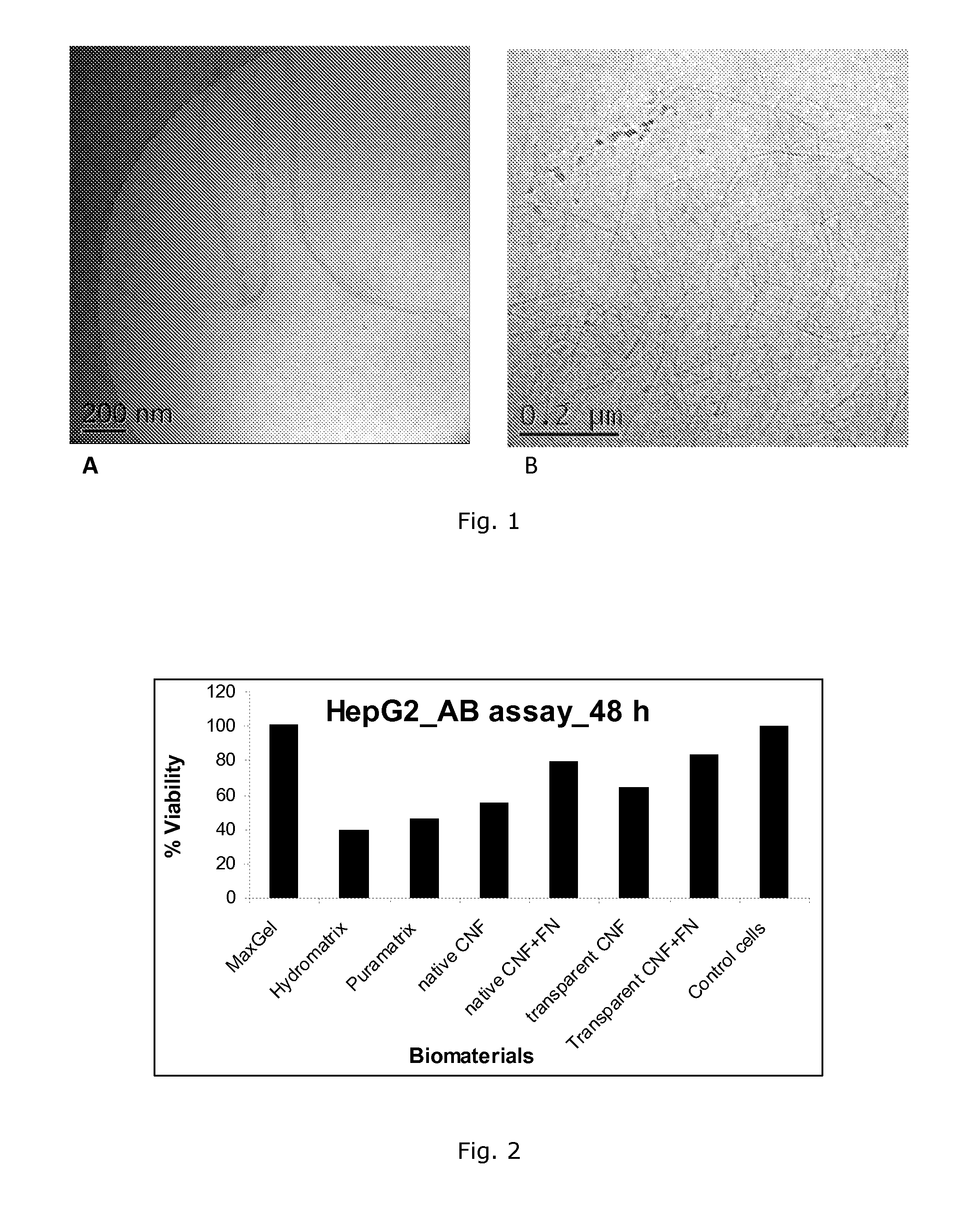
Comparison Of Cell Viability Of HepG2 Cells In Different Cell Culture Materials

[0107]Cellulose nanofiber hydrogels were placed in the bottom of a 96 well tissue culture plate and HepG2 cell suspension in maintenance growth media containing 25,000-50,000 cells per well were seeded either on top of the hydrogel or mixed with it. Hydrogel concentration ranges from 1-0.01%. The fluorescence intensity which indicates the cell viability and proliferation was measured as a function of days after culturing the cells on the cellulose nanofiber hydrogel in an incubator at 37 C in 5% CO2 and 95% relative humidity.

[0108]Three commercially available cell culture materials were used as reference 3D culture materials: MaxGel™ (Sigma-Aldrich), HydroMatrix™ (Sigma-Aldrich) and PuraMatrix™ (3DM Inc.). The experimental setup was identical for all the studied materials.

[0109]Viability of HepG2 cells was quantified by AlamarBlue™ Cell Viability Assay Kit (Biotium Inc., Hayward, Calif., USA) as presented...

example 2

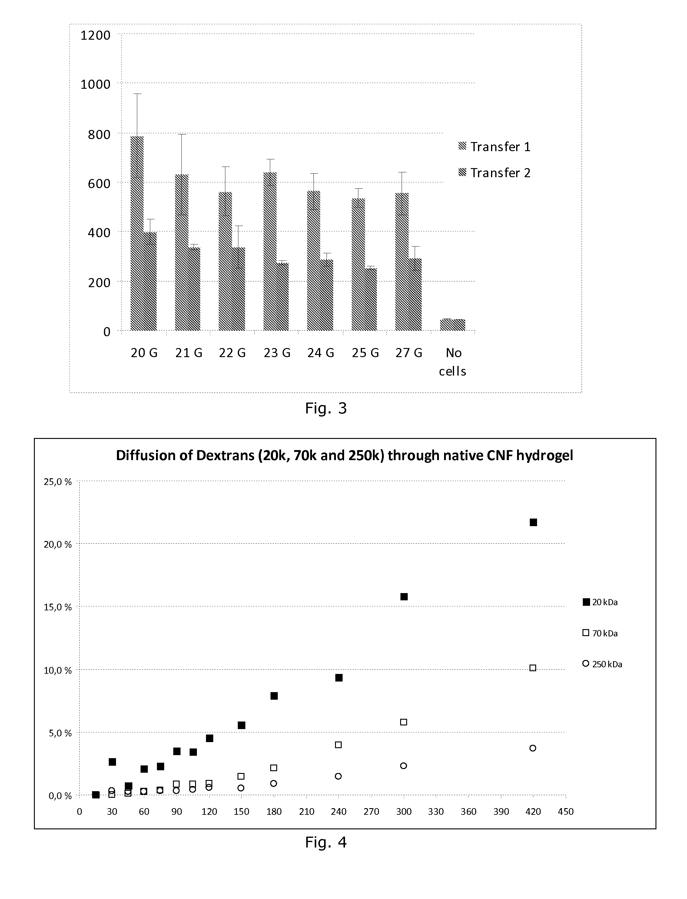
Transferring Of ARPE-19 Cells With A Syringe Needle

[0111]ARPE-19 cells (25 000 cells per well) were seeded and cultured in CNF matrix on the bottom of the 96 well plate. Viability of ARPE-19 cells after transferring the cells with a syringe needle of different sizes is presented in FIG. 3. The same phenomenon can be obtained also with other cell types like HepG2 and ES cells.

[0112]More detailed explanation of the Transferring of ARPE-19 cells with a syringe needle are follows: At the Transfer 1 in FIG. 3 the cells were incubated for 48 hours with 1.66% CNF, and after that the cells were transferred with a syringe (20G-27G) about 100 μl into a 96-well plate. After the transfer with the syringe the cells were incubated for 24 hours, and the viability of the cells in CNF was measured.

[0113]At the Transfer 2, the cells incubated for 24 hours in 1.66% CNF were transferred with a 27G syringe (about 2 ml) into a fresh medium. The cells were incubated for 24 hours after the transfer, after ...

example 3

Stem Cells

Live / Dead Staining Of hES Cell-Derived Hepatic Progenitor Cells

[0115]Human ES cell-derived hepatic progenitor cells were imbedded in native CNF hydrogel (FIG. 15) and cultured for 7 days with and without collagen IV. No background was detected. Human ES cell-derived hepatic progenitor cells were imbedded in transparent CNF hydrogel and cultured for 7 days with and without collagen IV. No background was detected, which makes this material extremely easy to use in this context. Usually other materials used e.g. MatriGel and MaxGel have a significant fluorescent background, and therefore it is difficult to work with those matrices. The ES cells are possible to keep in CNF hydrogel, they survive and thus this material is able to keep them alive. In addition, ES cells form also 3D structure, which has not been observed earlier with any other material.


PUM

PropertyMeasurementUnit
diameteraaaaaaaaaa
diameteraaaaaaaaaa
diameteraaaaaaaaaa

Abstract

The present invention relates material that is useful in culturing and transferring cells as well as delivering cells. The material comprises plant derived cellulose nanofibers or derivatives thereof, wherein the cellulose nanofibers are in a form of a hydrogel or membrane. The invention also provides methods for producing these materials and compositions and uses thereof.

Description

FIELD OF THE INVENTION[0001]The invention relates to plant derived cell culture and cell delivery compositions comprising cellulose nanofibers and / or derivatives thereof.BACKGROUND OF THE INVENTION[0002]Health care remains at the foremost frontiers for scientific research. The need to discover and develop cost-effective and safer medications is ever increasing. The ability to accurately model the cellular organization within a particular tissue or organ is of paramount importance. A close replica of the in vivo system to in vitro would require cell growth in three dimensions (3D). The “cross-talk” achieved between the cells in a 3D cell culture in vitro is a close mimic of cell growth under physiological conditions. Indeed, 3D cell culture have assumed significance in efforts directed towards regenerative medicine, better understanding of chronic diseases and providing superior in vitro model system for screening drugs and toxicological assays. Its emergence is thus being aptly tout...

Claims


Application Information

Patent Timeline
Patent Type & Authority Applications(United States)
IPC IPC(8): C12N5/0735C12N5/079C12N5/071
CPCC12N5/0606C12N5/067C12N5/0621A61K9/06A61K47/38A61L27/20A61L27/3637A61L27/38A61L27/52C08J3/075C08L1/02C12N5/0068C12N11/04C12N11/12C12N2533/78C08L97/02C08J2389/00C08J2301/02C12N5/0062C12N1/22B82Y5/00A61K47/26C12N5/0671C12N5/0672
Inventor YLIPERTTULA, MARJOLAUREN, PATRICKBHATTACHARYA, MADHUSHREELOU, YANRULAUKKANEN, ANTTI
Owner UPM-KYMMENE OYJ
Why Eureka
Try Eureka