Tissue adhesive substrates
a technology of tissue adhesive and substrate, which is applied in the field of tissue adhesive substrate, can solve the problems of poor tissue, inability to produce high-quality array tomography data, and inability to meet the requirements of the array tomography data,
- Summary
- Abstract
- Description
- Claims
- Application Information
AI Technical Summary
Benefits of technology
Problems solved by technology
Method used
Image
Examples
example 1
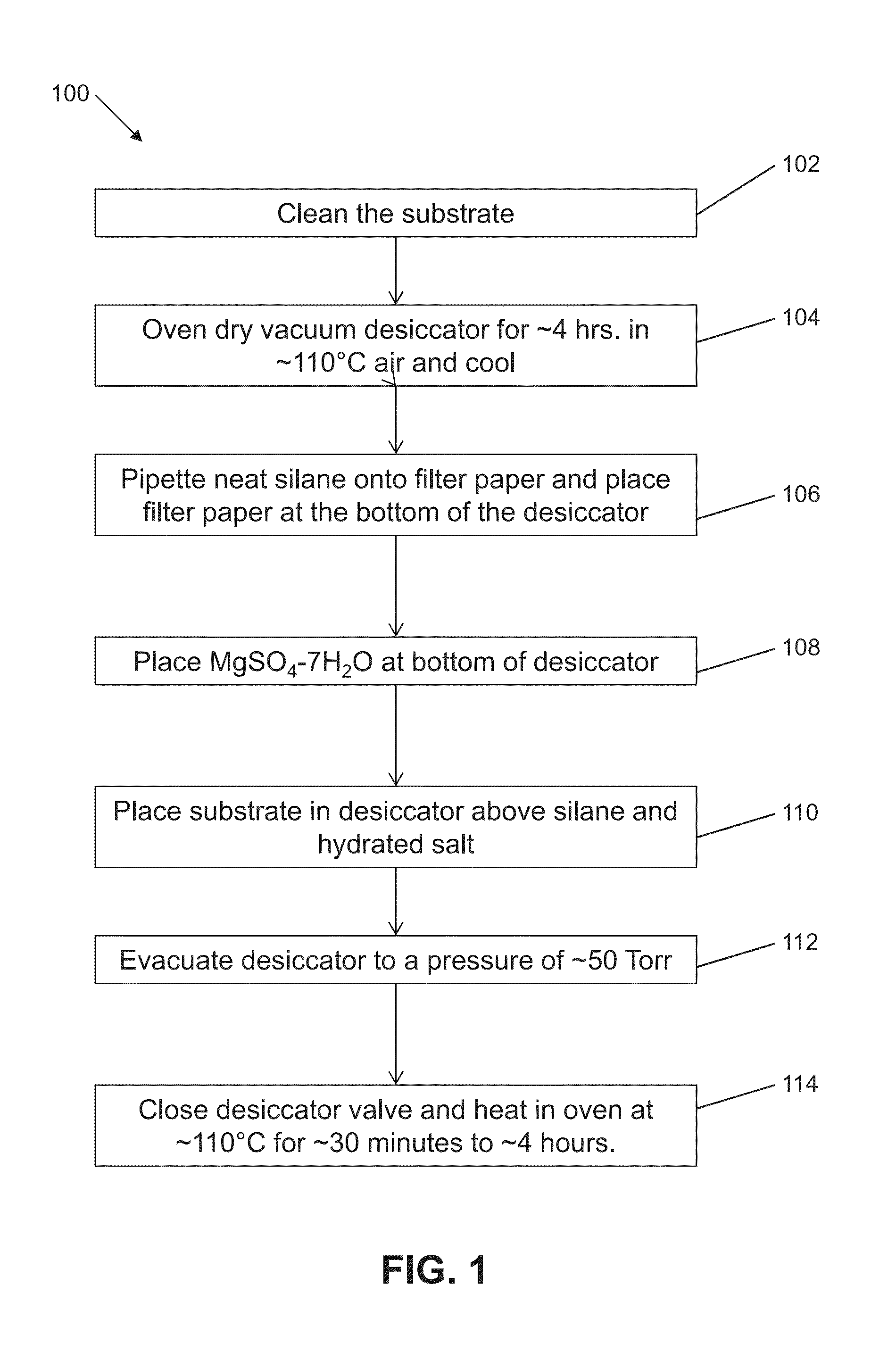
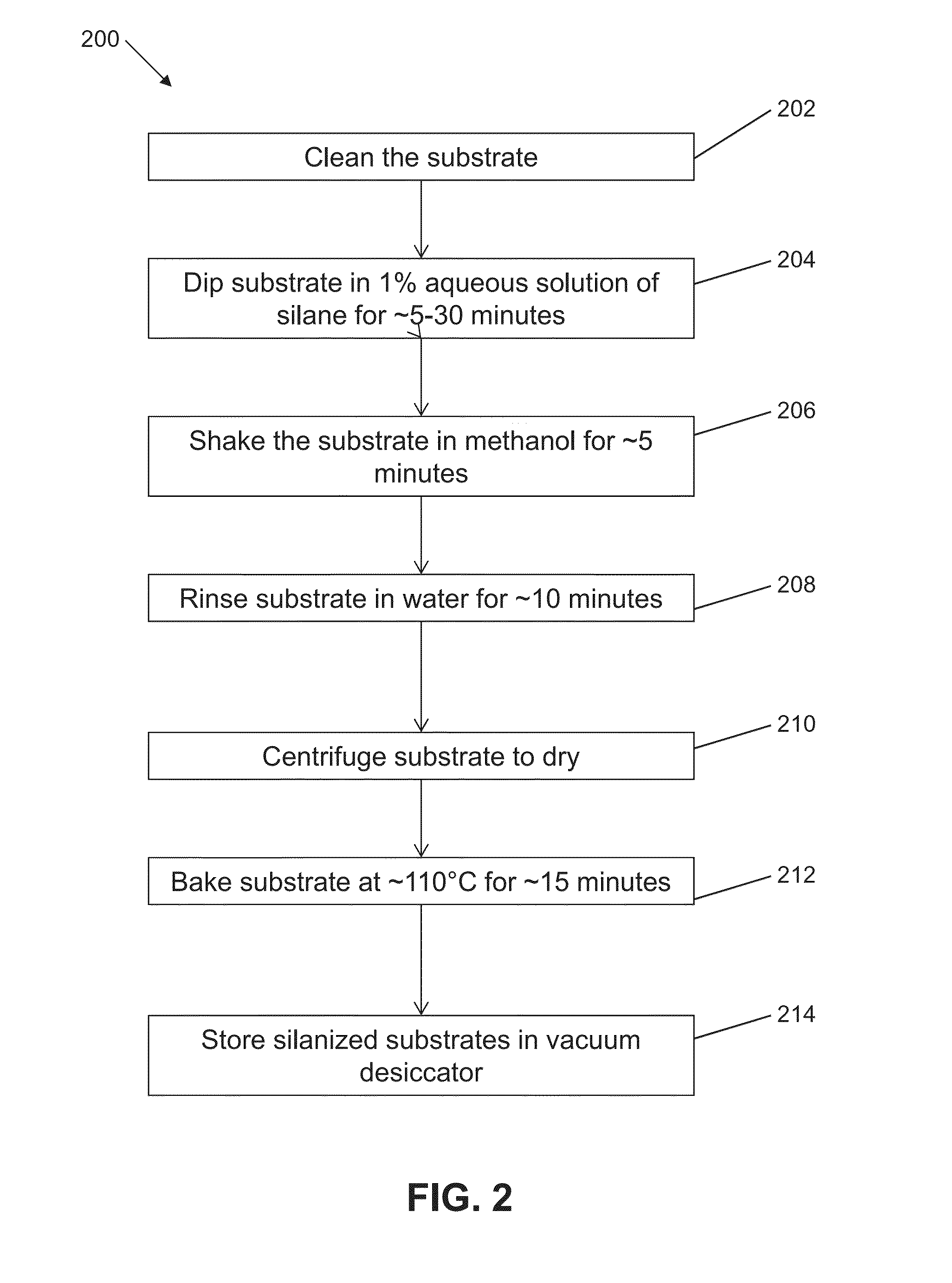
[0069]Embedded tissue sections were adhered to subbed coverslips that were prepared according to Micheva, K D, O'Rourke, N, Busse, B, Smith, S J, (2010). Array tomography: Production of arrays. Cold Spring Harb Protoc doi:10.1101 / pdb.prot5524 (Treatment 1). Embedded tissue sections were also adhered to commercially-available silanized Schott Nexterion A+ glass substrates (Treatment 2). Embedded tissue sections were also adhered to glass coverslips treated with aminopropyltrimethoxysilane, which was deposited by vapor deposition for 4 hours (Treatment 3).
[0070]Tissue sections adhered to Treatment 1 substrates and Treatment 2 substrates began to show damage after 4 elution cycles (80 minutes total in elution solution). Tissue sections adhered to Treatment 3 substrates repeatedly showed no damage after 12 elution cycles (240 minutes total in elution buffer). These results are shown in FIG. 6. FIGS. 7A to 7I depict phase-contrast micrographs of tissue sections on different substrates af...
example 2
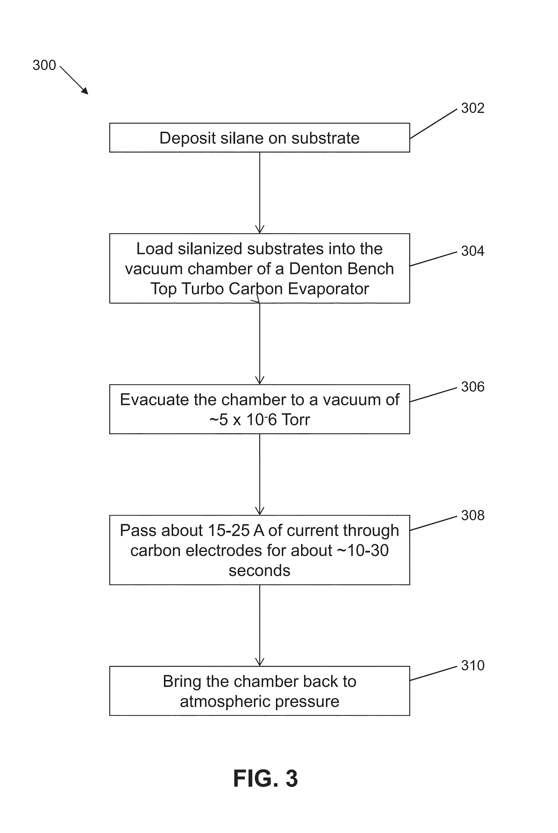
[0071]We compared tissue section retention on carbon-coated silanized coverslips to carbon-coated subbed and carbon-coated commercially silanized glass. Embedded tissue sections were adhered to the various carbon-coated substrates and treated with various numbers of elution cycles, as tabulated in FIG. 8. Phase-contrast micrographs for tissue sections adhered to these substrates after repeated elution cycles are depicted in FIGS. 9A to 9J. Phase-contrast micrographs of tissue sections on surface treatments 4 through 13 before repeated elution cycles are depicted in Panel-a of FIGS. 9A to 9J, and micrographs of the tissue sections after repeated elution cycles are depicted in Panel-b of FIGS. 9A to 9J. Enlargements of a region annotated in Panels-b are shown in Panels-c for each treatment. Tissue sections adhered to carbon-coated subbed substrates (Treatment 4) began to show damage after 8 elution cycles (160 minutes total in elution solution), while tissue sections mounted on carbon...
example 3
[0072]An expanded set of silanes were tested in an additional experiment. The table depicted in FIG. 10 lists the different tissue adhesive substrates to which embedded tissue sections were mounted, and the condition of the tissue section and / or substrate after repeated elution cycles.
[0073]A variety of silanes, having hydroxyl-methoxy- or ethoxy-reactive groups and a variety of terminal groups, including charged, polar, aryl and alkyl moieties, when deposited with the vapor procedure described above onto substrates that have been cleaned per the protocol, act to adhere a vapor-deposited carbon layer to a glass or silica substrate. This may work to allow embedded tissue to be stably adhered to the substrate against the equivalent of >100 cycles of antibody elution.
[0074]Adhesion of embedded tissue directly to a silanized surface through multiple (e.g., about 20) rounds of the elution procedure was observed only for amino-terminated compounds. Strikingly, the substrate prepared per t...
PUM
 Abstract
Description
Claims
Application Information
 - R&D
- Intellectual Property
- Life Sciences
- Materials
- Tech Scout
- Unparalleled Data Quality
- Higher Quality Content
- 60% Fewer Hallucinations
Browse by: Latest US Patents, China's latest patents, Technical Efficacy Thesaurus, Application Domain, Technology Topic, Popular Technical Reports.
© 2025 PatSnap. All rights reserved.Legal|Privacy policy|Modern Slavery Act Transparency Statement|Sitemap|About US| Contact US: help@patsnap.com


