Iteration of optical time reversal by ultrasonic encoding in biological tissue

a biological tissue and ultrasonic encoding technology, applied in the field of time-reversed ultrasonic encoding optical focusing, can solve the problems of limited application of light imaging techniques such as optical imaging within biological tissues and other heterogeneous media, and current optical imaging techniques, such as optical coherence tomography, are limited, and other well-known techniques, such as confocal microscopy and multi-photon microscopy, have even more restricted penetration paths

Active Publication Date: 2014-01-09
CALIFORNIA INST OF TECH
View PDF4 Cites 59 Cited by
  • Summary
  • Abstract
  • Description
  • Claims
  • Application Information

AI Technical Summary

Benefits of technology

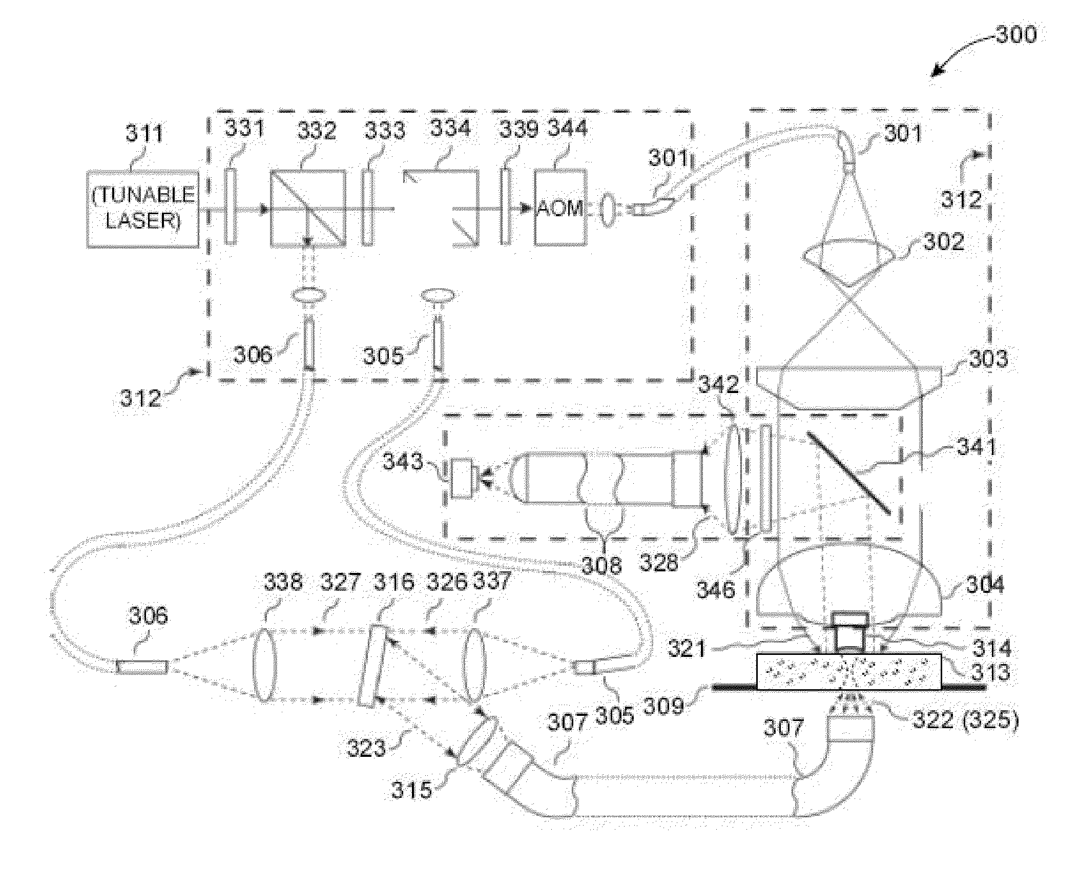
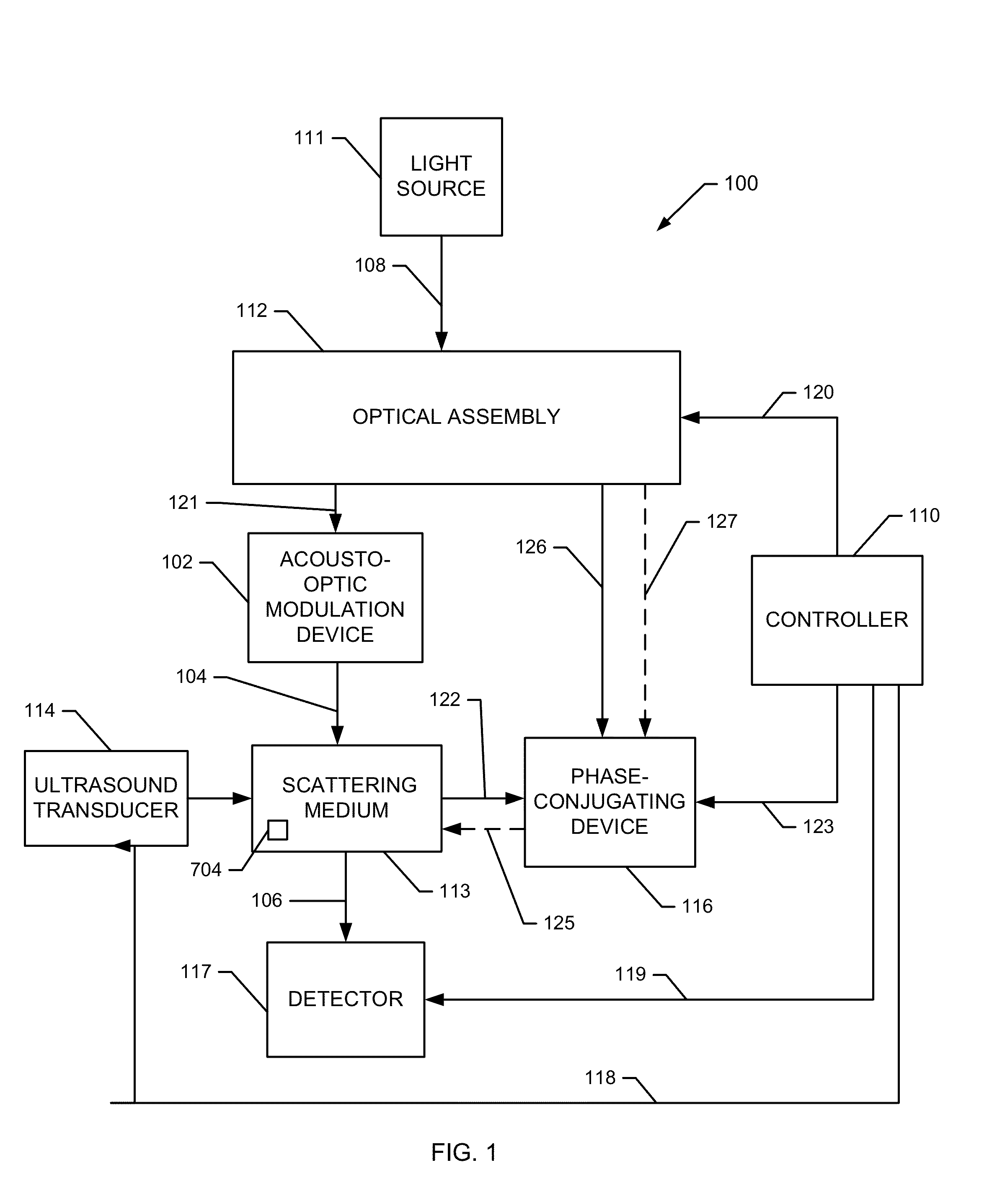
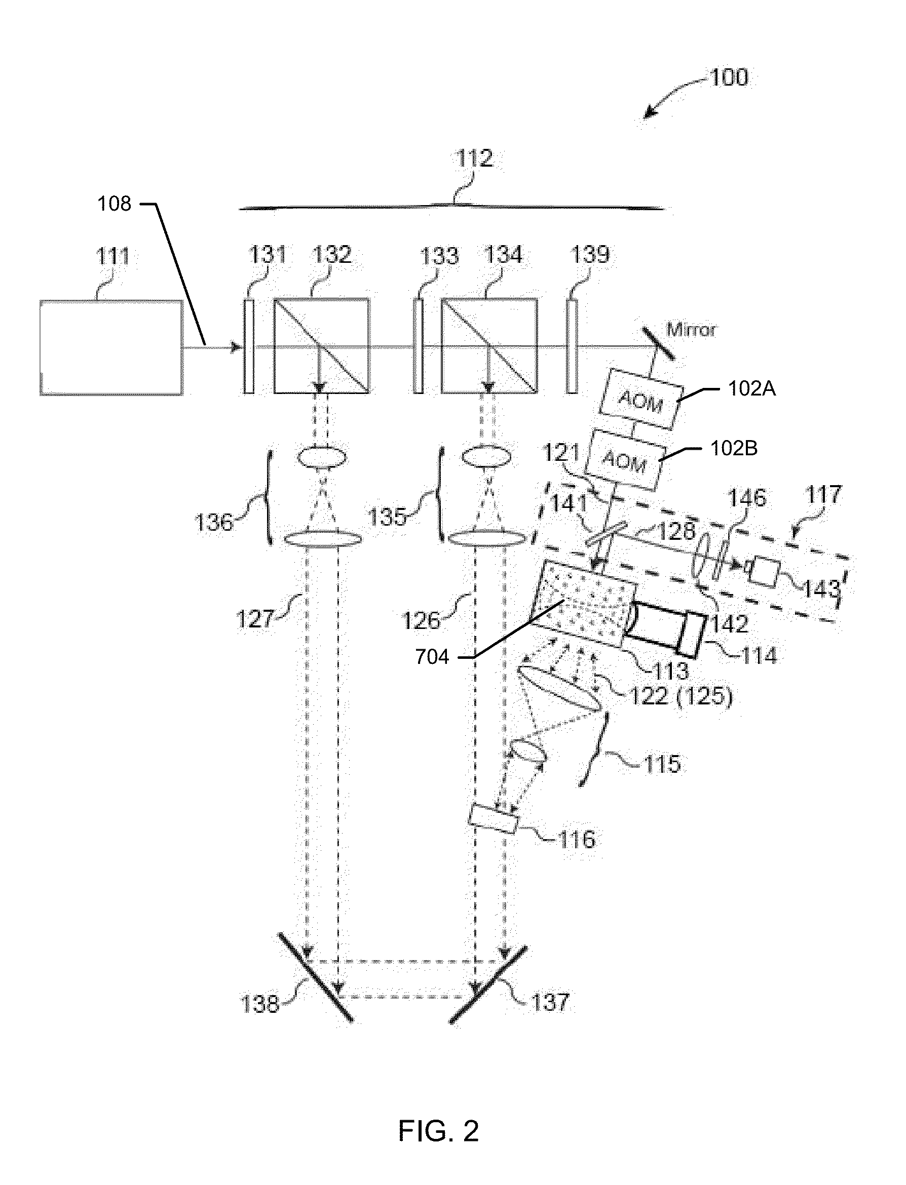
[0013]In an additional aspect, a system for focusing light within a scattering medium is provided that includes a moveable ultrasound transducer, a coherent light source, an optical assembly, a phase-conjugating device, and a controller. The moveable ultrasound transducer is operatively connected to the scattering medium to produce at least one focused ultrasound wave within each of a sequence of focus regions situated within the scattering medium. The optical assembly is operatively connected to the coherent light source, the scattering medium, and the phase-conjugating device. The optical assembly receives one or more coherent light beams from the coherent light source, delivers a sample beam into the scattering medium, and delivers a reference beam and a reconstruction beam to the phase-conjugating device. The phase-conjugating device is operatively connected to the scattering medium. The phase-conjugating device receives an ultrasound-encoded portion of the

Problems solved by technology

Despite the advantages of light as a means of probing and monitoring structures and processes, the application of light imaging techniques such as optical imaging is limited within biological tissues and other heterogeneous media.
Current optical imaging techniques, such as optical coherence tomography, are limited to a depth of approximately one optical transport mean free path, typically about 1 millimeter within biological tissues.
Other well-known techniques, such as confocal microscopy and multi-photon microscopy, have even more restricted penetration paths.
However, multiple scattering bodies such as cells, tissues, and organs within scattering media such as biological tissues i

Method used


Image

  • Iteration of optical time reversal by ultrasonic encoding in biological tissue
  • Iteration of optical time reversal by ultrasonic encoding in biological tissue
  • Iteration of optical time reversal by ultrasonic encoding in biological tissue

Examples


example 1

Digital Ultrasonically Encoded (Due) Optical Focusing Using Prototype Iterative Feedback System

[0188]To demonstrate the feasibility of using an iterative feedback method to shape the phase distribution across a wavefront of light to enhance transmission through a scattering medium, the following experiment was conducted.

[0189]A prototype system, shown schematically in FIG. 8, was used to demonstrate DUE optical focusing an iterative feedback focusing method as described herein above. The prototype system 800 includes a spatial light modulator (SLM) 802, divided into 20×20 independently controlled segments, to shape the incident wavefront 804 delivered by a coherent light source 806. The SLM response was modulated by control signals 808 received from a computer 810; the SLM response was calibrated to provide a linear phase shift of 2π over 191 grayscale values for each segment. To simulate light transmission through a scattering medium, the phase-shifted wavefronts 812 were delivered...

example 2

Assessment of Effectiveness of Digital Ultrasonically Encoded (Due) Optical Focusing Using Prototype Iterative Feedback System

[0195]To demonstrate the effectiveness of using an iterative feedback method to shape the phase distribution across a wavefront of light to enhance transmission through a scattering medium, the following experiment was conducted.

[0196]The prototype system of Example 1, shown schematically in FIG. 8, was used to implement DUE optical focusing the genetic optimization algorithm as described in Example 1. To assess the effectiveness of DUE optical focusing, a bar 836 containing fluorescent quantum dots was embedded within the clear gelatin medium 818 within the focal region of the system. The bar was situated in a horizontal orientation (perpendicular to the plane of the page as illustrated in FIG. 8) and had a 1 mm×1 mm square cross-sectional profile. Referring again to FIG. 8, a CCD 836 outfitted with a longpass filter 838 was used to measure the intensity of ...

example 3

Sensitivity of Phase Distribution Optimization Procedure Using Simulated WFS System

[0199]To assess the sensitivity of the optimization of phase distribution to the characteristics of the prototype DUE optical focusing system 800, the following experiment was conducted.

[0200]The genetic optimization algorithm and DUE optical focusing system 800 described previously in Example 1 were simulated to assess the sensitivity of the light intensity generated within the media 818 in the focus region to the number of elements in the array of the A simulation of the optimization of the phase distribution of the light modulator (SLM) 802.

[0201]Without being limited to any particular theory, it is known in the art that the expected increase in intensity η is equal to the ratio of the number of independent SLM segments N to the number of speckle grains in the ultrasound focus M, according to Eqn. (XII):

η=N+1MEqn.(XII)

[0202]A simulation was developed to model the expected increase in intensity. To ...


PUM


Abstract

Iterating an optical phase conjugation of ultrasonically-modulated diffuse light emitted by a scattering medium includes illuminating the scattering medium with a light beam from a coherent light source, modulating the diffuse light transmitted through the scattering medium with an ultrasonic wave focused on a region of interest within the scattering medium, fixing a hologram, retro-reflectively illuminating the scattering medium using a phase-conjugated copy of the diffuse light that was ultrasonically modulated, moving the ultrasonic focus, and iterating until light is focused on the final target.

Description

CROSS REFERENCE TO RELATED APPLICATIONS[0001]This application is a continuation-in-part of U.S. application Ser. No. 13 / 574,994 filed on Aug. 14, 2012 and entitled “OPTICAL TIME REVERSAL BY ULTRASONIC ENCODING IN BIOLOGICAL TISSUE”, which is a National Stage Entry of PCT application PCT / US2011 / 022253 filed on Jan. 24, 2011, which claims the benefit of U.S. Provisional Application 61 / 298,086 filed Jan. 25, 2010, each of which is hereby incorporated by reference herein in their entirety. This application also claims the benefit of U.S. Provisional Application 61 / 700,771 filed Sep. 13, 2012 and entitled “ITERATION OF OPTICAL TIME REVERSAL BY ULTRASONIC ENCODING IN BIOLOGICAL TISSUE”, which is also incorporated herein by reference in its entirety.STATEMENT REGARDING FEDERALLY SPONSORED RESEARCH & DEVELOPMENT[0002]This disclosure was made with government support under grants R01 CA094267, R01 CA106728, and DP1 EB016986 awarded by the U.S. National Institutes of Health. The government has...

Claims


Application Information

Patent Timeline
IPC IPC(8): G02F1/33G03H1/12G03H1/04
CPCG02F1/33G03H1/04G03H1/12G03H2001/0434G03H1/0005G03H1/22G03H2001/0083G03H2210/63G03H2260/36G03H1/0465
Inventor WANG, LIHONGXU, XIAO
Owner CALIFORNIA INST OF TECH
Why Eureka
Try Eureka