Methods and compositions for treatment of disease or injury of the nervous system

a technology of the nervous system and composition, applied in the direction of drug composition, peptide/protein ingredient, metabolic disorder, etc., can solve the problem of no method available to reverse glial scars for brain repair

Active Publication Date: 2014-01-23
PENN STATE RES FOUND
View PDF3 Cites 24 Cited by
  • Summary
  • Abstract
  • Description
  • Claims
  • Application Information

AI Technical Summary

Benefits of technology

[0147]After injecting retrovirus expressing NeuroD1-IRES-GFP, many NeuroD1-infected cells along the injection site were immunopositive for neuronal markers doublecortin (DCX), neuronal nuclei (NeuN), or β3-tubulin (Tuj1), and showed extensive neurites, FIGS. 4B-C and FIG. 5C-D. FIG. 4B shows NeuroD1-IRES-GFP infected cells were immunopositive for DCX along the injection site (14 DPI). FIG. 4C, NeuroD1-IRES-GFP infected cells were also immunopositive for NeuN and extended long neurites (21 DPI). Scale bar, 20 μm for 4B-C. The total number of NeuroD1-transduced neurons after single injection in the cortex is quantified and 175±13 neurons were found around the injection site (14 DPI, n=3 animals, 6-8 sections for each mouse brain). FIG. 5C shows low magnification images showing NeuroD1-infected cells (14 DPI) positive for DCX in cortical area and above hippocampal area, scale bar, 100 μM. FIG. 5D shows a high magnification image showing a NeuroD1-transduced cell immunopositive for Tuj1 but not GFAP, scale bar, 20 μm.
[0147]After injecting retrovirus expressing NeuroD1-IRES-GFP, many NeuroD1-infected cells along the injection site were immunopositive for neuronal markers doublecortin (DCX), neuronal nuclei (NeuN), or β3-tubulin (Tuj1), and showed extensive neurites, FIGS. 4B-C and FIG. 5C-D. FIG. 4B shows NeuroD1-IRES-GFP infected cells were immunopositive for DCX along the injection site (14 DPI). FIG. 4C, NeuroD1-IRES-GFP infected cells were also immunopositive for NeuN and extended long neurites (21 DPI). Scale bar, 20 μm for 4B-C. The total number of NeuroD1-transduced neurons after single injection in the cortex is quantified and 175±13 neurons were found around the injection site (14 DPI, n=3 animals, 6-8 sections for each mouse brain). FIG. 5C shows low magnification images showing NeuroD1-infected cells (14 DPI) positive for DCX in cortical area and above hippocampal area, scale bar, 100 μM. FIG. 5D shows a high magnification image showing a NeuroD1-transduced cell immunopositive for Tuj1 but not GFAP, scale bar, 20 μm.
[0147]After injecting retrovirus expressing NeuroD1-IRES-GFP, many NeuroD1-infected cells along the injection site were immunopositive for neuronal markers doublecortin (DCX), neuronal nuclei (NeuN), or β3-tubulin (Tuj1), and showed extensive neurites, FIGS. 4B-C and FIG. 5C-D. FIG. 4B shows NeuroD1-IRES-GFP infected cells were immunopositive for DCX along the injection site (14 DPI). FIG. 4C, NeuroD1-IRES-GFP infected cells were also immunopositive for NeuN and extended long neurites (21 DPI). Scale bar, 20 μm for 4B-C. The total number of NeuroD1-transduced neurons after single injection in the cortex is quantified and 175±13 neurons were found around the injection site (14 DPI, n=3 animals, 6-8 sections for each mouse brain). FIG. 5C shows low magnification images showing NeuroD1-infected cells (14 DPI) positive for DCX in cortical area and above hippocampal area, scale bar, 100 μM. FIG. 5D shows a high magnification image showing a NeuroD1-transduced cell immunopositive for Tuj1 but not GFAP, scale bar, 20 μm.
[0148]To investigate whether NeuroD1-transduced neurons were converted from reactive astrocytes, NeuroD1-transduced cortical tissue was immunostained with GFAP and DCX at 2-3 DPI in an attempt to capture a possible transitional stage. During this early expression period, a mixture of NeuroD1-infected cells that were labeled by either DCX or GFAP was observed. Surprisingly, some NeuroD1-infected cells were even labeled by both GFAP and DCX, suggesting that these cells were transitioning from astrocytes into neurons, FIG. 4D, arrow. FIG. 4D shows transitional stage between GFAP-positive astrocytes and DCX-labeled neurons revealed 3 days after injecting NeuroD1 retrovirus. The arrow points to a NeuroD1-infected cell with substantial GFAP signal and weak DCX signal, whereas the arrowhead points to a NeuroD1-infected cell mainly stained by DCX but little GFAP signal. Scale bar, 5 μm.

Problems solved by technology

Currently there is no method available to reverse glial scar for brain repair.

Method used


Image

  • Methods and compositions for treatment of disease or injury of the nervous system
  • Methods and compositions for treatment of disease or injury of the nervous system
  • Methods and compositions for treatment of disease or injury of the nervous system

Examples


example 1

Human Cortical Astrocytes

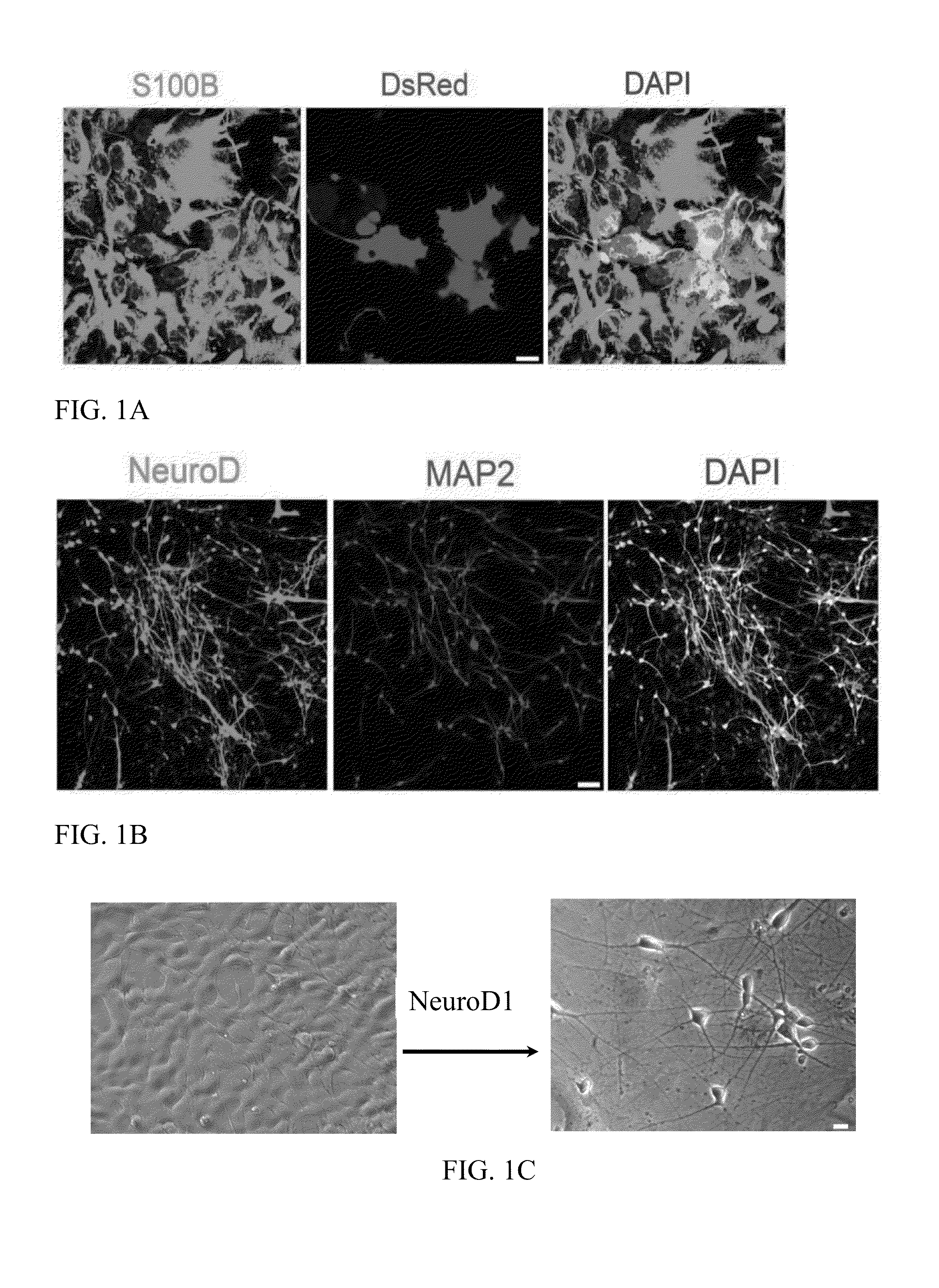
[0118]Human cortical astrocytes (HA1800) were purchased from ScienCell (California). Cells were subcultured when they were over 90% confluent. For subculture, cells were trypsinized by TrypLE™ Select (Invitrogen), centrifuged for 5 min at 1,000 rpm, re-suspended, and plated in a medium consisting of DMEM / F12 (Gibco), 10% fetal bovine serum (Gibco), penicillin / streptomycin (Gibco), 3.5 mM glucose (Sigma), and supplemented with B27 (Gibco), 10 ng / mL epidermal growth factor (EGF, Invitrogen), and 10 ng / mL fibroblast growth factor 2 (FGF2, Invitrogen). The astrocytes were cultured on poly-D-lysine (Sigma) coated coverslips (12 mm) at a density of 50,000 cells per coverslip in 24-well plates (BD Biosciences).

example 2

Retrovirus Production

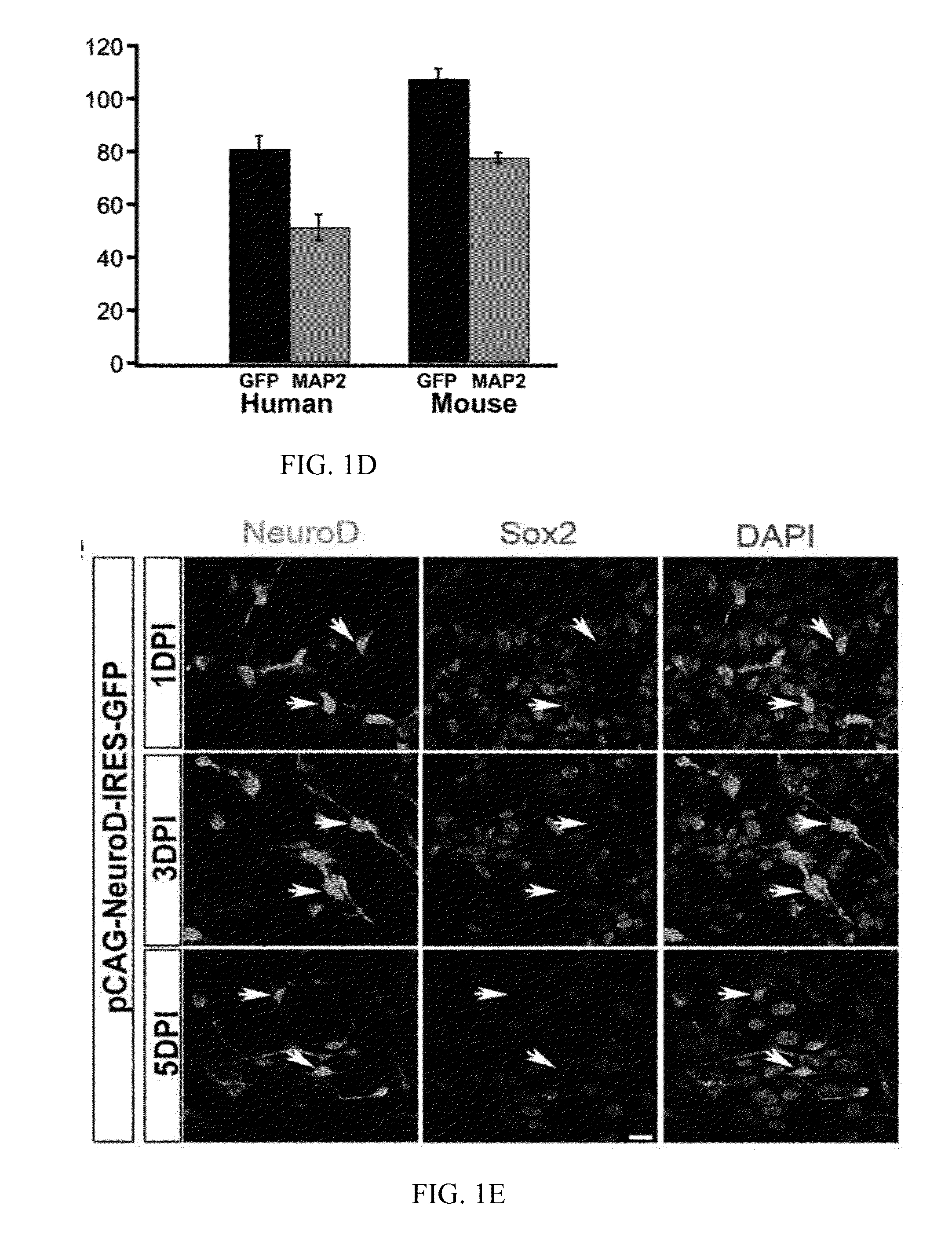
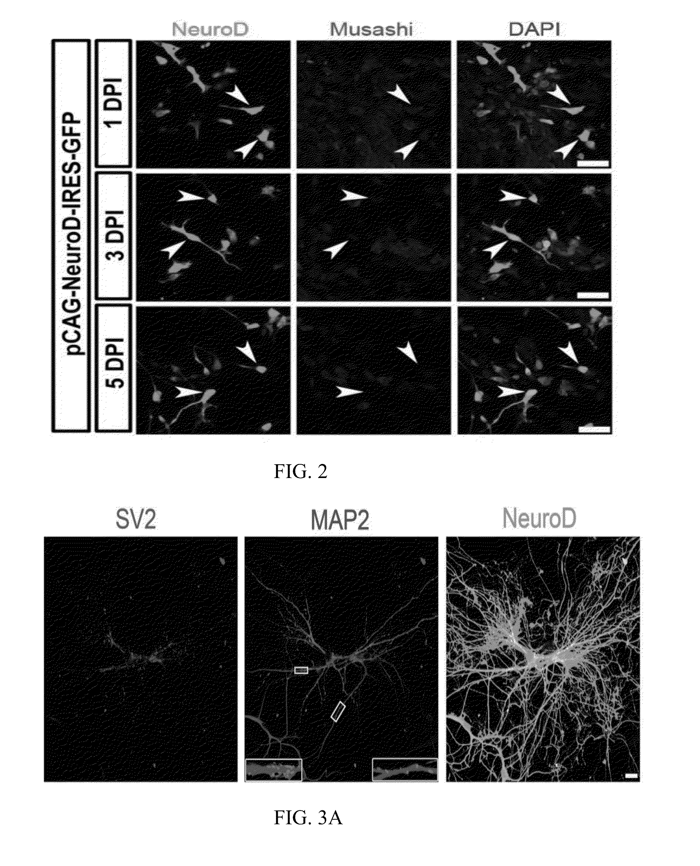
[0119]The pCAG-IRES-DsRed plasmid (Heinrich, C. et al., Directing astroglia from the cerebral cortex into subtype specific functional neurons. PLoS Biol 8 (5), e1000373 (2010)) containing the DsRed fluorescent reporter gene was used as the test plasmid. The mouse NeuroD1 gene was subcloned from the pAd NeuroD-1-nGFP (Addgene) and inserted into a pCAG-GFP-IRES-GFP retroviral vector (Zhao, C., Teng, E. M., Summers, R. G., Jr., Ming, G. L., & Gage, F. H., Distinct morphological stages of dentate granule neuron maturation in the adult mouse hippocampus. J Neurosci 26 (1), 3-11 (2006)) to generate the pCAG-NeuroD1-IRES-GFP plasmid, encoding the NeuroD1 protein of SEQ ID NO: 4. The restriction enzymes BamH I and Age I were used for subcloning. Viral particles were packaged in gpg helperfree HEK (Human embryonic kidney) cells to generate VSV-G (vesicular stomatitis virus glycoprotein)-pseudotyped retroviruses encoding neurogenic factors. The titers of viral particles w...

example 3

Culture Conditions for Trans-Differentiation of Human Astrocytes into Neurons

[0120]Twenty-four hours after infection of human cortical astrocytes with retrovirus encoding DsRed or encoding NeuroD1, the culture medium was completely replaced by a differentiation medium including DMEM / F12 (Gibco), 0.5% FBS (Gibco), 3.5 mM glucose (Sigma), penicillin / streptomycin (Gibco), and N2 supplement (Gibco). Brain-Derived Neurotrophic Factor (BDNF, 20 ng / mL, Invitrogen) was added to the cultures every four days during the differentiation to promote synaptic maturation.


PUM

PropertyMeasurementUnit
flow rateaaaaaaaaaa
volumeaaaaaaaaaa
speedaaaaaaaaaa

Abstract

Methods for producing new neurons in the brain in vivo are provided according to aspects of the present invention which include introducing NeuroD1 into a glial cell, particularly into a reactive astrocyte or NG2 cell, thereby “converting” the reactive glial cell to a neuron. Methods of producing a neuronal phenotype in a glial cell are provided according to aspects of the present invention which include expressing exogenous NeuroD1 in the glial cell, wherein expressing exogenous NeuroD1 includes delivering an expression vector, such as a viral expression vector, including a nucleic acid encoding the exogenous NeuroD1 to the glial cell.

Description

REFERENCE TO RELATED APPLICATION[0001]This application claims priority from U.S. Provisional Patent Application Ser. Nos. 61 / 673,471, filed Jul. 19, 2012 and 61 / 762,506, filed Feb. 8, 2013, the entire content of which is incorporated herein by reference.GOVERNMENT SUPPORT[0002]This invention was made with government support under Grant No. MH083911, awarded by the National Institutes of Health. The Government has certain rights in the invention.FIELD OF THE INVENTION[0003]General aspects of the invention relate to the in situ conversion of glial cells to functional neuronal cells in the central nervous system (CNS), and the methods to transforming glial cells to neuronal cells both in vitro and in vivo.BACKGROUND OF THE INVENTION[0004]The central nervous system in mammals is largely unable to regenerate itself following injury. Neurons are often killed or injured as a result of an injury or neurological condition, such as a disease or other pathology. It is well known that glial cel...

Claims


Application Information

Patent Timeline
Patent Type & Authority Applications(United States)
IPC IPC(8): A61K48/00A61K38/17C12N15/86
CPCA61K48/005C12N15/86A61K38/1709A01K2227/105A01K2267/0356C12N2740/13043A61P25/00
Inventor CHEN, GONGGUO, ZIYUANWU, ZHENG
Owner PENN STATE RES FOUND
Why Eureka
Try Eureka