Kinase substrate sensor

a substrate sensor and kinase technology, applied in the field of cytoplasmic protein complexes, can solve the problems of requiring lysis to corrupt the normal cellular context, requiring high throughput screening, and tedious methods

Active Publication Date: 2014-01-30
VLAAMS INTERUNIVERSITAIR INST VOOR BIOTECHNOLOGIE VZW +1
View PDF1 Cites 4 Cited by
  • Summary
  • Abstract
  • Description
  • Claims
  • Application Information

AI Technical Summary

Benefits of technology

[0011]There is still a need for a sensitive identification system for compound-compound interactions that can study these interactions

Problems solved by technology
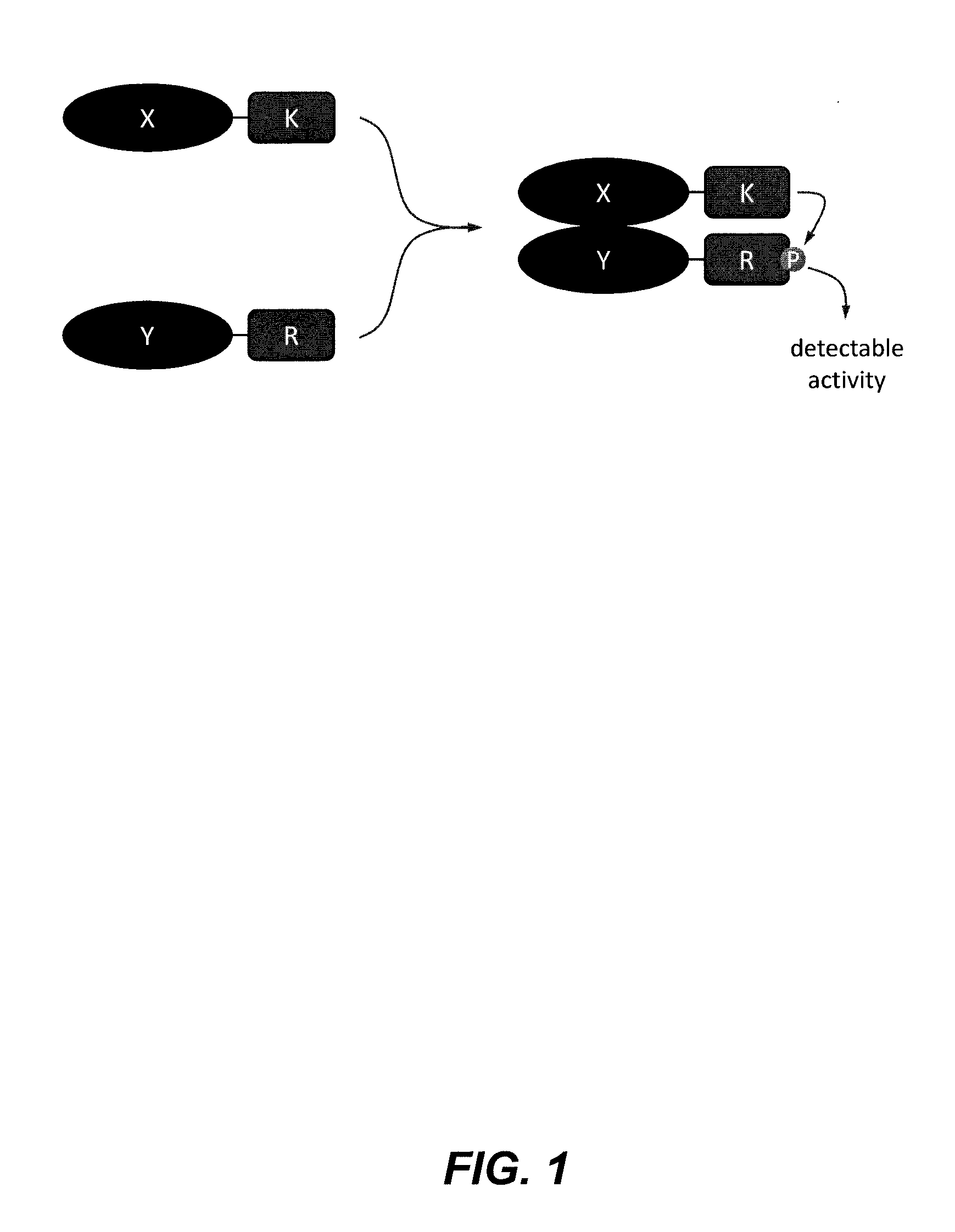
However, these methods are tedious and do not allow high throughput screening.
Moreover, they require lysis corrupting the normal cellular context.
Proteins with intrinsic transcription activation properties may cause false positives.
Moreover, interactions that are dependent upon secondary modifications of the protein such as phosphorylation cannot be easily detected.
However, all these methods suffer from the difficulties that are inherent at the phage display metho

Method used


Image

  • Kinase substrate sensor
  • Kinase substrate sensor
  • Kinase substrate sensor

Examples


example 1

Detection of the Interaction Between HIV1 Reverse Transcriptase (RT) Subunits

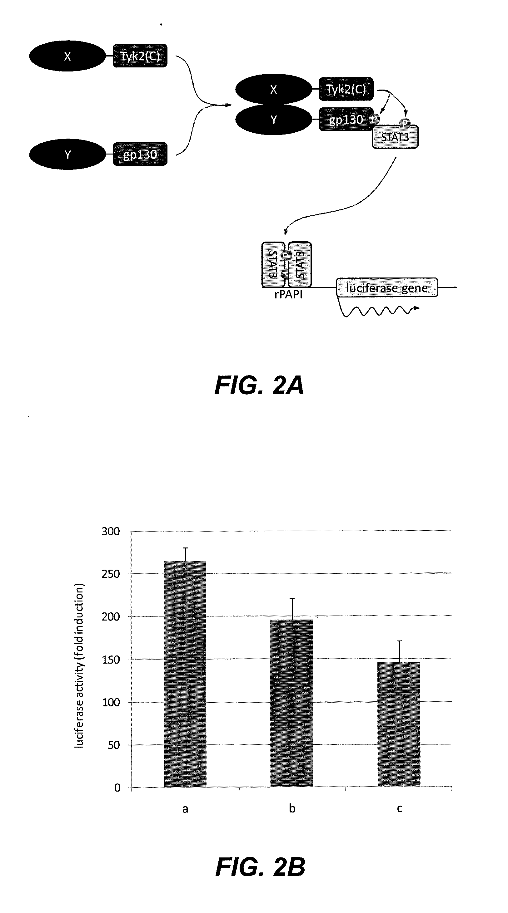
[0062]In order to determine the functionality of the assay, the interaction between HIV1 subunits that form homo- and heterodimers was tested by transfecting the following combinations of plasmids (100 ng of the Tyk2(C) fusion construct, 1 μg of the gp130 fusion construct and 50 ng of the luciferase reporter construct) according to the methods described above:[0063]a) pMet7-HA-Tyk2(C)-RTp66+pMG2-RTp51+pXP2d2-rPAPI-luciferase[0064]b) pMet7-HA-Tyk2(C)-RTp66+pMG2-RTp66+pXP2d2-rPAPI-luciferase[0065]c) pMet7-HA-Tyk2(C)-RTp51+pMG2-RTp51+pXP2d2-rPAPI-luciferase

[0066]Background signal for each Tyk2(C) fusion polypeptide was determined by transfecting the plasmid that encodes it together with a plasmid encoding an unfused gp130 fragment (pMG1) and the luciferase reporter plasmid (pMet7-HA-Tyk2(C)-RTp66+pMG1+pXP2d2-rPAPI-luciferase in a) and b); pMet7-HA-Tyk2(C)-RTp51+pMG1+pXP2d2-rPAPI-luciferase in c)). The fold ind...

example 2

Interaction Between Nuclear Proteins

[0067]To determine whether the method can detect interactions between nuclear proteins, we tested the interaction between HIV 1 Integrase (IN) and human LEDGF and between human p53 and MDM2, proteins with a nuclear localization. Cells were transfected with the following combinations of plasmids (250 ng of the Tyk2(C) fusion construct, 500 ng of the gp1 30 fusion construct and 50 ng of the luciferase reporter construct) according to the methods described above:[0068]a) pMet7-HA-Tyk2(C)-LEDGF+pMG2-IN+pXP2d2-rPAPI-luciferase[0069]b) pMet7-HA-Tyk2(C)-MDM2+pMG1-p53+pXP2d2-rPAPI-luciferase

[0070]Background signal for each Tyk2(C) fusion polypeptide was determined by transfecting the plasmid that encodes it together with a plasmid encoding an unfused gp130 fragment (pMG1) and the luciferase reporter plasmid (pMet7-HA-Tyk2(C)-LEDGF+pMG1+pXP2d2-rPAPI-luciferase in a); pMet7-HA-Tyk2(C)-MDM2+pMG1+pXP2d2-rPAPI-luciferase in b)). The fold induction for each tes...

example 3

Dose-Dependent Disruption of the Interaction Between Human p53 and MDM2 by Nutlin-3

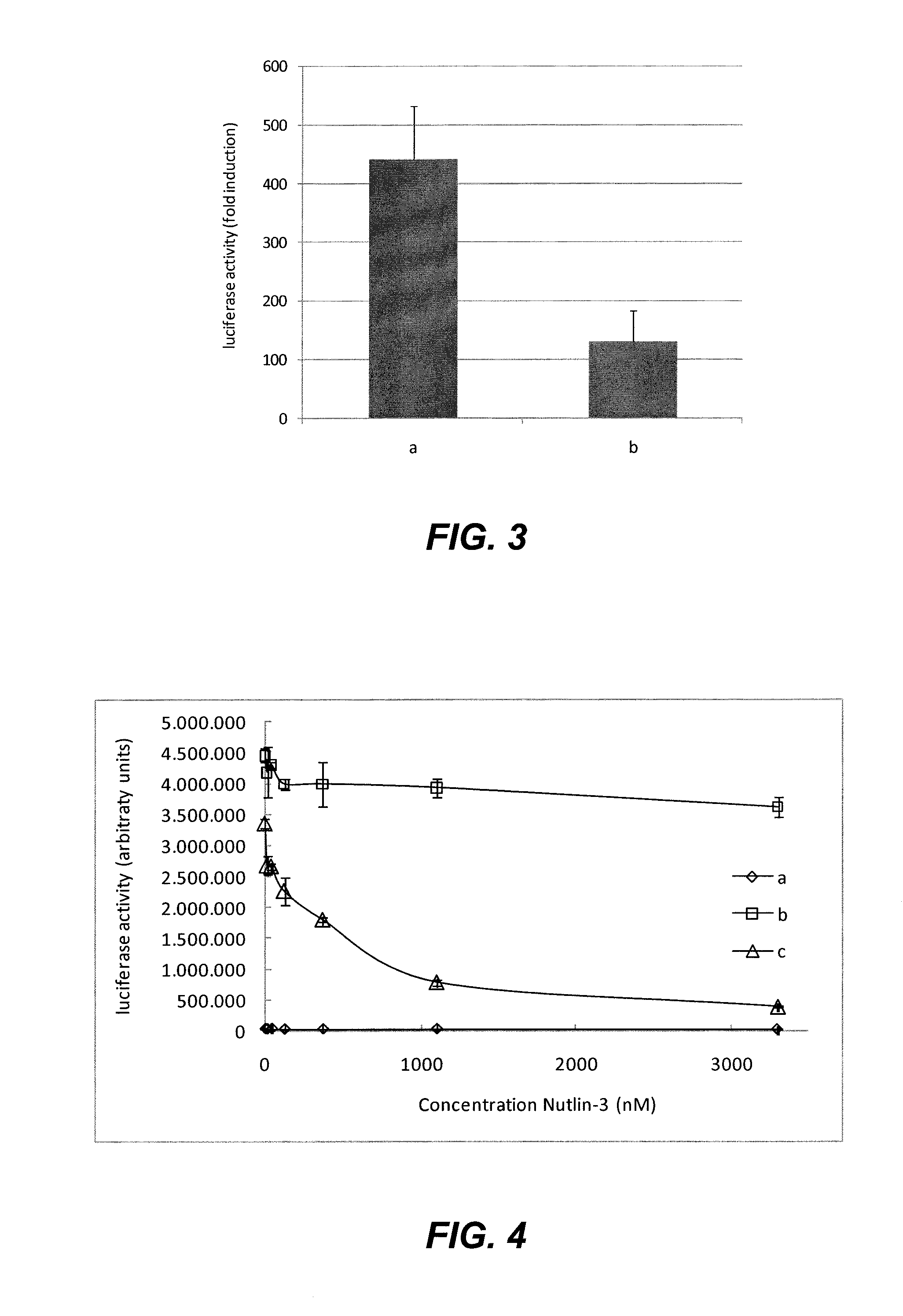
[0071]To show that the method can detect modulation of protein-protein interactions by small molecules, we analyzed the interaction between p53 and MDM2, which has been reported to be disrupted by Nutlin-3, a member of the nutlin family of potential novel anti-cancer compounds (Vassilev et al., 2004). Cells were transfected with the following combinations of plasmids (100 ng of the Tyk2(C) fusion construct, 1000 ng of the gp130 fusion construct and 50 ng of the luciferase reporter construct) according to the methods described above:[0072]a) pMet7-HA-Tyk2(C)-p53(N)+pMG1+pXP2d2-rPAPI-luciferase[0073]b) pMet7-HA-Tyk2(C)-p53(N)+pMG1-EFHA1+pXP2d2-rPAPI-luciferase[0074]c) pMet7-HA-Tyk2(C)-p53(N)+pMG2-MDM2+pXP2 d2-rPAPI-luciferase

[0075]After transfection, cells were treated with 0-14-41-123-370-1111-3333 nM Nutlin-3, final concentration. The results shown in FIG. 4 show a dose-dependent decrease of the signa...


PUM

PropertyMeasurementUnit
Interactionaaaaaaaaaa

Abstract

The disclosure relates to a cytoplasmic protein complex comprising: (a) a first recombinant fusion protein comprising a kinase, fused to a first interaction polypeptide; and (b) a second recombinant fusion protein comprising a domain comprising a reporter phosphorylation site, whereby the domain is fused to a second interaction polypeptide. The disclosure relates further to a method to detect compound-compound-interaction using the cytoplasmic protein complex, and to cells comprising such cytoplasmic protein complex.

Description

CROSS-REFERENCE TO RELATED APPLICATIONS[0001]This application is a national phase entry under 35 U.S.C. §371 of International Patent Application PCT / EP2012 / 053463, filed Feb. 29, 2012, designating the United States of America and published in English as International Patent Publication WO 2012 / 117031 A1 on Sep. 7, 2012, which claims the benefit under Article 8 of the Patent Cooperation Treaty and under 35 U.S.C. §119(e) to Great Britain Patent Application Serial No. 1103453.5, filed Mar. 1, 2011, and to U.S. Provisional Patent Application Ser. No. 61 / 464,285, filed Mar. 1, 2011.TECHNICAL FIELD[0002]The disclosure relates to a cytoplasmic protein complex comprising (a) a first recombinant fusion protein comprising a kinase, fused to a first interaction polypeptide and (b) a second recombinant fusion protein comprising a domain comprising a reporter phosphorylation site, wherein the domain is fused to a second interaction polypeptide. It relates further to a method of detecting compou...

Claims


Application Information

Patent Timeline
IPC IPC(8): C12Q1/48C12N9/12
CPCC12N9/12C12Q1/485C12N15/1055C12N15/62G01N33/542C07K2319/00
Inventor TAVERNIER, JANLIEVENS, SAMUEL
Owner VLAAMS INTERUNIVERSITAIR INST VOOR BIOTECHNOLOGIE VZW
Why Eureka
Try Eureka