Complex in which Anti-cotinine antibody is bound to conjugate of cotinine and binding substance, and use thereof
- Summary
- Abstract
- Description
- Claims
- Application Information
AI Technical Summary
Benefits of technology
Problems solved by technology
Method used
Image
Examples
example 1
Preparation of Gene of Anti-Cotinine Rabbit / Human Chimeric IgG
[0083](1-1) Amplification of Antibody Variable Region from Anti-Cotinine Rabbit scFv
[0084]To amplify an antibody variable region (VL and VH) of a rabbit, a polymerase chain reaction (PCR) was performed using an anti-cotinine rabbit scFv gene (see U.S. Pat. No. 8,008,448), as a template, 60 pmole of forward and reverse primers (SEQ ID NOs: 11 and 12) for VL, respectively, and 60 pmole of forward and reverse primers (SEQ ID NOs: 13 and 14) for VH, respectively.
[0085]Specifically, to perform a PCR reaction, 1 μL of cDNA (about 0.5 μg) which was synthesized in U.S. Pat. No. 8,008,448, 60 pmol of a forward primer and a reverse primer, respectively, 10 μL of 10×PCR buffer, 8 μL of 2.5 mM dNTP mixture, and 0.5 μL of Taq polymerase were mixed, and then, 100 μL of distilled water was added thereto. The resultant mixture was denatured at 94° C. for 10 minutes, proceeded with 30 thermal cycles of 94° C. for 15 seconds, 56° C. for 30...
example 2
Expression and Purification for In Vitro Analysis of Anti-Cotinine Rabbit / Human Chimeric IgG
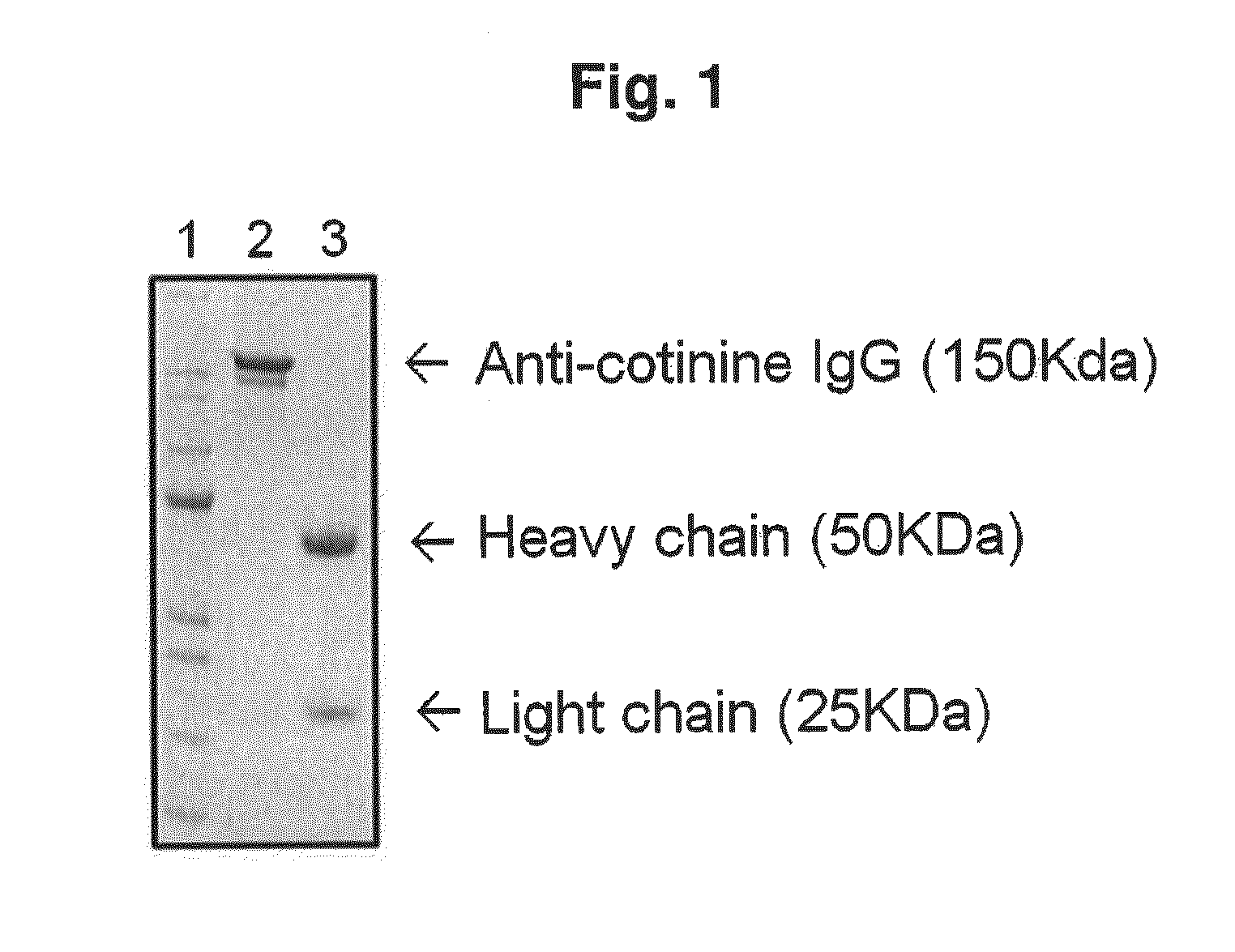
[0098]A mammalian cell, CHO DG 44 (Invitrogen, USA), was transfected with an expression vector DNA including an anti-cotinine rabbit / human chimeric IgG gene. The transfected cell was cultured in a condition of 37° C. and 135 rpm in CD OptiCHO™ expression medium (GIBCO), including 100 U / mL of penicillin and 100 g / mL of streptomycin (GIBCO, USA), to which 500 μg / mL of G418 was added. The supernatant of the culture medium was concentrated through Labscale TFF system (Millipore, USA), and then purified with a protein A column (Repligen Co., USA). The purified IgG of 150 KDa was determined by Coomassie staining (see FIG. 1) and used in a subsequent experiment (Experimental Example).
[0099]As shown in FIG. 1, it could be understood that lane 1 shows a size marker, lane 2 shows unreduced anti-cotinine IgG (150 KDa), and lane 3 shows a light chain (25 KDa) and a heavy chain (50 KDa) which were as redu...
example 3
Preparation of Conjugate of Cotinine and Binding Material
[0100](3-1) Conjugate of Cotinine and Peptide
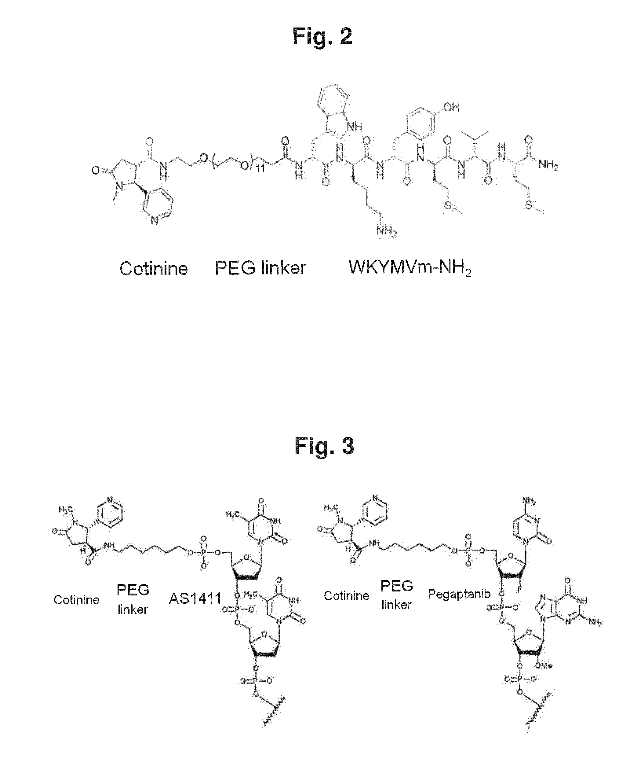
[0101]A WKYMVm-NH2 peptide (WKYMVm-NH2, SEQ ID NO: 1) and a wkymvm-NH2 peptide (wkymvm-NH2, SEQ ID NO: 2) were used as peptides. WKYMVm-NH2 and wkymvm-NH2 were synthesized in an ASP48S automatic peptide synthesizer by a solid phase peptide synthesis method. Then, the peptides were purified through reverse phase HPLC using Vydac Everest C18 column (250 mm×22 mm, 10 μm) (>95% purity), and the size of peptides were determined through LC / MS (Agilent HP1100 series) (>95% purity).
[0102]Meanwhile, cotinine-WKYMVm-NH2 and cotinine-wkymvm-NH2, which were conjugates of cotinine and a peptide, were synthesized by performing the same method as above except that a PEG linker and cotinine were introduced at the last process for synthesizing a peptide.
[0103]A method for preparing the conjugate of cotinine and a peptide was performed according to the following steps of: firstly synthesizing a basic...
PUM
| Property | Measurement | Unit |
|---|---|---|
| Cytotoxicity | aaaaa | aaaaa |
| Distribution | aaaaa | aaaaa |
Abstract
Description
Claims
Application Information
 - R&D
- Intellectual Property
- Life Sciences
- Materials
- Tech Scout
- Unparalleled Data Quality
- Higher Quality Content
- 60% Fewer Hallucinations
Browse by: Latest US Patents, China's latest patents, Technical Efficacy Thesaurus, Application Domain, Technology Topic, Popular Technical Reports.
© 2025 PatSnap. All rights reserved.Legal|Privacy policy|Modern Slavery Act Transparency Statement|Sitemap|About US| Contact US: help@patsnap.com


