Biofunctional nanofibers for analyte separation in microchannels

a microchannel and biofunctional technology, applied in the direction of diaphragms, fluid speed measurement, chemical vapor deposition coating, etc., can solve the problem of limited diffusion transport and other problems

Inactive Publication Date: 2014-03-27
CORNELL UNIVERSITY
View PDF3 Cites 17 Cited by
  • Summary
  • Abstract
  • Description
  • Claims
  • Application Information

AI Technical Summary

Problems solved by technology

Diffusion limited transport is a draw-back for many microfluidic devices, in particular those that require fast mixing or high chemical reaction rate at the interfaces of the fluid flow and the channel walls.

Method used


Image

  • Biofunctional nanofibers for analyte separation in microchannels
  • Biofunctional nanofibers for analyte separation in microchannels
  • Biofunctional nanofibers for analyte separation in microchannels

Examples


example 1

6.1. Example 1

Electrospun Nanofibers for Microfluidic Analytical Systems

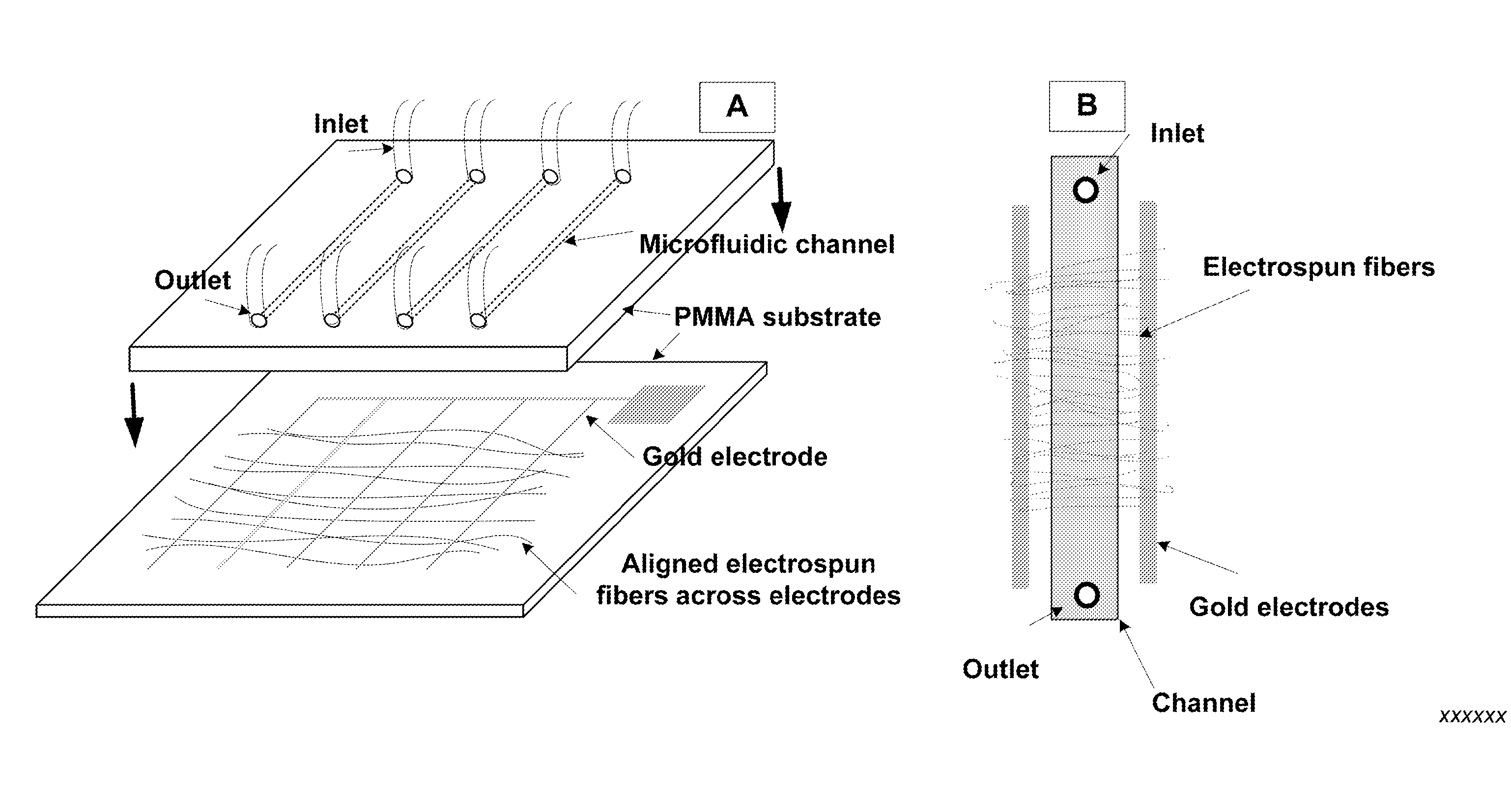
[0222]In this example, poly(vinyl alcohol) (PVA) blend nanofibers formulated to create variations in fiber surface chemistry were electrospun to form patterns around gold microelectrodes on a poly(methyl methacrylate) (PMMA) chip surface. These nanofiber patterns were integrated into polymer-based microfluidic channels to create a functionalized microfluidic system with potential applications in bioanalysis. Spinning conditions and microelectrodes were optimized to enable an alignment of the nanofibers across the microfluidic channel. X-ray photoelectron spectroscopy (XPS) and Fourier transform infrared spectroscopy (FTIR) were used to characterize the electrospun fibers and the results demonstrated that functional nanofibers were successfully spun from the polymers. Nanofibers spun into the microfluidic channel maintained their morphologies during fluid flow at linear velocities of 3.4 and 13.6 mm / s Nanofibers ...

example 2

6.2. Example 2

Demonstration of Biosensing by Nanofibers in Microfluidic Channels

6.2.1. Background

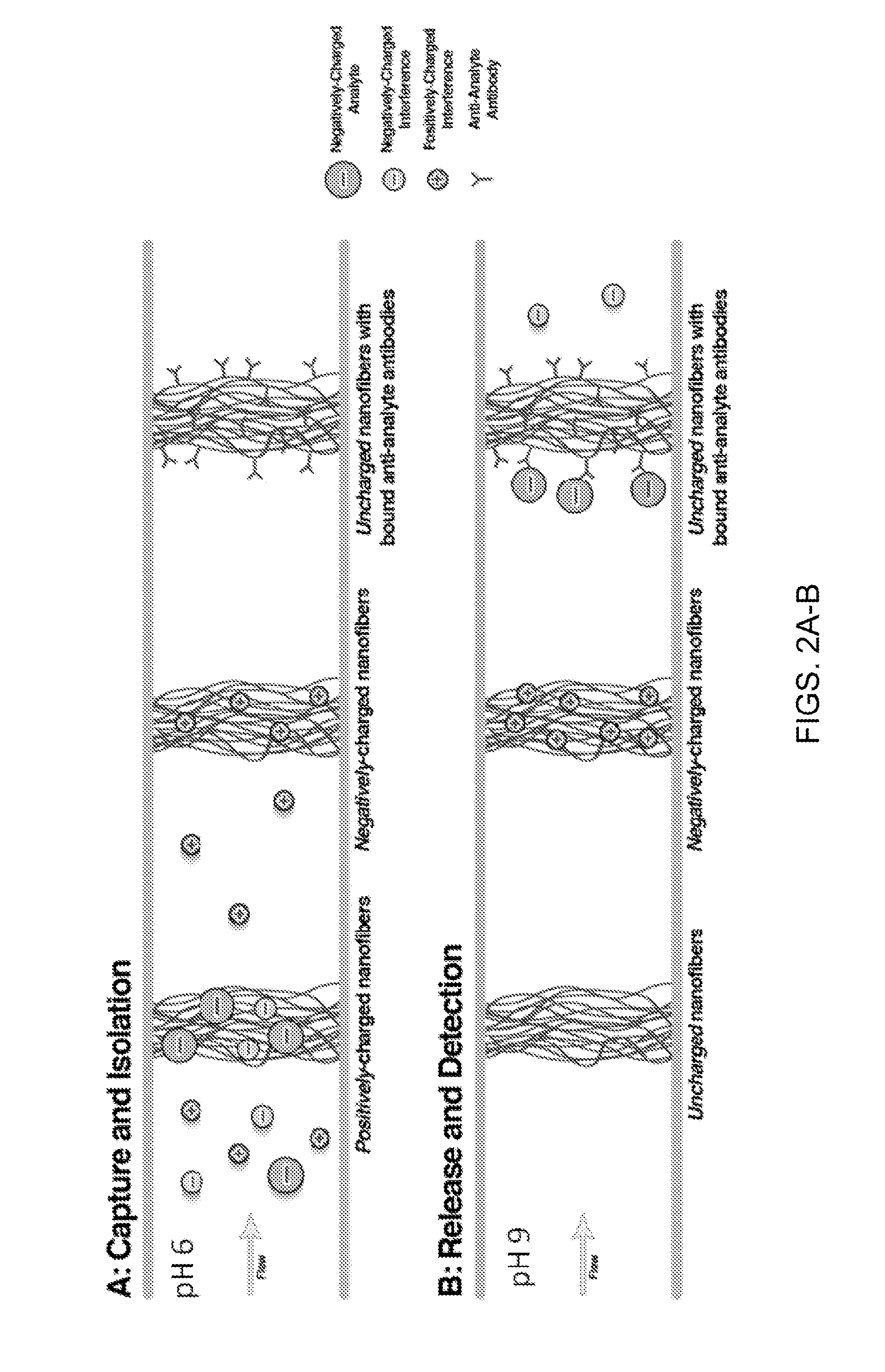
[0257]Food- and environmental safety, biosecurity and clinical diagnostics all rely on the ability to detect pathogens, toxins, or clinical markers at low concentrations, accurately and reliably. Simple, over-the-counter biosensors have been developed for some particular cases: the home pregnancy test, and the glucometer for diabetic patients. However, for most detection challenges that we face as a society lengthy and expensive laboratory procedures are required. The costs of the tests and time until results are obtained are insurmountable obstacles. For safety and security-related tests and for diagnostics in resource-limited settings rapid, inexpensive and easy to use tests can have a huge impact.

[0258]Over the last three decades, researchers have developed sensing technology that is capable of detecting single cells and even single molecules. However, their detection can only be acco...

example 3

6.3. Example 3

Nanofibers for Use in Microfluidic Channels in In Vitro Models for Cancer Cell Migration Studies and with Relevant Chemical and Biological Functionality for MicroTAS Systems

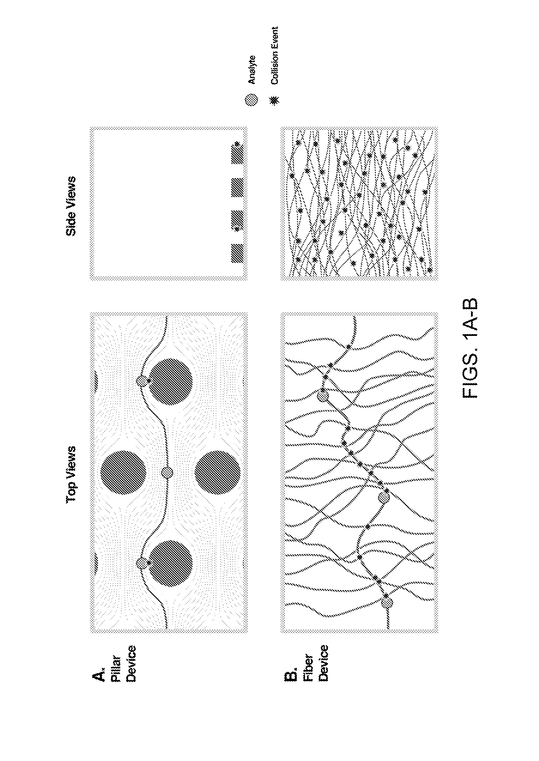
[0262]This example describes the development of nanofibers from materials compatible with microfluidic in vitro models for cancer cell migration studies and with relevant chemical and biological functionality for microTAS systems. Nanofibers can provide increased surface area and surface functionality patterned at specific locations within channels. Nanofibers have quantifiable and modifiable mechanical, chemical and biological properties that enhance the range of variables addressable in microfluidic devices.

6.3.1. Experimental Design

[0263]A range of fiber chemical, biological and physical properties has the properties necessary to serve as ECM for specific cell growth within a microfluidic in vitro model, and selectively capture components of a mixed analyte, immobilize proteins or antibodies, and...


PUM

PropertyMeasurementUnit
diameteraaaaaaaaaa
thicknessaaaaaaaaaa
pHaaaaaaaaaa

Abstract

A method is provided for producing, in a substrate, an enclosed channel or enclosed cavity comprising at least one functional nanofiber, the method comprising the steps of providing a first substrate and a second substrate; forming a channel or cavity on the first substrate or the second substrate; electrospinning at least one functional nanofiber on the first substrate; assembling the first and second substrates, wherein the first substrate is placed over the second substrate, or the second substrate is placed over the first substrate; and bonding the first substrate and the second substrate to form the substrate, thereby forming an enclosed channel or enclosed cavity comprising the at least one functional nanofiber in the substrate. An enclosed channel or cavity comprising at least one functional electrospun nanofiber is also provided. A microfluidic device is also provided comprising an enclosed channel or cavity comprising at least one functional electrospun nanofiber.

Description

CROSS-REFERENCE TO RELATED APPLICATIONS[0001]This application claims priority to and the benefit of co-pending U.S. provisional patent application Ser. No. 61 / 467,197, entitled Biofunctional Nanofibers for Analyte Separation in Microfluidic Channels, filed Mar. 24, 2011, which is incorporated herein by reference in its entirety.STATEMENT REGARDING FEDERALLY SPONSORED RESEARCH OR DEVELOPMENT[0002]The disclosed invention was made with government support under grant no. 0852900 from the Division of Chemical, Bioengineering, Environmental, and Transport Systems of the National Science Foundation. The government has rights in this invention.1. TECHNICAL FIELD[0003]The present invention relates to methods for producing microscale channels or cavities comprising functional nanofibers. The invention further relates to microfluidic devices and other microscale devices comprising microscale channels or cavities that comprise functional nanofibers. The invention also relates to microfluidic de...

Claims


Application Information

Patent Timeline
Patent Type & Authority Applications(United States)
IPC IPC(8): B81B1/00B81C1/00
CPCB81C1/00119B81B1/006B01L3/502707B01L2300/0681B01L2300/0816B81B1/00
Inventor BAEUMNER, ANTJE J.FREY, MARGARET W.CHO, DAEHWAN
Owner CORNELL UNIVERSITY
Why Eureka
Try Eureka