
Fenestration through foreign material

a technology of foreign material and fenestration, which is applied in the field of creating a channel through foreign material, can solve the problems of difficult access to the left side of the heart, difficulty in delivering blood to the brain, organs, tissues, and low oxygen levels in arterial blood that supplies brain, heart failure or death, etc., and achieves the effect of reducing procedural complexity, reducing recovery times for patients, and reducing oxygen levels

Active Publication Date: 2014-04-10
BOSTON SCI MEDICAL DEVICE LTD
View PDF3 Cites 12 Cited by
  • Summary
  • Abstract
  • Description
  • Claims
  • Application Information

AI Technical Summary

Benefits of technology

The patent describes using radiofrequency electrical energy to create channels in foreign material within the body of a patient, such as to treat heart defects. This method can reduce the risk of complications and minimize the risk of accidental puncture or patent text describes a method for creating channels in foreign material within a patient's body, such as for the treatment of heart defects. The method can use radiofrequency electrical energy to create the channel without needing to mechanically tear the foreign material. The technical effect of this method is that it can provide a safer and more effective way to treat heart defects and other applications where foreign material needs to be cleared from the body.

Problems solved by technology

This may lead to lower oxygen levels in the arterial blood that supplies the brain, organs, and tissues and may additionally lead to other complications such as risk of embolisms, or even heart failure or death.
However, once the septal defect is sealed by the foreign material, it may be difficult to access the left side of the heart using conventional techniques that are often used to puncture through the septum mechanically.
This is partially due to the fact that the natural path of the mechanical apparatus through the septum may now be obstructed by the foreign material.
More specifically, the foreign material included in an occluder may be difficult to cross using conventional mechanical puncturing techniques, for example with a percutaneous mechanical needle, because of the relatively large forces required.
Also, the relatively large forces exerted onto the needle may pose an additional risk that the needle will pass through the foreign material suddenly in an uncontrolled manner and damage adjacent tissues within the left side of the heart.

Method used


Image

  • Fenestration through foreign material
  • Fenestration through foreign material
  • Fenestration through foreign material

Examples


example 1a

[0096]In a first example, an embodiment of a proposed method is used to create a channel 512, seen in FIG. 5B, within a septal patch 510 made of foreign material, the septal patch 510 defining a material first surface 514 and a substantially opposed material second surface 516. The channel 512 extends between the material first and second surfaces 514 and 516. The septal patch 510 extends across an aperture 518 defined by the septum 520 of the heart 500 of the patient, for example an atrial septum or a ventricular septum. For example, the septal patch 510 covers the aperture and extends in a plane outside of the septum 520. In other examples, the septal patch 510 extends inside the aperture 518. Some of these procedures may involve patients that have had a septal defect repaired with the septal patch 510. In some cases, such patients may suffer from one or more conditions which require access to the left side of the heart for treatment to be performed. In such situations, access to ...

example 1b

[0098]In another example, with reference to FIGS. 5C-5I, an embodiment of a proposed method is used in a procedure to create a channel within foreign material that is positioned within a patient's body. More specifically, the proposed method may be performed in patients that have a septal defect 518′ within the septum 520 of the heart 500 (as shown more clearly in FIG. 5C), where the septal defect 518′ has been previously repaired with foreign material. For example, the foreign material may be included within an occluder 1510 positioned within the septum 520, as shown in FIG. 5D. The septal occluder 1510 prevents blood from shunting between the left side and the right side of the heart 500 that occurs when the septal defect 518′ is present. For example, the septal defect 518′ may be in the form of an aperture 518 within the septum 520 of the heart 500, for example within an atrial septum as shown. Alternatively, the defect may be within the ventricular septum. The occluder 1510 may ...

example 2a

[0118]With reference now to FIGS. 6A, 6B, 7A and 7B, methods for in-situ creation of a channel through a stent-graft are illustrated. In the illustrated embodiments, a stent-graft 606, composed of a foreign material, has been placed to cover an aneurysm 604 in an abdominal aorta 600. As shown in these figures, stent-graft 606 occludes the renal arteries ostia 605.

[0119]This positioning of the stent-graft 606 is typically necessitated by an inadequate, i.e. too short, proximal neck of the abdominal aorta 600. One of the greatest challenges of stent-grafting an abdominal aortic aneurysms 604 is to obtain a long proximal attachment site to ensure a good seal without occluding the renal or supra-aortic vessels. If a long proximal site is unavailable, the ostia of the renal and / or supra-aortic vessels may become occluded by the stent-graft 606. The present invention provides a method for creating a transluminal in-situ channel in order to restore blood flow to any vessels that do become ...


PUM


Abstract

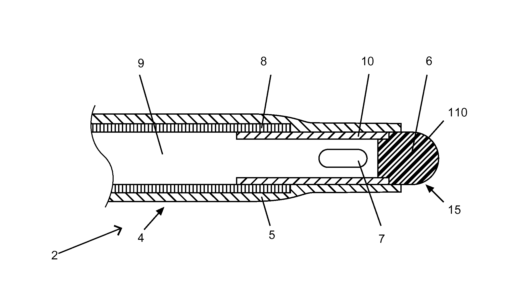
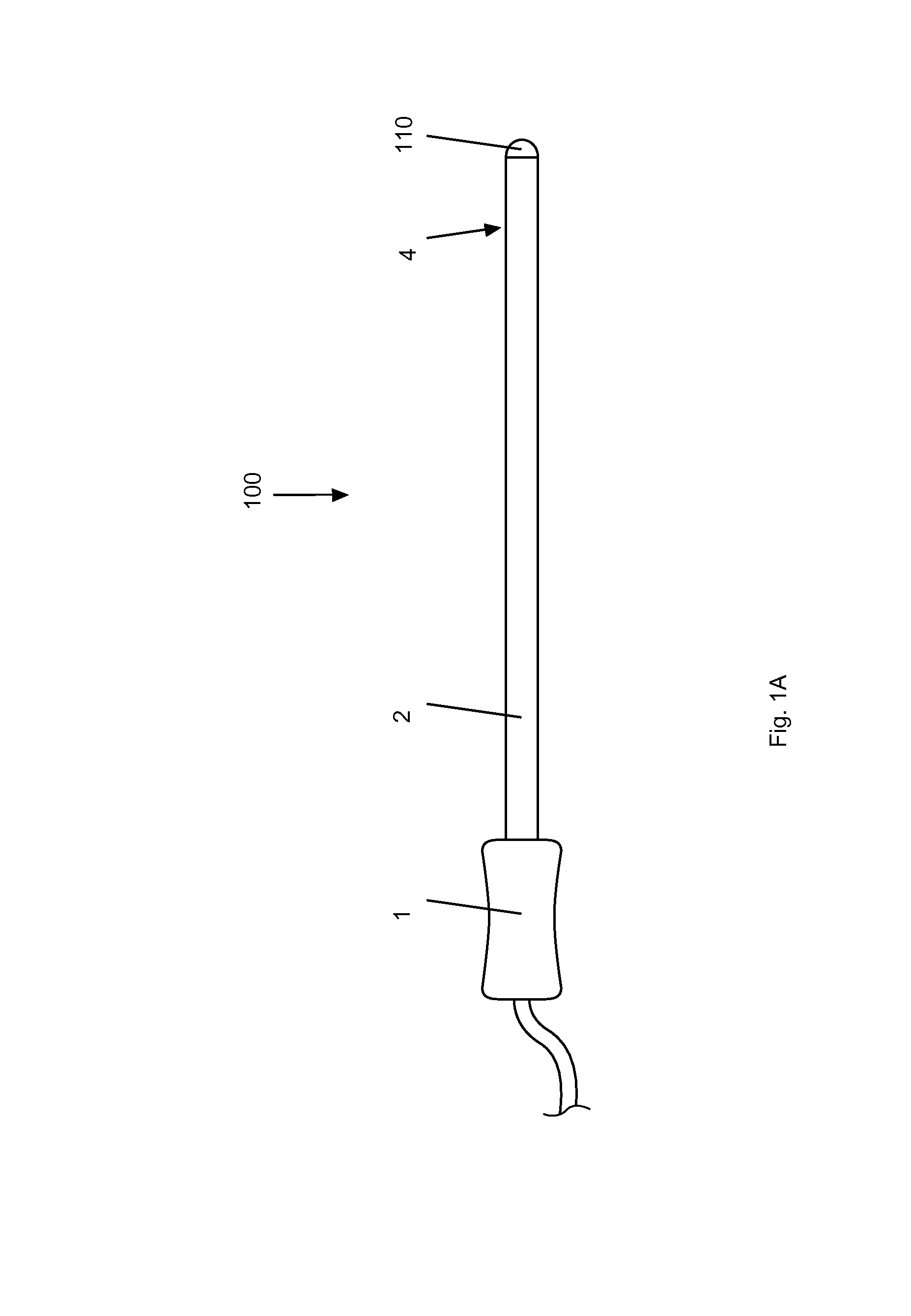
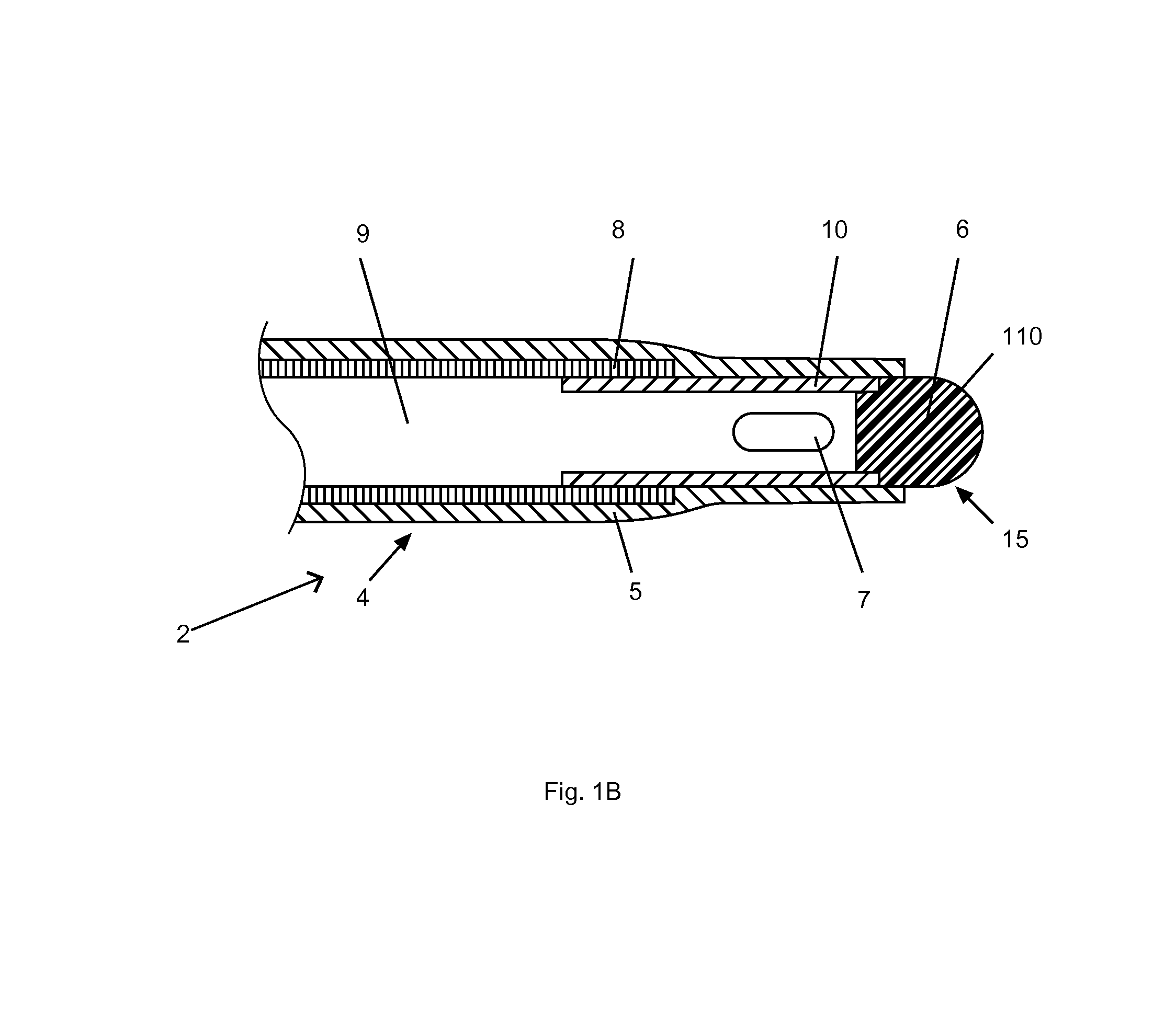
Described herein is a method for creating a channel through a foreign material located in a septum of a heart at the site of a septal defect. The foreign material defines a material first surface and a substantially opposed material second surface, and the channel extends through the foreign material at least partially between the material first and second surfaces. The method uses an apparatus including an electrode and includes the steps of: positioning the electrode substantially adjacent to the material first surface; energizing the electrode with a radiofrequency current; and using the electrode energized with the radiofrequency current to deliver energy into the foreign material to create the channel.

Description

CROSS-REFERENCES TO RELATED APPLICATIONS[0001]This application is a continuation in part of U.S. patent application Ser. No. 13 / 286,041, filed on 31 Oct. 2011, which is a continuation-in-part of U.S. patent application Ser. No. 11 / 905,448, now U.S. Pat. No. 8,048,071, filed on Oct. 1, 2007, which claims the benefit of U.S. provisional patent application Ser. No. 60 / 827,466 filed on 29 Sep. 2006. U.S. patent application Ser. No. 13 / 286,041, further claims the benefit of U.S. provisional application No. 61 / 448,578, filed on Mar. 2, 2011. All of these US patent applications and provisional patent applications are hereby incorporated by reference in their entirety.FIELD OF THE INVENTION[0002]The present invention relates generally to methods and devices usable to deliver energy within the body of a patient. More specifically, the present invention is concerned with a method for creating a channel through foreign material.SUMMARY OF THE INVENTION[0003]There are certain situations in whic...

Claims


Application Information

Patent Timeline
Patent Type & Authority Applications(United States)
IPC IPC(8): A61B18/18
CPCA61B18/18A61B8/12A61B18/1492A61B2018/00083A61B2018/00214A61B2018/00702A61B2018/00827A61B2018/00875A61B2018/00898A61B2018/00351A61F2002/061A61B2034/2055A61B2090/3788A61B90/02A61B34/76A61B2090/065A61B2090/3966
Inventor BIADILLAH, YOUSSEFHARTLEY, AMANDADAVIES, GARETHVISRAM, NAHEEDJUZKIW, TARASURBANSKI, JOHN PAUL
Owner BOSTON SCI MEDICAL DEVICE LTD
Learn More