
Intraoperative and blood derived adhesives

a technology applied in the field of intraoperative and blood derived adhesives, can solve the problems of limited commercially available adhesives, weak adhesive properties of materials, toxic and difficult deformation,

Inactive Publication Date: 2014-04-24
THE JOHN HOPKINS UNIV SCHOOL OF MEDICINE
View PDF0 Cites 0 Cited by
  • Summary
  • Abstract
  • Description
  • Claims
  • Application Information

AI Technical Summary

Benefits of technology

This patent describes a new type of hydrogel composition that can be used to fill voids in a subject. The hydrogel is made by combining a blood product and an amine-reacting proteoglycan, which can modify the pH, create cross-links, and enhance the stability of the hydrogel. This composition can be used to treat damaged or missing tissues and organs in the body, providing a promising avenue for regenerative medicine. The technical effects of the patent include improved stability and flexibility of the hydrogel, as well as the ability to fill voids and promote tissue regeneration.

Problems solved by technology

Commercially available adhesives are limited either by toxicity or lack of desired adhesive strength.
For example, cyanoacrylates adhere strongly to tissue but are toxic and difficult to degrade.
On the other end of the spectrum, fibrin glue is highly compatible and supports cell growth, but the material exhibits weak adhesive properties and degrades rapidly.
Therefore, a need exists for a biomaterial that is cytocompatible, has moderate tissue adhesive strength, and has a tunable degradation rate.
However, fibrin glues lack the desired adhesive strength and degrade rapidly.
However, these intraoperative biologics are difficult to place in the defect spaces because of their (liquid) consistencies.

Method used


Image

  • Intraoperative and blood derived adhesives
  • Intraoperative and blood derived adhesives
  • Intraoperative and blood derived adhesives

Examples


example 1

[0202]The method of CS—NHS synthesis using carbodiimide was as known in the art. The imide derivative significantly improved efficacy and biocompatibility. A CS-amine to act as the amine donor also was synthesized. For example, an about 3:3:1 ratio of CS, succinimide and diimide, respectively, can be reacted in a small volume of saline for a short period of time. A suitable ratio of the three reagents can be about 75:100:38, as a design choice.

[0203]In another embodiment, CS (750 mg) was dissolved in 6 ml: PBS (phosphate buffered saline). 1-Ethyl-3-[3-dimethylamino-propyl]carbodiimide (EDC, 1.572 g, 8.2 mmol) was dissolved in 1.5 ml PBS. A 3.3 mmol solution of N-hydroxysuccinimide (NHS) was made by dissolving 380 mg in 1.5 ml PBS. The NHS solution and the EDC were added to the CS solution, vortexed, and allowed to react for 10 minutes at 37° C. The reaction was then chilled for 30 minutes at −80° C. and precipitated with ethanol. The solution was then centrifuged for 5 min and the s...

example 2

[0212]In this example, a chemically modified glycosaminoglycan (GAG) was used in combination with blood to create a tissue adhesive with tunable physical and biological properties. The mechanical properties and biological composition of the adhesive were varied to control physical and biological interactions. Finally, the ability of stem cells to form new tissue in the hydrogel was evaluated.

[0213]Cell spreading and cell migration through materials is important for regenerating the surrounding tissue and facilitating tissue integration. Commonly, integrin-binding peptides (IBP) such as YRGDS or proteins containing such peptides (i.e., fibronectin and collagen) are added to scaffolds to improve cell spreading and cell migration. However, purified extracellular matrix proteins are relatively expensive and can be difficult to incorporate into scaffolds at high concentrations. Blood, on the other hand, is rich in many proteins with IBP motifs, is readily available, and is naturally in a...

example 3

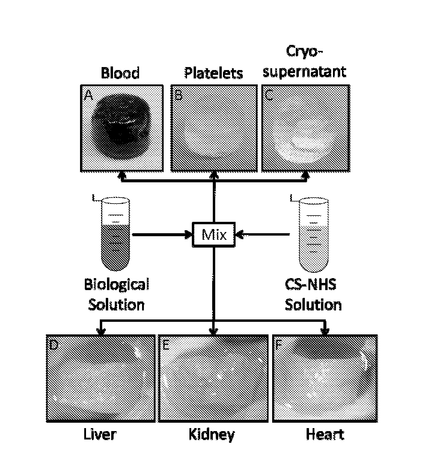
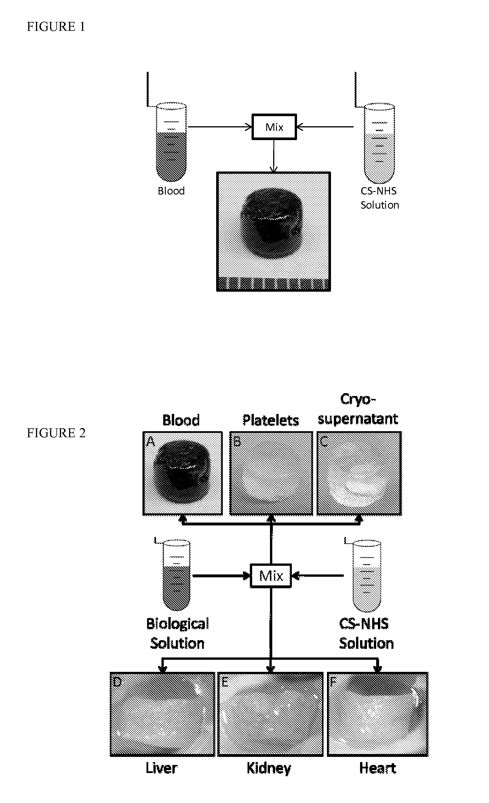
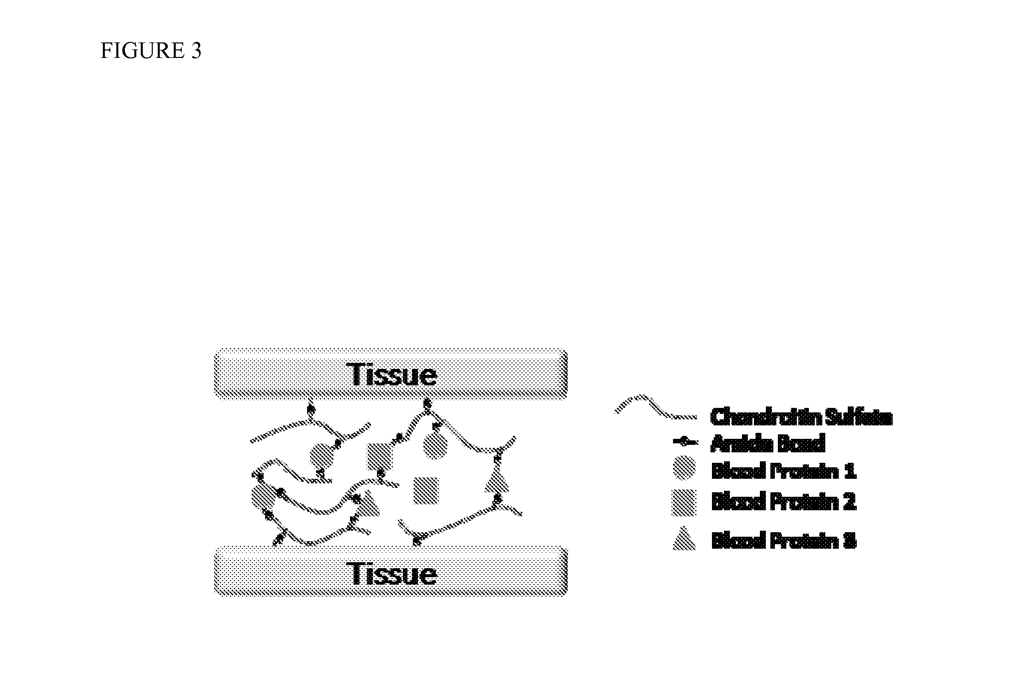
[0218]Mechanism of gelation and adhesion. A two component, in situ forming biomaterial was created by combining chemically modified chondroitin sulfate (CS—NHS) with blood (FIG. 1). The CS—NHS component reacts with primary amines found on proteins in blood and tissues. Hence, the material is a bioadhesive due to reaction of the CS—NHS with proteins in tissue that covalently anchors the hydrogel to the tissue. Gelation is initiated once the two components are mixed together, and the crosslinks are stabilized via amide bonds (FIG. 2). Thus, a hydrogel forms within a minute and becomes progressively stiffer over a period of 10 minutes. In addition to whole blood, it was observed that the CS—NHS molecule can react with proteins in bone marrow and plasma to form adhesive hydrogels. Therefore, any biological solution containing an adequate number of primary amine bearing polymers (i.e., proteins) can potentially form a bioadhesive when combined with CS—NHS.


PUM

PropertyMeasurementUnit
v/vaaaaaaaaaa
v/vaaaaaaaaaa
v/vaaaaaaaaaa

Abstract

The invention features the production of an amine-reactive proteoglycan, specifically chondroitin sulfate or hyaluronic acid. This material can be provided in powder (solid) or liquid form and combined with blood derivatives including serum, platelets, platelet rich plasma, bone marrow, or with other tissue products to form hydrogels. The properties (physical and biological) are different for each of these hydrogels and can be further manipulated by controlling the conditions under which the hydrogels are formed. Such properties include the biodegradability of the hydrogel, the compressibility, the adhesive strength, the presence of pharmaceutical agents or therapeutic cells, and resiliency.

Description

REFERENCE TO RELATED APPLICATIONS[0001]This application claims the benefit of U.S. Provisional Patent Application No. 61 / 470,763, filed on Apr. 1, 2011, which is hereby incorporated by reference for all purposes as if fully set forth herein.BACKGROUND OF THE INVENTION[0002]Clinically there exists a need for adhesive biomaterials that bond tissues and support tissue growth. Commercially available adhesives are limited either by toxicity or lack of desired adhesive strength. Additionally, properties such as degradation should be tunable as rate of healing varies in different tissues. For example, cyanoacrylates adhere strongly to tissue but are toxic and difficult to degrade. On the other end of the spectrum, fibrin glue is highly compatible and supports cell growth, but the material exhibits weak adhesive properties and degrades rapidly. Therefore, a need exists for a biomaterial that is cytocompatible, has moderate tissue adhesive strength, and has a tunable degradation rate.[0003]B...

Claims


Application Information

Patent Timeline
Patent Type & Authority Applications(United States)
IPC IPC(8): A61L24/08A61L24/00
CPCA61L24/0005A61L24/08A61K31/727A61K31/728A61K31/737A61K35/14A61K9/06A61K47/46A61L27/20A61L27/3616A61L27/52A61L2400/06C08L5/08
Inventor ELISSEEFF, JENNIFER H.STREHIN, IOSSIF
Owner THE JOHN HOPKINS UNIV SCHOOL OF MEDICINE
Learn More