Breast Reconstruction Device and Methods

a breast and breast technology, applied in the field of breast reconstruction devices and methods, can solve the problems of reducing the surgical efficiency of breast reconstruction, showing any promise, and increasing the risk of complications, so as to eliminate any hope of robust neo-vascularization and inhibit angiogenesis

Inactive Publication Date: 2014-05-15
BIOSTRUXS
View PDF0 Cites 19 Cited by
  • Summary
  • Abstract
  • Description
  • Claims
  • Application Information

AI Technical Summary

Problems solved by technology

No new design innovation on the horizon has shown any promise to prevent this complication from occurring in some patients.
The fundamental problem appears to be caused by a nonspecific inflammatory response to a foreign body introduced into human tissue.
Unfortunately, as seen in the above statistics, revision augmentation cases will have an even higher rate of eventual contractures.
Subsequent surgeries will be more difficult, and in many cases will require autologous tissue flaps to preserve the patient's breasts.
It is difficult to envision that this device would work on post-mastectomy cases, because there is only skin left without breast in the setting of adjuvant cancer treatments that inhibit new tissue and blood vessel formation.
However, tissue resorption often occurs when non-vascularized grafts are transferred in human autograft transplantation.
This makes it difficult to compensate for resorption by overgrafting with larger volumes.
This artifact makes cancer surveillance difficult with mammography.
As a consequence, fat grafting for breast augmentation may make future cancer detection difficult.
Fat necrosis also causes concerns when it occurs in the reconstructed breast.
Fat grafting certainly does not produce enough volume to make an entire breast, therefore it is not a viable option for breast reconstruction.
It is difficult to envision that this and similar procedures would effectively provide enough volume to replace one or two entire breasts, as needed for mastectomy cases.
It is constructed from only polyglycolic acid, is inert, nonantigenic, noncollagenous, and does not enhance any secondary infection.
Despite these successes, major roadblocks still exist in translational research.
As a consequence, tissue engineering on scaffolds is limited in size by the lack of arterial and venous structures which do not grow as well as capillaries.
Of course, this concept would not work for organogenesis.
However, cells would perish without new blood vessels, and this idea did not materialize into practical use since its issued patent in 1998.
In the case of breast reconstruction, immediate reconstruction after mastectomy is a particularly challenging situation for tissue engineering.
Furthermore, a significant number of post-mastectomy patients also need postoperative radiation to the chest and therefore the new “breast.” Radiation effectively eliminates any hope for robust neo-vascularization, not just for the moment but for the rest of the patient's life.
However, some major disadvantages have prevented omental flaps from wide clinical acceptance.
The shape of the resulting reconstructed breast can widely vary due to lack of structural support.
The size of the omentum is variable in individuals, and there is no reliable method prior to surgery to accurately estimate its size.
Usually, the omentum is too small to adequately reconstruct both breasts and sometimes is too small to reconstruct even only one breast.

Method used


Image

  • Breast Reconstruction Device and Methods
  • Breast Reconstruction Device and Methods
  • Breast Reconstruction Device and Methods

Examples


example 1

Rat Study

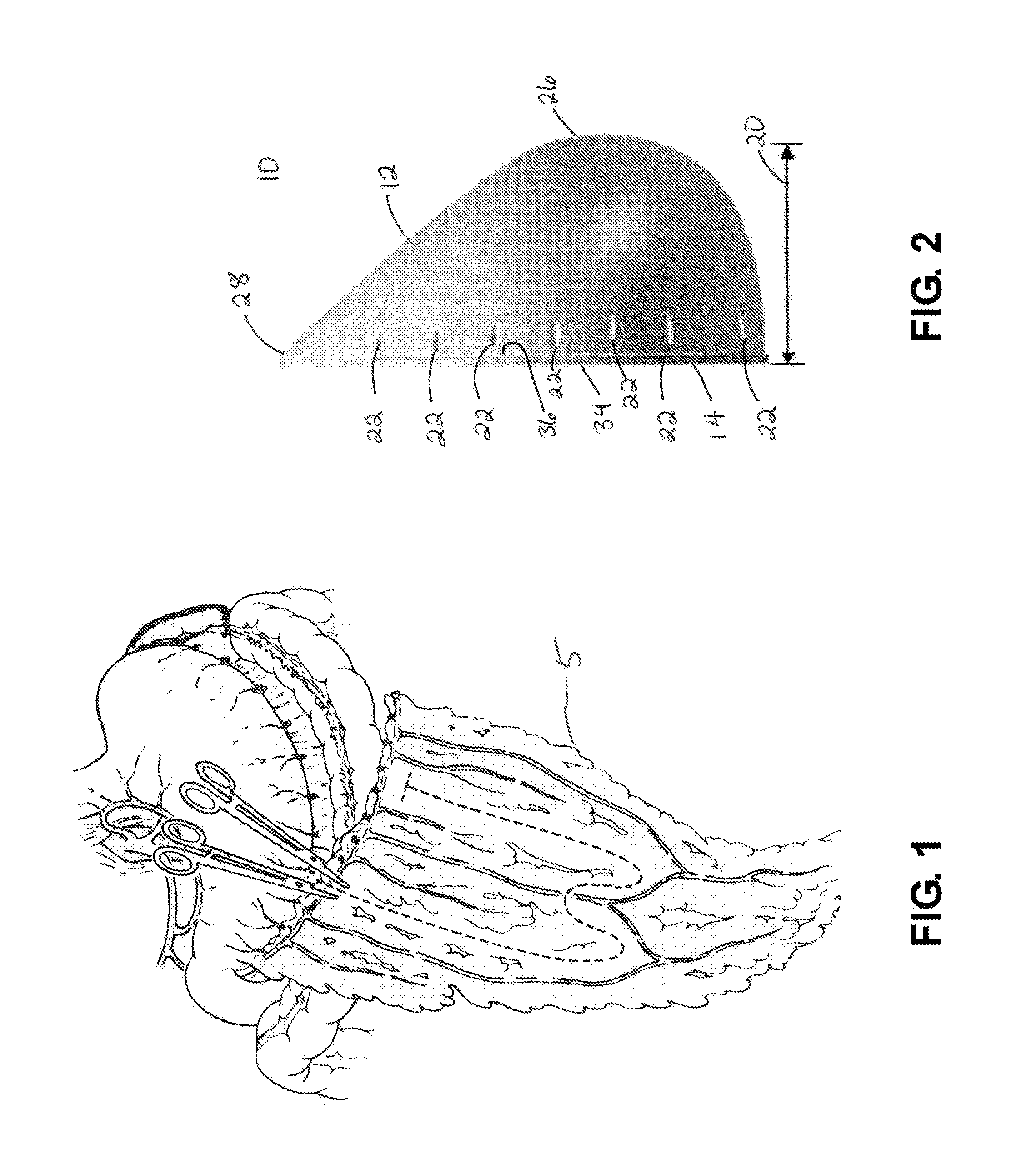
[0101]Preliminary experiments were carried out in Sprague Dawley female rats at approximately 3.5 months in age. Under general anesthesia, an incision was made in the inguinal region of the rat, and a portion of the rat's fat tissue was harvested. This fat tissue was manually mixed with PuraMatrix (Becton Dickinson) in 10 percent sucrose solution. The fat tissue matrix or mixture was placed inside a biodegradable mesh pocket fabricated of Dexon mesh (U.S. Surgical Corporation), and secured shut with sutures. A midline laparotomy incision was made in the same individual rat, its omentum was identified and wrapped around the mesh pocket, and secured with sutures. The rats tolerated the surgery well, and recovered without any complications. Four weeks later, the rats were sacrificed. The mesh pockets with fat inside were placed in paraffin, and Hematoxylin & Eosin, H&E, stained slides were generated.

[0102]The results demonstrated that the fat tissue inside the mesh pocket surv...

example 2

Pig Study

[0104]For a pig study, a 9-month old Yucatan female pig weighing 50 kg was used. Under general anesthesia, an incision was made in the subcutaneous abdominal region of the pig, and a portion of the pig's fat tissue was harvested. A midline laparotomy incision was made, the omentum identified, and placed inside the scaffold. The fat tissue matrix was placed inside the implant between the folds of the omental, and the base and body of the implant was secured shut with sutures. The implant remained inside the pig's abdominal cavity, attached to the omentum blood supply. The laparotomy incision was closed with running sutures.

[0105]The pig was observed during recovery daily by veterinary staff and exhibited no signs of complications. Four weeks later, the pig was sacrificed. The time period of four weeks was chosen because it usually takes at least two weeks to develop histological evidence of fat necrosis. The implant with omentum and fat inside was retrieved. In the breast, f...


PUM

PropertyMeasurementUnit
median volumeaaaaaaaaaa
thicknessaaaaaaaaaa

Abstract

A novel human breast implant and method for using the same comprising a bioabsorbable implant into which native, autologous vascularized tissue and autologous fat is placed and propagated within a patient's chest as a breast implant.

Description

RELATED APPLICATIONS[0001]This continuation claims priority pursuant to 35 U.S.C. §120 to patent application Ser. No. 13 / 098,304, filed Apr. 29, 2011, a U.S. Non-Provisional Patent Application that claims priority to U.S. Provisional Application Ser. No. 61 / 329,496, filed Apr. 29, 2010, entitled Breast Reconstruction Device and Methods, and is related to the subject matter of the Assignee's U.S. Pat. No. 7,846,728, entitled Tissue Engineering In Vivo With Vascularized Scaffolds, filed Oct. 9, 2007, which claims priority to U.S. Provisional Application Ser. No. 60 / 851,686, entitled Tissue Engineering In Vivo With Vascularized Scaffolds, filed Oct. 13, 2006, the contents of which are each incorporated in their entirety herein.FIELD OF THE INVENTION[0002]The present invention relates to devices and related methods for organ reconstruction and, more particularly, to devices and methods for the reconstruction of breasts.BACKGROUND OF THE INVENTION[0003]Breast cancer is the most common fo...

Claims


Application Information

Patent Timeline
Patent Type & Authority Applications(United States)
IPC IPC(8): A61F2/12
CPCA61F2/12A61F2210/0004
Inventor BROOKS, MAI N.WATSON, JAMES P.
Owner BIOSTRUXS
Why Eureka
Try Eureka