
Anti-cd3 therapies

a technology of anti-cd3 and anti-cd4, applied in the field of anti-cd3 therapies, can solve the problems of affecting other cells, unable to be specific, and cell cytotoxicity, and achieve the effect of reducing severity and/or

Inactive Publication Date: 2014-05-22
MAYO FOUND FOR MEDICAL EDUCATION & RES
View PDF2 Cites 3 Cited by
  • Summary
  • Abstract
  • Description
  • Claims
  • Application Information

AI Technical Summary

Benefits of technology

The patent describes a method for using an anti-CD3γε antibody to induce immunosuppression without causing a cytokine release syndrome or internalizing the antibodies. This antibody can trigger the death of T cells through apoptosis, reducing the severity and allowing recovery from autoimmune diseases like multiple sclerosis. The method can also reduce the likelihood of transplant rejection in mammals by killing T cells without increasing production of cytokines. The antibody used can be a humanized version of an anti-CD3γε antibody.

Problems solved by technology

While non-immunolglobulin drugs can be efficient to cause T cell cytotoxicity, they fail to be specific and they also can affect other cells.
Anti-CD3ε antibodies can neutralize T cell function mainly by causing the death of T cells, but can also result in the undesired activation of some T cells that escape the death.
Administration of the preparation to a mammal can induce T cell death within the mammal.
Administration of the preparation to a mammal can induce T cell death within the mammal with no detectable T cell division.

Method used


Image

  • Anti-cd3 therapies
  • Anti-cd3 therapies
  • Anti-cd3 therapies

Examples


example 1

Effects of Anti-CD3γε Dimer Antibodies

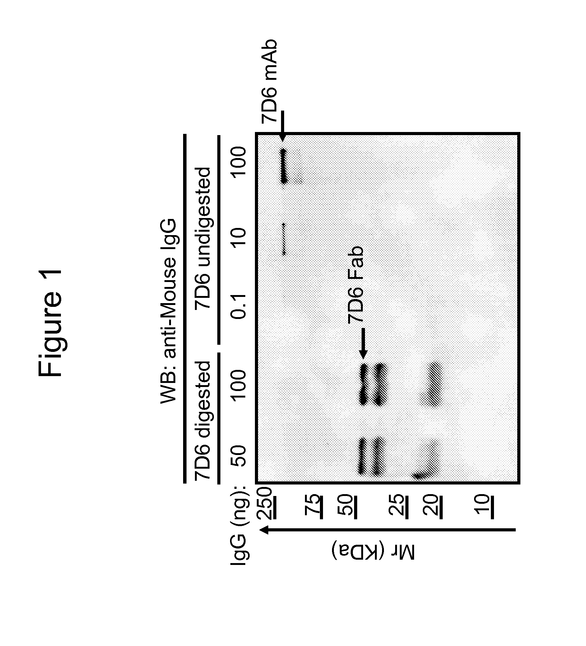
[0052]The monoclonal 7D6 antibody was generated by injecting T cells from a B6 mouse into 129 / Sv mice as described elsewhere (Coulie et al., Eur. J. Immunol., 21: 1703-1709 (1991)). Fab fragments were made by digesting purified 7D6 mAb with papain during 24 hours at 37° C. FIG. 1 demonstrates Western blot quality controls for 7D6 Fab preparations. Titrated amounts of 7D6 mAb or Fab preparations were analyzed by SDS-PAGE and Western blot with anti-mouse IgG. The Fab was about 50 kDa. The smaller molecular weight Ig species in the Fab lanes originated from intact Fab that falls apart during boiling and preparation for Western blot prep (as confirmed by Size Exclusion Chromatography (SEC) analysis). 7D6 digestion appeared free of undigested 7D6 mAb at the level of detection of the assay. Thus, a ratio of 7D6 Fab / mAb was established as being ≦1 μg / ng in the preparation.

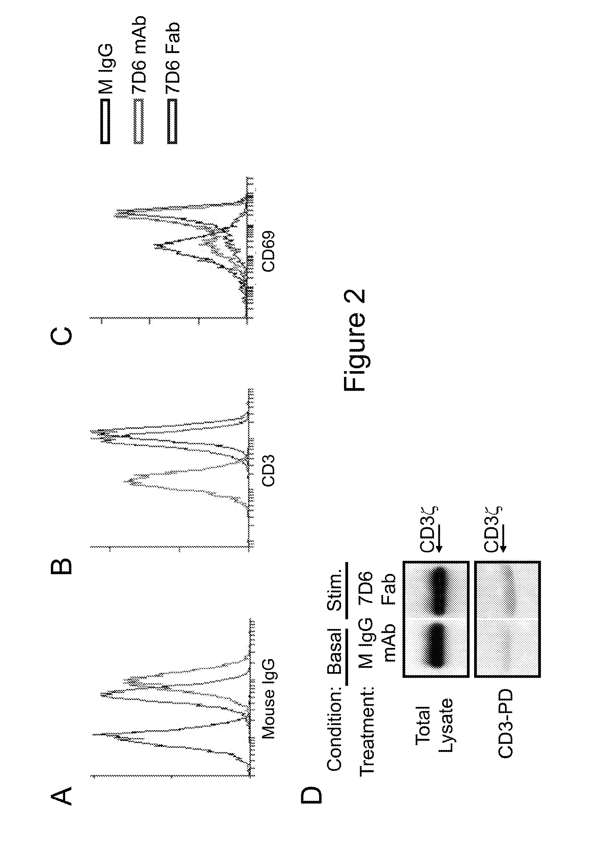
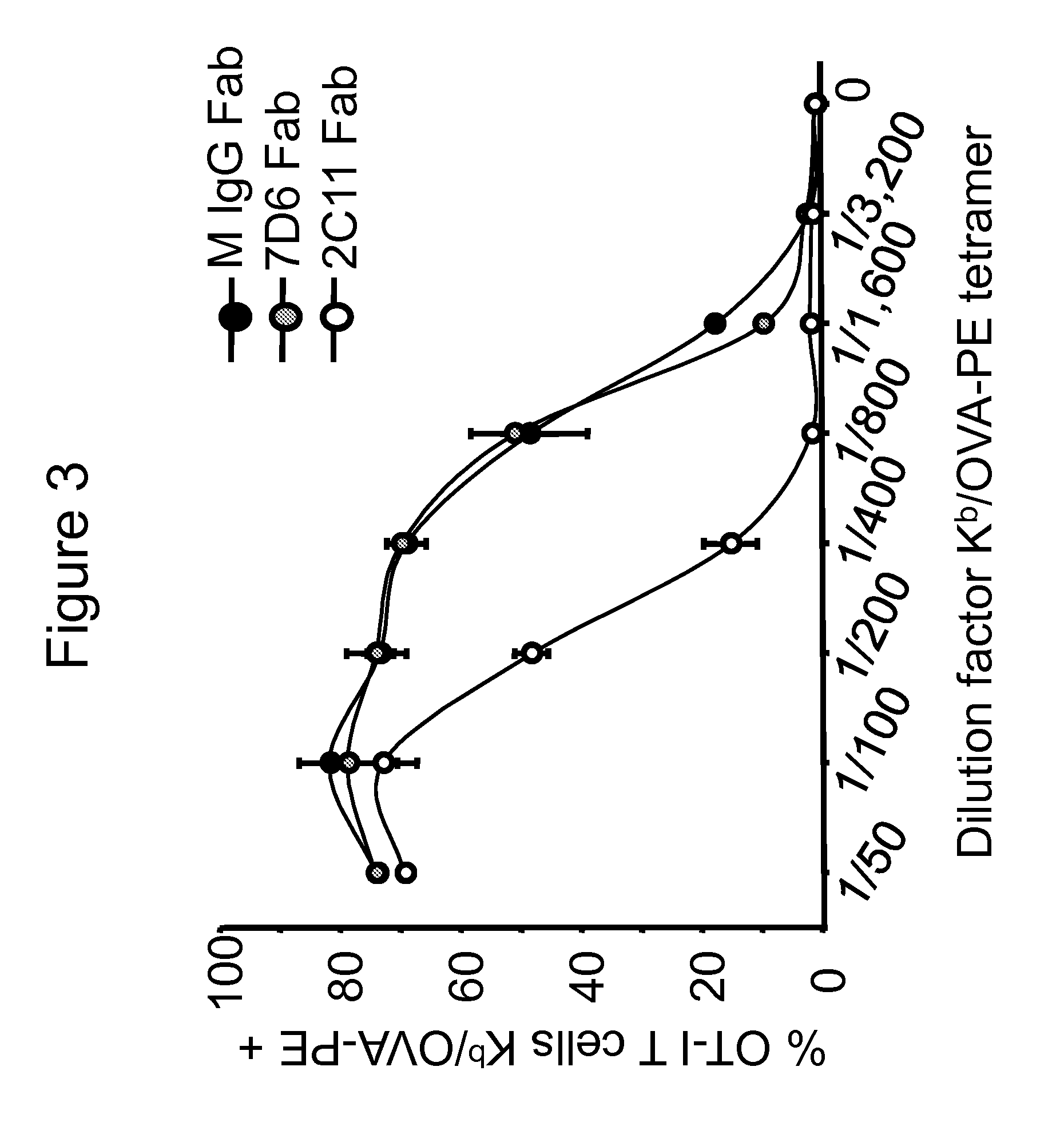
[0053]Next, the binding and stimulatory capacity of 7D6 Fab on T cells was tested...

example 2

Producing Humanized Anti-Human CD3γε Antibodies

[0070]A first nucleic acid construct is constructed to encode the extracellular domain of human CD3εfused to the transmembrane and cytoplasmic domains of mouse CD3ε, while a second nucleic acid construct is constructed to encode the extracellular domain of human CD3γ fused to the transmembrane and cytoplasmic domains of mouse CD3γ. The first and second nucleic acid constructs are engineered into a vector to produce a multicistronic vector. A transfection technique such as a retroviral transduction is used to introduce the multicistronic vector into mouse T cells such that the mouse T cells are capable of expressing the human / mouse chimeric CD3ε polypeptide and the human / mouse chimeric CD3γ polypeptide. A monoclonal antibody specific for the human CD3εextracellular domain (e.g., OKT3) is used to test for proper surface expression of the chimeric human / mouse CD3γε dimer.

[0071]Mouse T cells expressing the chimeric human / mouse CD3γε dimer a...


PUM

PropertyMeasurementUnit
Cell deathaaaaaaaaaa
Immunogenicityaaaaaaaaaa

Abstract

This document provides methods and materials related to anti-CD3 therapies. For example, anti-CD3γε antibody preparations as well as methods and materials for using anti-CD3γε antibody preparations to reduce a T cell-mediated immune response within a mammal are provided.

Description

CROSS-REFERENCE TO RELATED APPLICATION[0001]This application claims benefit of U.S. Provisional Application Ser. No. 61 / 496,886, filed Jun. 14, 2011. The disclosure of the prior application is considered part of (and is incorporated by reference in) the disclosure of this application.BACKGROUND[0002]1. Technical Field[0003]This document provides methods and materials related to anti-CD3 therapies. For example, this document provides anti-CD3γε antibody preparations as well as methods and materials for using anti-CD3γε antibody preparations to reduce a T cell-mediated immune response within a mammal.[0004]2. Background Information[0005]T cells can drive adaptive immune responses that can be pathogenic when directed against a body's own tissues and organs. In addition, T cells can be the main cellular component that drives acute organ rejection after transplantation. Different strategies for depleting / inactivating T cells have been pursued to treat patients either suffering from autoi...

Claims


Application Information

Patent Timeline
IPC IPC(8): A61K39/395
CPCC07K16/2809A61K2039/505A61K39/39566C07K2317/55C07K2317/73C07K2317/24A61P3/00A61P3/10
Inventor GIL PAGES, DIANASCHRUM, ADAM G.
Owner MAYO FOUND FOR MEDICAL EDUCATION & RES
Learn More