
Method for preparing corneal donor tissue for refractive eye surgery utilizing the femtosecond laser

a corneal donor and laser technology, applied in laser surgery, medical science, surgery, etc., can solve the problems of unpredictability of the final shape of the lenticule, unpredictability and dehydration of the lenticule material

Inactive Publication Date: 2014-06-05
CUMMING JAMES STUART
View PDF13 Cites 32 Cited by
  • Summary
  • Abstract
  • Description
  • Claims
  • Application Information

AI Technical Summary

Benefits of technology

The patent is about a method for making a corneal inlay that can be implanted into a patient's eye. The method allows for the creation of the inlay in large quantities and with a high degree of precision before being attached to the eye. The inlay should not change shape or clarity during the fabrication process or when placed in the eye. Overall, the method provides a way to make a reliable and effective corneal inlay for use in eye surgery.

Problems solved by technology

However, such materials suffer from unpredictability regarding the final shape of the lenticule, and therefore the refractive correction of the surgical procedure.
Since the corneal stroma is maintained in a partially dehydrated state, by the fluid barrier provided by the corneal epithelium, and the fluid pumping action provided by the corneal endothelium, the lenticule material is also subject to dehydration, which is not totally predictable.
Accordingly, a major problem, which is encountered when human corneal tissue is used to form the lenticule its unpredictability, as discussed above.
Specifically, it is not presently feasible to determine the refractive power of the resulting lenticule prior to its attachment to the eye of the recipient.
This problem is compounded by the fact that if a number of human corneal tissues are lathed to a particular thickness, they tend to exhibit different refractive powers.
These problems lead to substantial trial and error in present-day refractive surgical procedures, often requiring multiple attempts to form a lenticule, with the correct refractive power.
However, many patients complain of blurred vision, glare and halos resulting in difficulty in driving at night, and loss of depth perception; they are unable to tolerate these side effects.
While a number of multifocal intraocular lenses are commercially available and have been shown to allow good functional vision without the use of corrective lenses, they are also known to cause glare disability, and halos.
Additionally, since conical inlays utilize the monovision treatment with a Laser-Assisted in situ Keratomileusis (“LASIK”) procedure, they have the associate risks described above for monovision.
Moreover, a major concern with corneal inlays is the potential for deformation of the cornea (keratectasia), which can lead to a severe decrease in vision, which is very difficult to treat.
The cornea is subjected to the internal pressure of the eye, which, should the structural integrity of the cornea be reduced (e.g. by LASIK), can cause a bulging forward of the posterior conical segment with a resulting deformation of the anterior corneal surface, leading to conical ectasia.
The intraocular pressure causes the thin posterior cornea to bulge forward, distorting the anterior flap resulting in irregular astigmatism frequently leading to the need for a conical transplant.

Method used


Image

  • Method for preparing corneal donor tissue for refractive eye surgery utilizing the femtosecond laser
  • Method for preparing corneal donor tissue for refractive eye surgery utilizing the femtosecond laser
  • Method for preparing corneal donor tissue for refractive eye surgery utilizing the femtosecond laser

Examples


Embodiment Construction

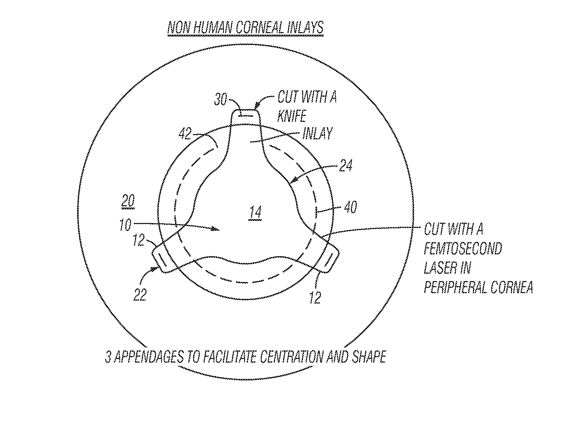
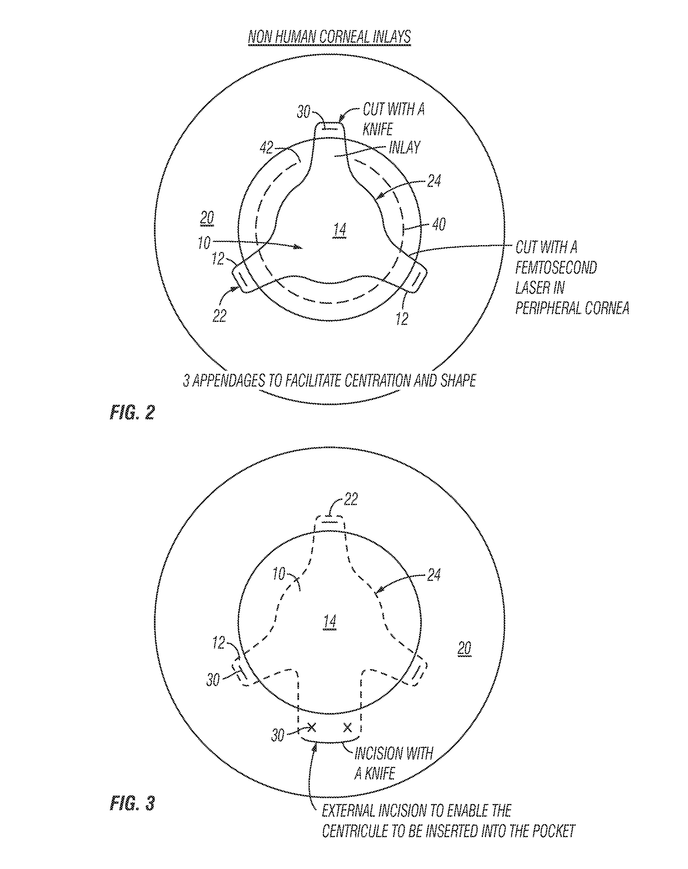
[0036]The above described drawing figures illustrate the described invention in at least one of its preferred, best mode embodiment, which is further defined in detail in the following description. Those having ordinary skill in the art may be able to make alterations and modifications to what is described herein without departing from its spirit and scope. Therefore, it should be understood that what is illustrated is set forth only for the purposes of example and should not be taken as a limitation on the scope of the present invention.

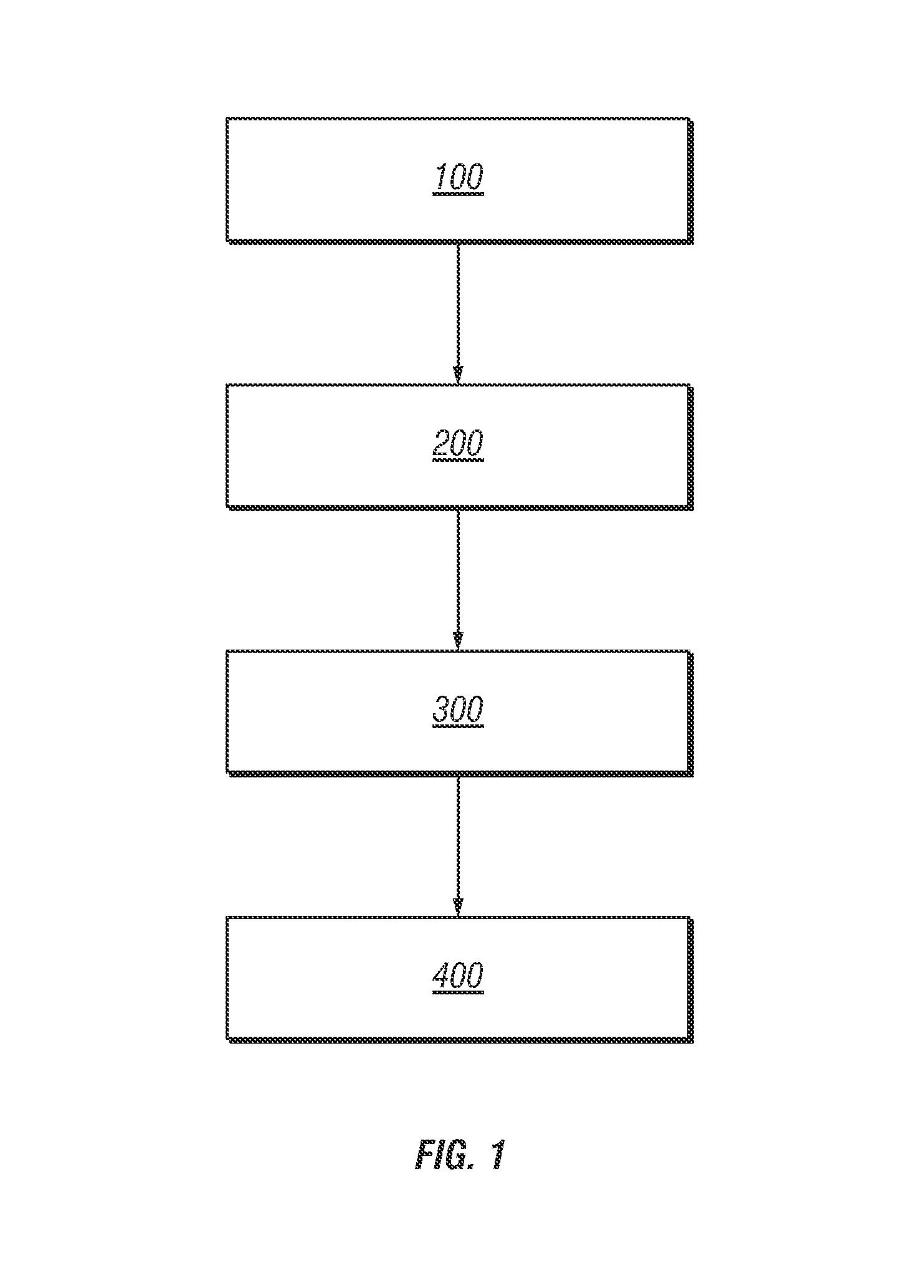
[0037]FIG. 1 illustrates a method for preparing cortical donor tissue for refractive eye surgery utilizing a femtosecond laser according to a preferred embodiment of the present invention.

[0038]Step 100: Donor corneal tissue is removed from a donor cornea. The donor cornea is preferably selected from a group of animals consisting of: pigs, cows, rabbits, cats, dogs, primates, cetacean dolphins, sharks, and warm blooded animals and chondrychthes. The...


PUM


Abstract

A method of accurately fabricating a non-human donor corneal tissue for implantation into a recipient human cornea, the method comprising the steps of: removing corneal tissue from a donor with a femtosecond laser; placing the corneal tissue in a fixative solution for a selected time interval to cross-link the collagen fibrils in the tissue and prevent swelling of the corneal tissue; and shaping the tissue to provide a conical inlay of a selected shape and thickness having one or more radial extensions. The method is such that the corneal inlay may be attached to the peripheral corneal and / or the sclera. The method is such that the corneal inlay may be stored for a period of up to two years prior to attachment.

Description

RELATED APPLICATIONS[0001]This application claims priority to U.S. Provisional Patent Application No. 61 / 288,265, filed on May 10, 2012, the entire contents and disclosure of which is hereby incorporated by reference.BACKGROUND OF THE INVENTION[0002]The correction of refractive errors of the eye by the attachment of donor material directly onto, or into, the cornea or by corneal transplant has been carried out in the prior art for several years, One technique is known as “epikeratophakia”, This technique involves the formation of a ring-shaped trough in the recipient corneal bed, and the placement and suturing of the donor material onto the optical center of the cornea with its edges fitting into the trough. This technique requires that the epithelium of the cornea grow over the resulting lenticule, which attaches the lenticule to the surface of the cornea.[0003]Another prior art procedure for refractive correction of the eye is a technique referred to as “intralamellar insertion”. ...

Claims


Application Information

Patent Timeline
IPC IPC(8): A61F9/008A61F2/16
CPCA61F2/142A61F9/00834A61F2/1451
Inventor CUMMING, JAMES STUART
Owner CUMMING JAMES STUART
Learn More