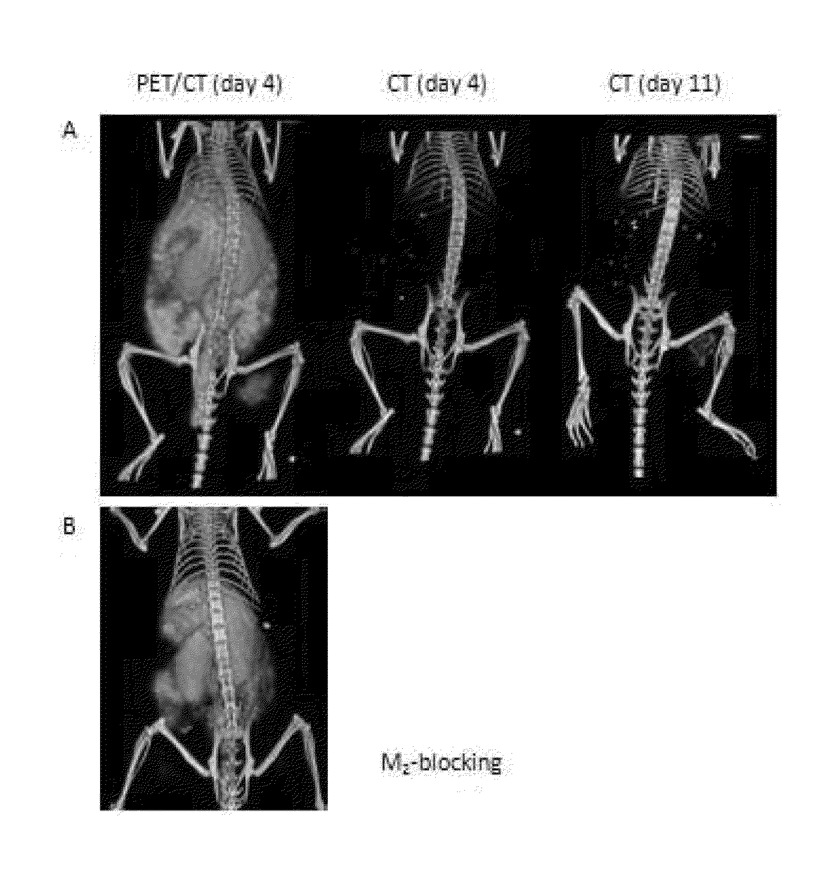

Methods for imaging bone precursor cells using dual-labeled imaging agents to detect mmp-9 positive cells

a bone precursor cell and imaging agent technology, applied in the field of cell biology, molecular biology, imaging, diagnostics, etc., can solve the problem of a single agent capable of carrying dual-contrast for both nuclear and ct or mri imaging modalities, and achieve the effect of reducing the difficulty of developmen

Inactive Publication Date: 2014-06-26
BAYLOR COLLEGE OF MEDICINE
View PDF0 Cites 4 Cited by
  • Summary
  • Abstract
  • Description
  • Claims
  • Application Information

AI Technical Summary

Benefits of technology

The patent describes methods and compositions for identifying and preventing the formation of heterotopic bone growth. Heterotopic bone growth can occur when there is an injury or other event that damages bones, resulting in the growth of bone in areas where it is not normally found. The invention provides detectable agents that can be used to identify markers of heterotopic bone growth, and therapeutic agents that can prevent or reduce the growth of heterotopic bone. These agents can be used in individuals at risk for developing heterotopic bone growth, such as individuals with certain genetic markers or who have undergone joint replacement surgery. The invention can help to improve the diagnosis and treatment of abnormal bone cell growth.

Problems solved by technology

The development of a single agent capable of carrying dual-contrast for both nuclear and CT or MRI imaging modalities proves challenging due to the inherent differences in measurement sensitivities between nuclear and all other conventional imaging modalities.

Method used


Image

  • Methods for imaging bone precursor cells using dual-labeled imaging agents to detect mmp-9 positive cells
  • Methods for imaging bone precursor cells using dual-labeled imaging agents to detect mmp-9 positive cells
  • Methods for imaging bone precursor cells using dual-labeled imaging agents to detect mmp-9 positive cells

Examples


example 1

Exemplary Experimental Procedures

[0112]Reagents

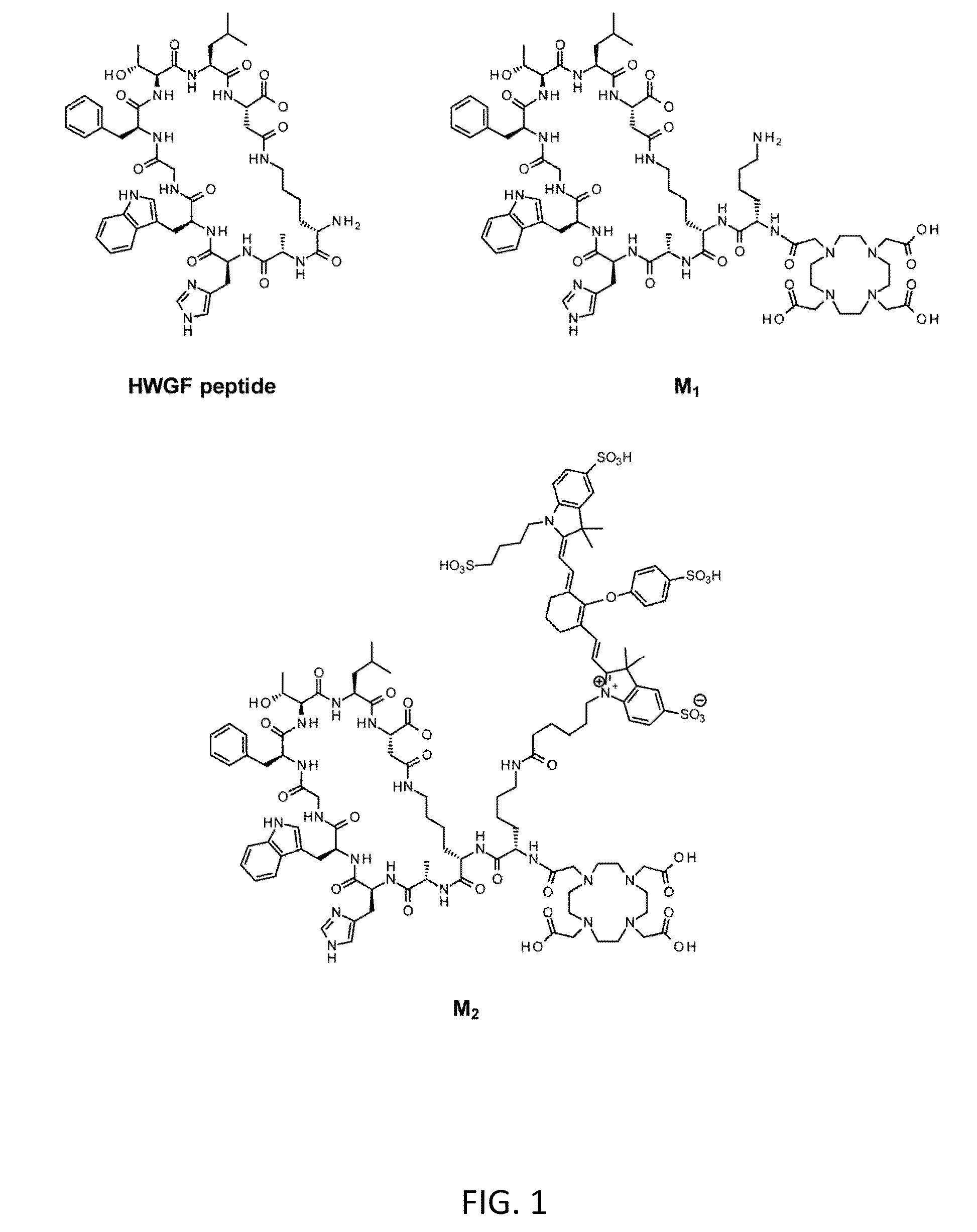
[0113]All reagents were purchased from commercial sources and used without further purification. Chelex-100 resin was purchased from Bio-Rad Laboratories (Richmond, Calif.) and used with all aqueous buffers to ensure metal-free conditions. The DOTA (1,4,7,10-tetraazacyclotetradecane-N′,N″,N″′,N″″-tetraacetic acid)-modified cyclic MMP-targeting peptide (lactam 2,10) DOTA-KKAHWGFTLD (M1) (SEQ ID NO:1) was synthesized by New England Peptide (Gardner, Mass.) according to standard Fmoc-protocols. A commercially-available 68Ge / 68Ga generator was purchased from Eckert & Ziegler (Berlin, Germany). Analytical high-performance liquid chromatography (HPLC) was performed on a Hitachi LaChrom system equipped with a 2.6 μm Kinetex C-18 column (Phenomenex, Torrance, Calif.) with a mobile phase of A=0.1% TFA in H2O, B=0.1% TFA in CH3CN; gradient, 0 min=10% B, 10 min=90% B; flow rate, 1 mL / min. Radio-thin-layer chromatography (radio-TLC) was carried out...

example 2

Conjugation of IRDye 800CW

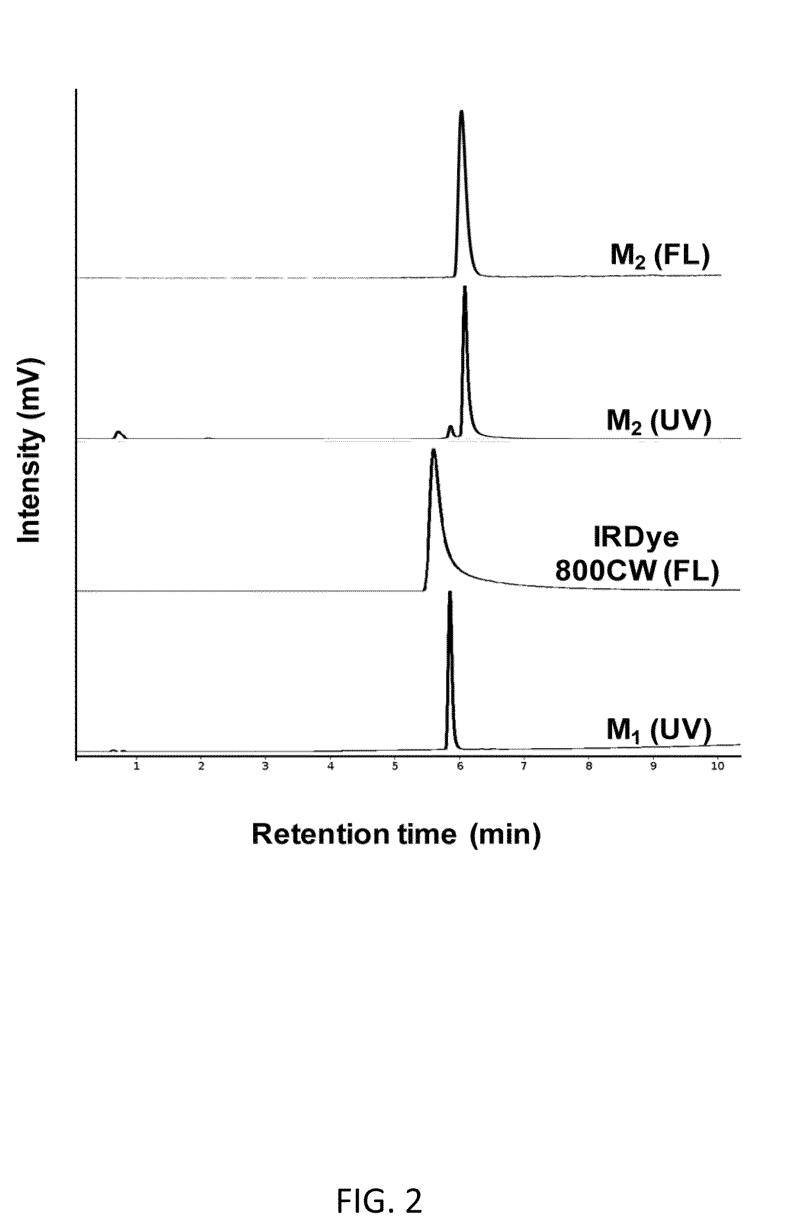
[0132]The dual-conjugate M2 was formed through attachment of IRDye 800CW to M1. Analysis of the spin column-purified sample by HPLC showed >90% purity with a small amount of unreacted M1 present in the final product and was used for all subsequent studies. FIG. 2 shows HPLC chromatograms with UV detection at 280 nm and fluorescence detection. Retention times of 5.9 min and 6.1 min were observed for M1 and M2 (at 280 nm), respectively, while fluorescence detection of IRDye 800CW had a retention time of 5.6 min. The HPLC data shows a single fluorescent peak for M2, confirming formation and purity of the dual-conjugate. Analysis by ESI-MS showed that the observed molecular weight (2555.9) was in excellent agreement with the calculated value (2555.92).

example 3

Radiochemistry

[0133]M2 was radiolabeled with 68Ga using the fractionation method and radiochemical purity (RCP) of >95% were achieved within 10 min, with 13 min selected as the optimal heating time. FIG. 3 shows that 68Ga-M2 is formed with high labeling efficiency over the range of peptide amounts investigated. Similar RCP were observed between the peptide amounts tested, therefore, 6 nmol (15 μg) was selected for radiolabeling experiments to achieve the highest specific activity and reaction conditions were optimized using 0.1 N NaOAc buffer. Co-injection of natGa-M2 and 68Ga-M2 on HPLC showed excellent correlation between the UV, fluorescent, and radiometric peaks (FIG. 4). The log P value for 68Ga-M2 was calculated to be −2.09±0.02.


PUM

PropertyMeasurementUnit
Injection velocityaaaaaaaaaa

Abstract

The present invention includes embodiments for methods and compositions that identify the presence of or risk for developing heterotopic ossification, particularly prior to mineralization of the bone. In particular embodiments, MMP-9 and / or MMP-2 agents comprising dual imaging moieties are used to identify patterns of MMP-9 and / or MMP-2 localization, respectively, that is then predictive of heterotopic ossification.

Description

CROSS-REFERENCE TO RELATED APPLICATIONS[0001]This Application claims the benefit of U.S. Provisional Application 61 / 483,600, filed on May 6, 2011, which is incorporated by reference herein in its entirety.STATEMENT REGARDING FEDERALLY SPONSORED RESEARCH OR DEVELOPMENT[0002]The invention was made with government support under R01EB005173 awarded by National Institute of Biomedical Imaging and Bioengineering and under W81XWH-07-1-0214 awarded by the Department of Defense and W911NF-09-1-0040 awarded by Defense Advanced Research Projects Agency. The government has certain rights in the invention.TECHNICAL FIELD[0003]The present invention includes at least the fields of cell biology, molecular biology, imaging, diagnostics, and medicine.BACKGROUND OF THE INVENTION[0004]Multimodality imaging provides complementary functional and anatomical information for diagnosis, treatment planning, and therapeutic monitoring. Clinical hybrid systems that combine functional imaging modalities such as ...

Claims


Application Information

Patent Timeline
IPC IPC(8): A61K49/00
CPCA61K49/0004A61K49/0002A61K49/0032A61K49/0056A61K51/088
Inventor DAVIS, ELIZABETH A.DAVIS, ALAN R.SEVICK-MURACA, EVA M.KWON, SUNKUK
Owner BAYLOR COLLEGE OF MEDICINE
Learn More