Autofocus method for imaging a biological sample and cartridge for use therein
Inactive Publication Date: 2014-07-03
ADVANCED ANIMAL DIAGNOSTICS
View PDF9 Cites 22 Cited by
Summary
Abstract
Description
Claims
Application Information
AI Technical Summary
This helps you quickly interpret patents by identifying the three key elements:
Problems solved by technology
Method used
Benefits of technology
Benefits of technology
The invention is a device that can detect substances of interest in a liquid sample. It consists of a support with a chamber for holding the sample, a carrier or agitator that moves or stirs the sample, and an anti-analyteantibody that binds to the substance of interest. The carrier or agitator brings the substance into close physical proximity with the antibody, so the antibody can bind to the substance without needing to rely on simple diffusion. This makes the device more efficient and allows for quicker detection of substances of interest.
Problems solved by technology
In research laboratories, Mycoplasma species are frequent contaminants in cell cultures.
Unfortunately, the most important group of antibiotics, the beta-lactams (which include both the penicillins and the cephalosporins) function by inhibiting cell wall synthesis.
Method used
Image
Click on the blue label to locate the original text in one second.
Reading with bidirectional positioning of images and text.
[0114]Antibodies that bind to Aflatoxin are produced as described in J. Langone and H. Van Vunakis, J. Natl. Cancer Inst. 56, 591-595 (1976), J. Groopman et al., Proc. Natl. Acad. Sci. USA 81, 7728-7731 (1984); G. Zhang and F. Chu, Experientia 45, 182-184 (1989), J. Gathumbi et al., Lett. Appl. Microbiol 32, 349-351 (2001), or variations thereof that will be apparent to those skilled in the art. In the alternative such antibodies are purchased from commercial sources such as Santa Cruz Biotechnology Inc., 2145 Delaware Avenue, Santa Cruz, Calif. 95060 USA.
[0115]Antibodies that bind to ergot alkaloids are produced as described in N. Hill et al., Antibody binding of circulating ergot alkaloids in cattle grazing tall fescue, Am. J. Vet. Res. 55, 419-424 (1994), or variations thereof that will be apparent to those skilled in the art.
[0116]Antibodies that bind to fumonisins are produced as desdribed in J. Azcona-Olivera et al., Applied and Environmental Microbiolog...
[0120]Antibodies of the Examples above are coupled to a polystyrene carrier or carrier segment as described in the Figures herein, or to a polystyrene chamber side wall portion or segment thereof as described in the Figures herein, by physical adsorption as described in W. Qian et al., Immobilization of antibodies on Ultraflat polystyrene surfaces, Clinical Chemistry 46, 1459-1463 (2000), or variations thereof that will be apparent to those skilled in the art.
[0121]Antibodies of the Examples above are covalently coupled to a polystyrene carrier or carrier segment as described in the Figures herein, or to a polystyrene chamber side wall portion or segment thereof as described in the Figures herein, by the method described in O Siiman et al., Covalently Bound Antibody on Polystyrene Latex Beads, Journal of Colloid and Interface Science, 234, 44-58 (2001), or variations thereof that will be apparent to those skilled in the art.
[0124]Preparation of Phosphate Buffer, 0.1 M, pH 5.0:
[0125]Phosphate Buffer Powder 0.1 M (Sigma Aldrich cat # P7994, lot #041M6108, 4.3 grams) was dissolved in distilled water (250 ml) and the pH was adjusted to 5.0 by the addition of concentrated Hydrochloric Acid.
[0126]Preparation of Spherotech Carboxy Magnetic Particle Stock Solution:
[0127]A tube was charged with 0.25 ml of Spherotech Carboxy Magnetic Particles (cat # CM-200-10, lot # AA01, 1.0% w / v, 21.4 microns, 10 ml). A magnet was applied to the bottom of the tube to pull the Carboxy Magnetic Particles to the bottom of the tube. The buffer was carefully pipetted off, the magnet removed, and the Carboxy Magnetic Particles were resuspended in 0.05 ml of Phosphate Buffer, 0.1 M, pH 5.0.
[0128]Preparation of N-(3-dimethylaminopropyl)-N-ethylcarbodiimide (EDC) Stock Solution:
[0129]A tube was charged with N-(3-dimethylaminopropyl)-N-ethylcar...
PUM
Abstract
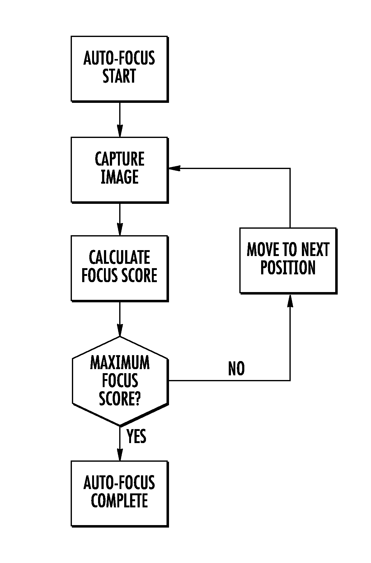
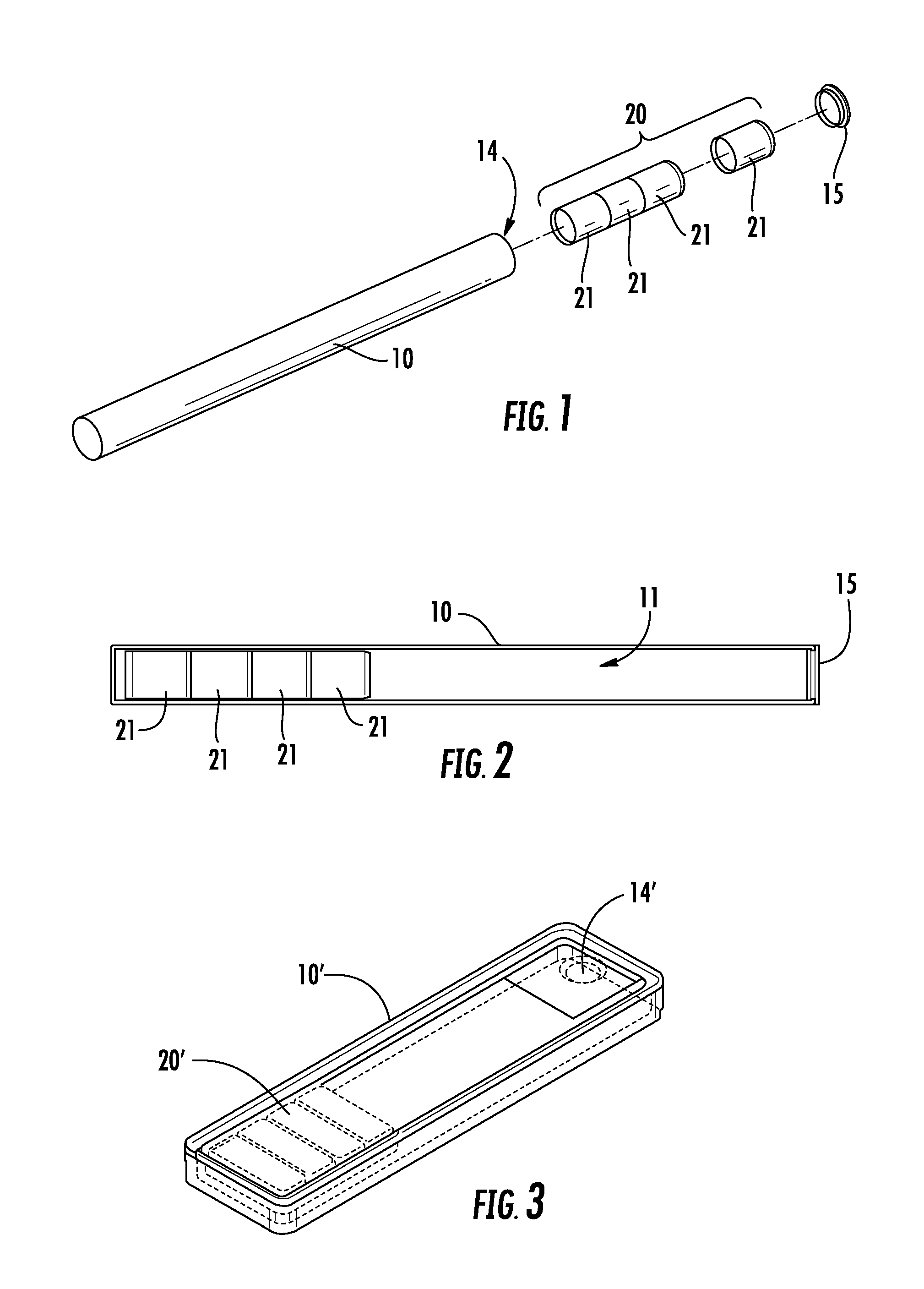
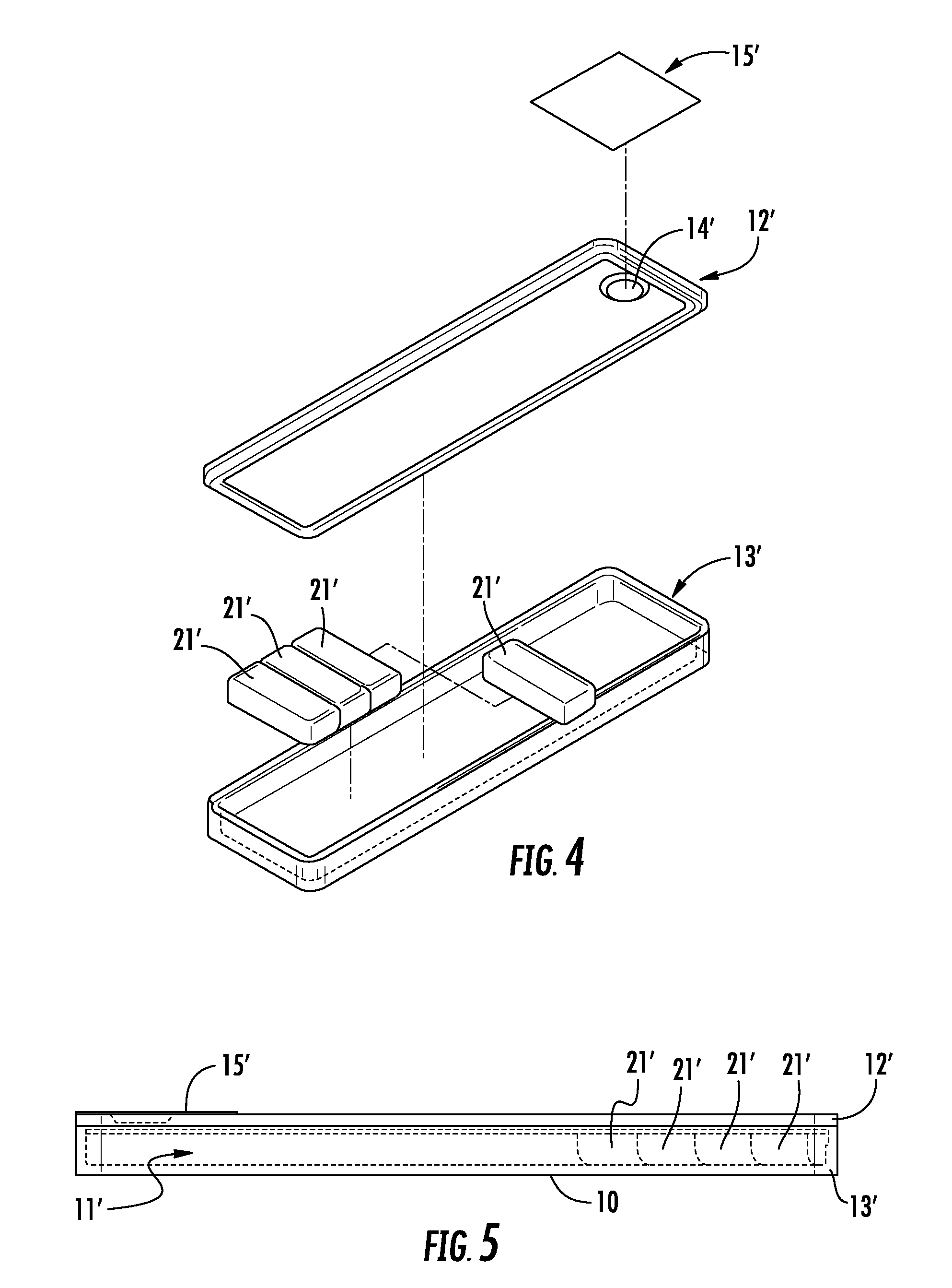
A method of automatically focusing a microscope on a specimen is carried out by capturing an image from each of a plurality of focal planes in or on said specimen, calculating a focus score for each of said images, selecting the focal plane corresponding to the image having the best focus score, and then repositioning said specimen relative to said microscope so that said microscope is focused on said selected focal plane, the method includes a plurality of exogenous targets in or on said specimen, which aids in focusing in the event particular objects of interest, such as cells / pathogens that may or may not be in the sample, are not present, or are present in low numbers. Automated microscopes and microscope cartridges useful in such methods are also described, along with methods of detecting pathogens in biological samples.
Description
RELATED APPLICATIONS[0001]This application claims the benefit of U.S. Provisional Applications No. 61 / 644,708, filed May 9, 2012 (Docket No. 9903-5PR), No. 61 / 667,691, filed Jul. 3, 2012 (Docket No. 9903-12PR), and No. 61 / 696,517, filed Sep. 4, 2012 (Docket No. 9903-13PR), the disclosures of which are incorporated by reference herein in their entirety.FIELD OF THE INVENTION[0002]The present invention concerns methods and apparatus for detecting analytes, including pathogens such as Mycoplasma species, in liquid samples such as biological fluids.BACKGROUND OF THE INVENTION[0003]The Mycoplasma are a wide-spread group of bacteria. Species such as M. pneumonia and M. genitalium cause disease in humans. Related species cause disease in plants. M. bovis is considered one of the more pathogenic species and causes pneumonia, mastitis, and arthritis in cattle. In research laboratories, Mycoplasma species are frequent contaminants in cell cultures.[0004]Mycoplasma are characterized by the abs...
Claims
Application Information
Patent Timeline
Application Date:The date an application was filed.
Publication Date:The date a patent or application was officially published.
First Publication Date:The earliest publication date of a patent with the same application number.
Issue Date:Publication date of the patent grant document.
PCT Entry Date:The Entry date of PCT National Phase.
Estimated Expiry Date:The statutory expiry date of a patent right according to the Patent Law, and it is the longest term of protection that the patent right can achieve without the termination of the patent right due to other reasons(Term extension factor has been taken into account ).
Invalid Date:Actual expiry date is based on effective date or publication date of legal transaction data of invalid patent.

