
Method for Estimating Flow Rates, Pressure Gradients, Coronary Flow Reserve, and Fractional Flow Reserve from Patient Specific Computed Tomography Angiogram-Based Contrast Distribution Data

a computed tomography and data comparison technology, applied in the field of cardiac imaging, can solve the problems of weakening the heart muscle, ischemia, heart attack, and reducing the flow rate of blood through,

Active Publication Date: 2014-07-03
THE JOHN HOPKINS UNIV SCHOOL OF MEDICINE
View PDF4 Cites 9 Cited by
  • Summary
  • Abstract
  • Description
  • Claims
  • Application Information

AI Technical Summary

Benefits of technology

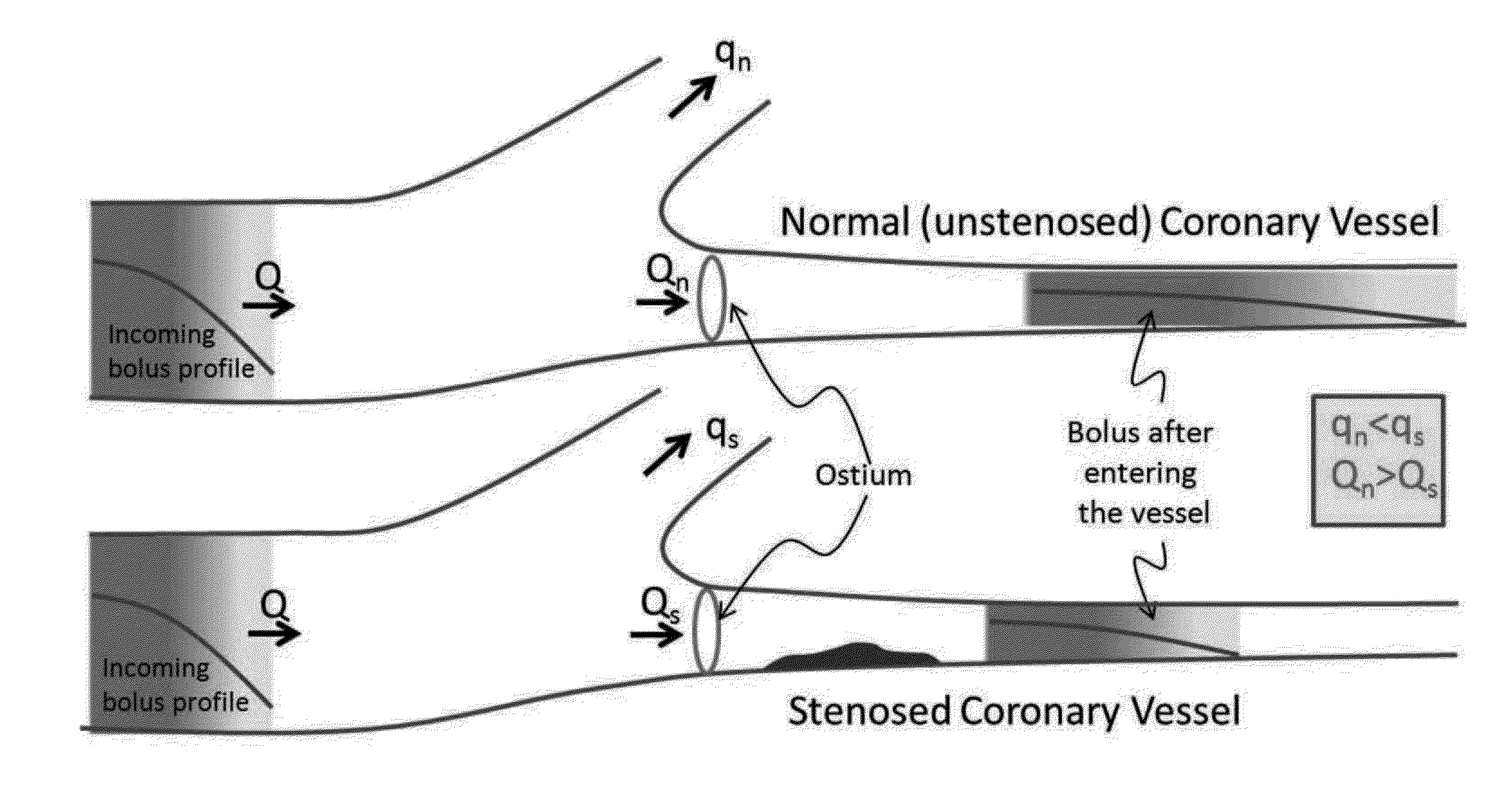
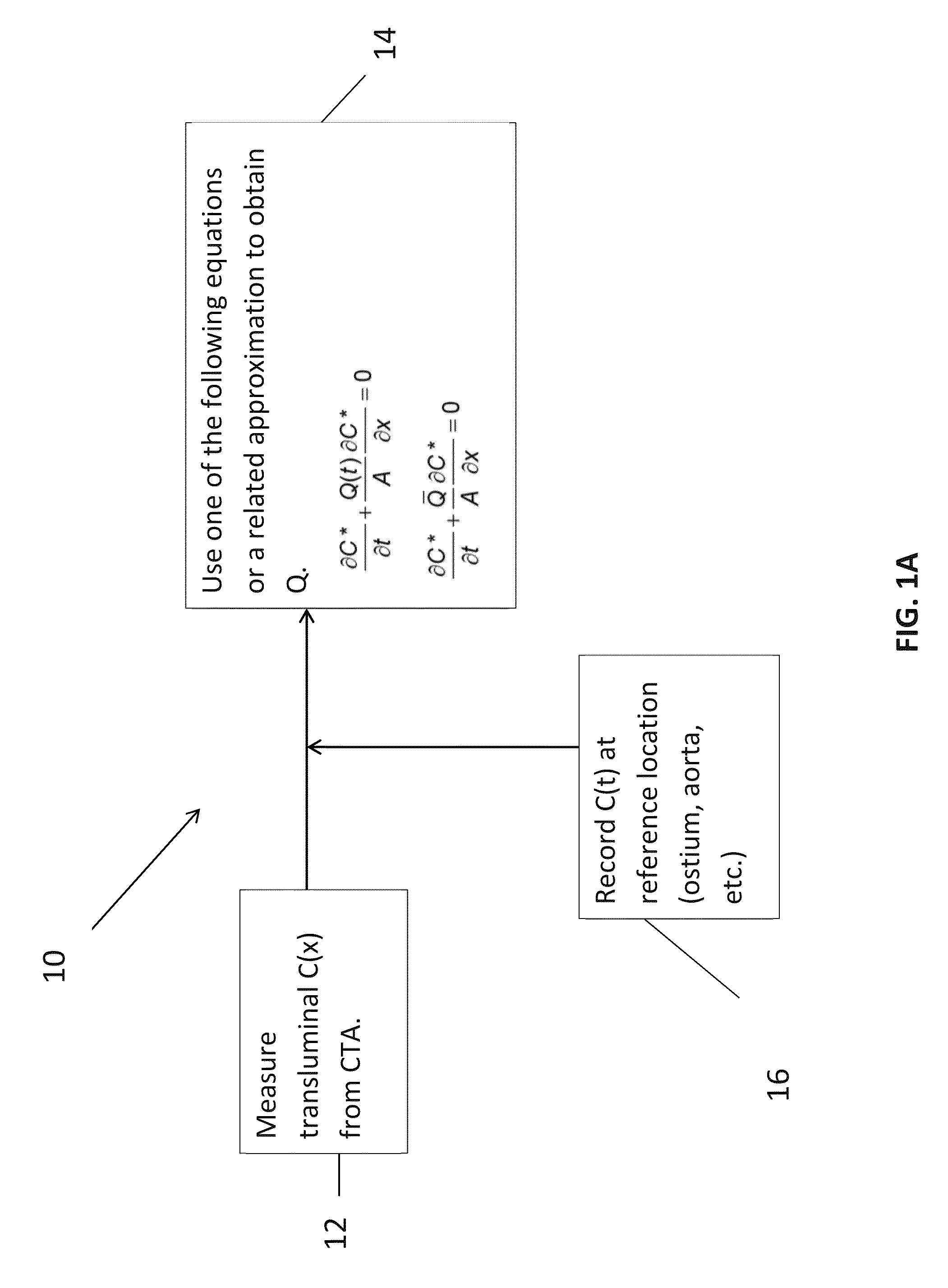
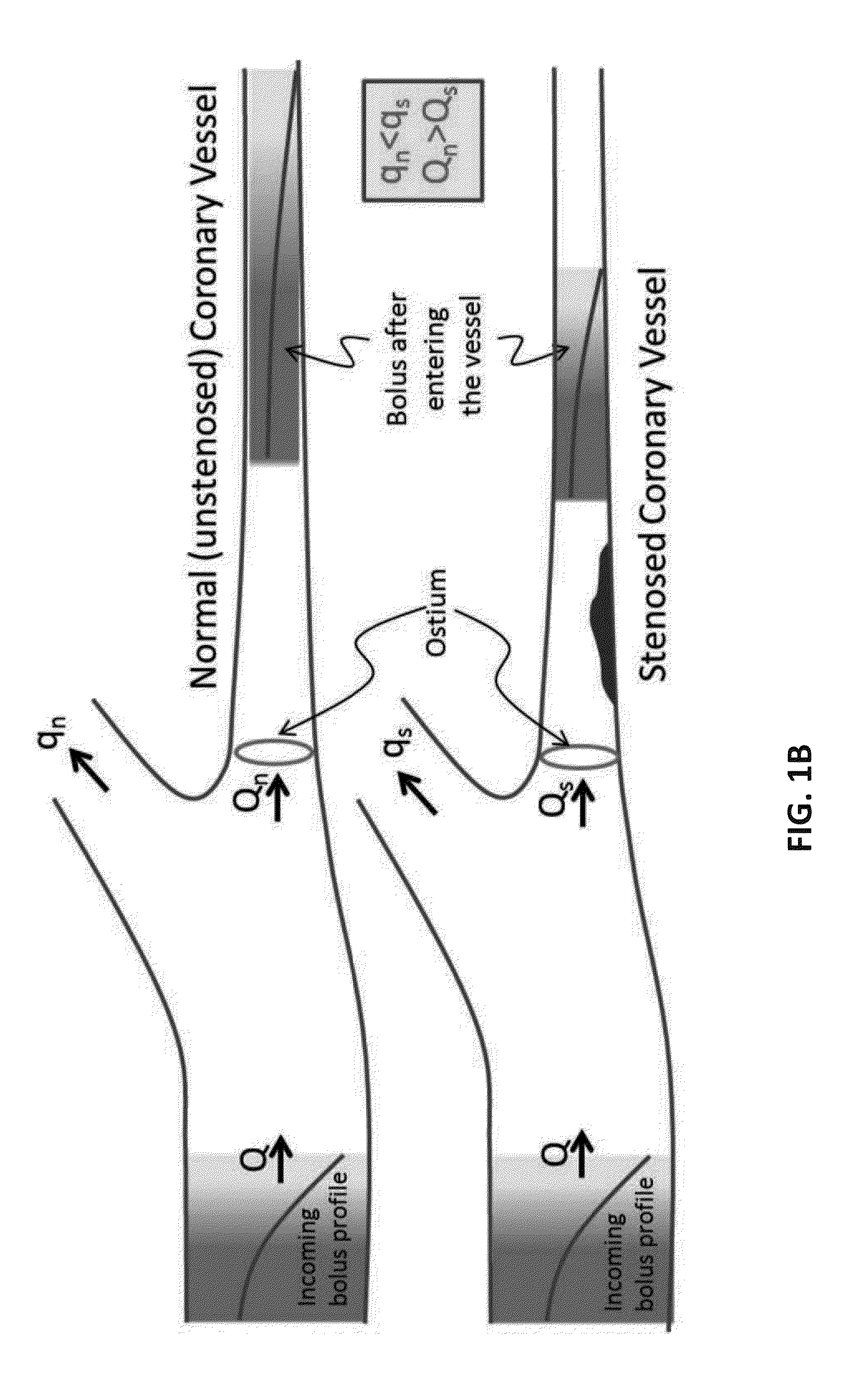
The present invention provides a method for determining the functional significance of a coronary artery stenosis through the estimation of the pressure gradient (PG) within the artery using a transluminal attenuation gradient (TAY) and the spatial concentration of a contrast agent at a given time. This method does not require a second measurement in stress condition and can be executed using a computer readable medium. The invention provides a more accurate and reliable method for the determination of the functional significance of a coronary artery stenosis which is useful in the diagnosis and treatment of cardiovascular disease.

Problems solved by technology

Excessive plaque build-up can lead to diminished blood flow through the coronary arteries and blow blood flow to the myocardium leading to chest pain, ischemia, and heart attack.
Coronary artery disease can also weaken the heart muscle and contribute to heart failure, a condition where the heart's efficiency as a pump is compromised.
This state can lead to electrical disorders of the heart that increase the possibility for sudden cardiac death.
Unfortunately, these non-invasive tests do not provide data related to the size of a coronary lesion or its specific effect on coronary blood flow, lesion pressure gradients and fractional flow reserve.
While this procedure provides an accurate measure of FFR for determining the functional severity of the coronary stenosis, it is only obtained after the risk and cost of an invasive procedure have already been assumed.
This approach requires a high level of sophistication, is computationally expensive, and requires that patient-specific data be transmitted out of the hospital environment to a third party vendor.
It is expensive and can take several days to obtain results.
Additionally, recent data testing this approach to predict actual FFR in a multicenter trial were very disappointing.

Method used


Image

  • Method for Estimating Flow Rates, Pressure Gradients, Coronary Flow Reserve, and Fractional Flow Reserve from Patient Specific Computed Tomography Angiogram-Based Contrast Distribution Data
  • Method for Estimating Flow Rates, Pressure Gradients, Coronary Flow Reserve, and Fractional Flow Reserve from Patient Specific Computed Tomography Angiogram-Based Contrast Distribution Data
  • Method for Estimating Flow Rates, Pressure Gradients, Coronary Flow Reserve, and Fractional Flow Reserve from Patient Specific Computed Tomography Angiogram-Based Contrast Distribution Data

Examples


Embodiment Construction

[0045]The presently disclosed subject matter now will be described more fully hereinafter with reference to the accompanying Drawings, in which some, but not all embodiments of the inventions are shown. Like numbers refer to like elements throughout. The presently disclosed subject matter may be embodied in many different forms and should not be construed as limited to the embodiments set forth herein; rather, these embodiments are provided so that this disclosure will satisfy applicable legal requirements. Indeed, many modifications and other embodiments of the presently disclosed subject matter set forth herein will come to mind to one skilled in the art to which the presently disclosed subject matter pertains having the benefit of the teachings presented in the foregoing descriptions and the associated Drawings. Therefore, it is to be understood that the presently disclosed subject matter is not to be limited to the specific embodiments disclosed and that modifications and other ...


PUM


Abstract

An embodiment in accordance with the present invention provides a method for non-invasively determining the functional severity of coronary artery stenosis. The method includes gathering patient-specific data related to concentration of a contrast agent within a coronary artery of a patient using a coronary computed tomography angiography scan (CCTA). The patient-specific data is used to calculate a patient-specific transluminal attenuation gradient for the coronary artery of the patient. The patient specific transluminal attenuation gradient is used to determine an estimate of a coronary flow velocity, pressure gradient, loss coefficient, coronary flow reserve, and / or fractional flow reserve for the patient. Coronary flow velocity, pressure gradient, loss coefficient, coronary flow reserve, and fractional flow reserve can then be used to estimate the functional severity of coronary artery stenosis.

Description

CROSS REFERENCE TO RELATED APPLICATIONS[0001]This application is a continuation of U.S. patent application Ser. No. 13 / 868,697 filed on Apr. 23, 2013, which claims the benefit of U.S. Provisional Patent Application No. 61 / 705,422 filed on Sep. 25, 2012, and International Application No. PCT / US2013 / 037804 filed on Apr. 23, 2013, all of which are incorporated by reference, herein, in their entirety.FIELD OF THE INVENTION[0002]The present invention relates generally to cardiology. More particularly, the present invention relates to a computed tomography cardiac imaging based method for determining flow rates, pressure gradients and coronary and fractional flow reserve.BACKGROUND OF THE INVENTION[0003]The coronary arteries supply the myocardium, or muscle of the heart with oxygen and nutrients. Over time the coronary arteries can become blocked with cholesterol and other material known as plaque. Coronary artery disease results from this buildup of plaque within the walls of the coronar...

Claims


Application Information

Patent Timeline
Patent Type & Authority Applications(United States)
IPC IPC(8): A61B6/00A61B6/03
CPCA61B6/507A61B6/03A61B6/5217A61B6/504A61B6/481A61B5/0044A61B5/02007A61B5/026A61B6/032A61B6/503A61B6/5205F04C2270/041G06T7/0016G06T2207/10076G06T2207/10081G06T2207/30076G06T2207/30104
Inventor MITTAL, RAJATLARDO, ALBERT C.SEO, JUNG HEE
Owner THE JOHN HOPKINS UNIV SCHOOL OF MEDICINE
Learn More