Method for detecting nucleosomes

a nucleosome and nucleosome technology, applied in the field of nucleosome detection, can solve the problems of inability to accurately detect nucleosomes in complex biological media,

Active Publication Date: 2014-07-24
SINGAPORE VOLITION PTE LTD +1
View PDF1 Cites 3 Cited by
  • Summary
  • Abstract
  • Description
  • Claims
  • Application Information

AI Technical Summary

Problems solved by technology

However, we have found that the results of nucleosome ELISA assays of the current art do not agree with each other.
Furthermore, although most circulating DNA in serum or plasma is reported to exist as mono-nucleosomes and oligo-nucleosomes (Holdenrieder et al, 2001), measured levels of nucleosomes and DNA in serum or plasma do not agree well.
It will be clear to those skilled in the art that this method requires relatively pure nucleosome samples and is not suitable for the direct measurement of histone PTMs in complex biological media such as blood, plasma or serum.
One disadvantage of immunohistochemical methods for clinical use is that tissue sample collection is invasive involving surgery or biopsy.
Another disadvantage of immunohistochemistry methods is that they are unsuited for early diagnosis or for screening diagnostics as a reasonable expectation of the disease must usually already exist before a biopsy or tissue resection is made.
However, cell free histone variants in nucleosomes have not been measured in blood.
There are currently no methods for the detection or measurement of histone variants in intact cell free nucleosomes.
The disadvantages of these methods are that they are labour intensive and / or require large amounts of good quality extracted DNA (Allen et al 2004).
Liquid Chromatography with mass spectrometry is considered the gold standard for global DNA methylation measurement but it is costly, and the DNA must be digested to the single nucleotide level prior to analysis (Vasser et al, 2009).
A disadvantage of these methods is that they require extraction of DNA.
Current methods for the detection of global DNA methylation involve extraction or purification of the DNA and are not suitable for rapid, high throughput, low cost, minimally-invasive diagnostic methods.
Such analysis cannot be carried out directly in complex biological media such as tissue lysate, blood, plasma or serum.

Method used


Image

  • Method for detecting nucleosomes
  • Method for detecting nucleosomes
  • Method for detecting nucleosomes

Examples


example 1

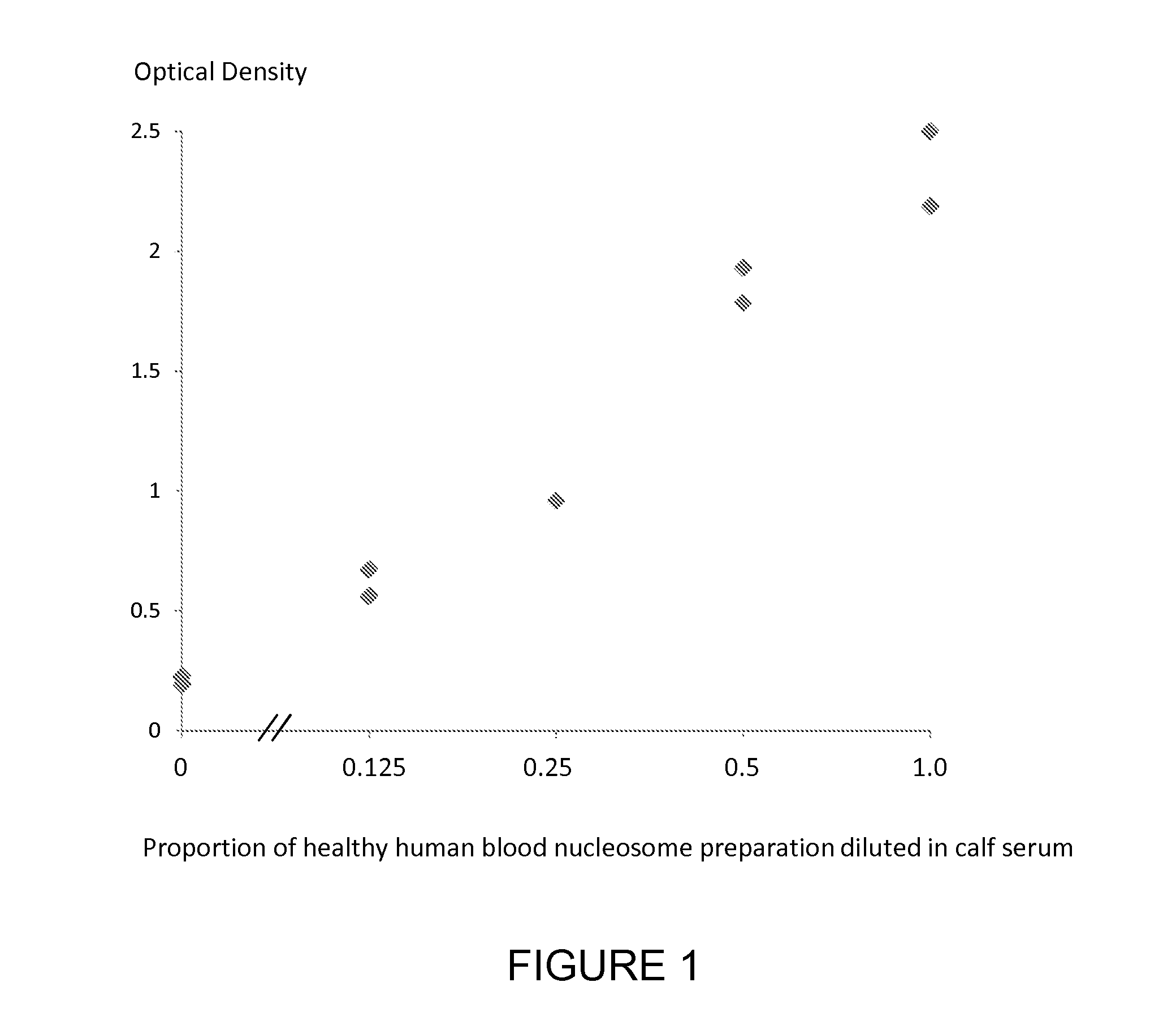
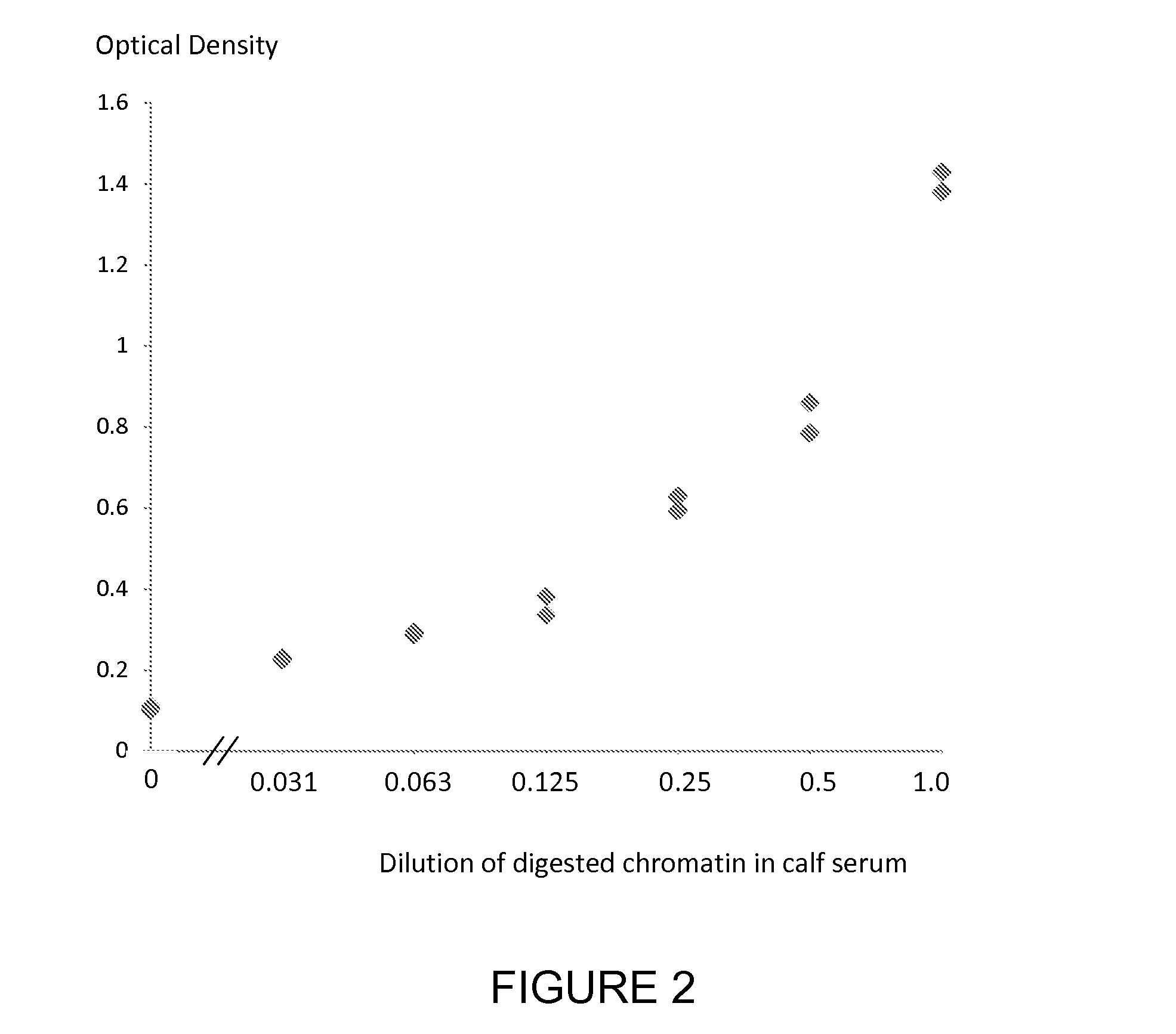
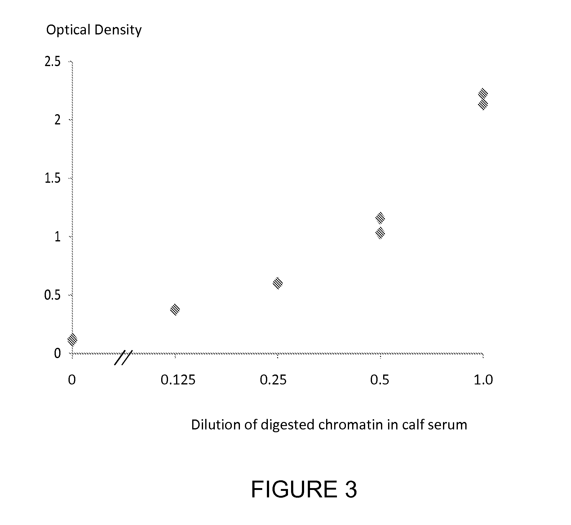
[0181]A nucleosome preparation (produced either by digestion of chromatin extracted from cancer cell lines or produced according to the method of *Holdenrieder et al, 2001) was serially diluted into fetal calf serum and assayed using individual ELISA methods for the nucleosome associated histone variants mH2A1.1, mH2A2, H2AZ and the nucleosome associated nucleotides 5-methylcytosine, 5-hydroxymethylcytosine and thymine and the nucleosome associated PTM P-H2AX(Ser139) using a solid phase anti-histone capture antibody that binds intact nucleosomes and does not bind histone H2 and an appropriate biotinylated anti-histone variant or anti-histone PTM or anti-nucleotide detection antibody. Neat fetal calf serum was also run in the ELISA as a control sample containing no cell free nucleosomes. The assay method was as follows: A solution of anti-histone antibody in 0.1 M phosphate buffer pH 7.4 was added to microtitre wells (100 μL / well) and incubated overnight at 4° C. to coat the wells wi...

example 2

[0182]We used two nucleosome ELISA methods of the current art to measure the circulating cell free nucleosome content of serum and plasma blood samples taken from 20 healthy subjects. The first current ELISA method (ELISA 1) was the Roche Cell Death ELISA and the other (ELISA 2) an ELISA employing a monoclonal anti-histone capture antibody and a monoclonal anti-histone-DNA complex detection antibody. The nucleosome levels detected by both current nucleosome ELISA methods were lower in normal plasma than in normal serum.

[0183]We also measured the individual levels of nucleosomes containing three nucleosome associated histone variants, a histone PTM and a nucleotide in the same samples. The results showed that the serum samples taken from healthy subjects have uniformly low levels of nucleosomes containing histone variants or PTM or nucleotides. The measured plasma results were higher for all 20 healthy subjects. Thus the results for the method of the invention (plasma higher than ser...

example 3

[0184]We used two nucleosome ELISA methods of the current art to measure the circulating cell free nucleosome content of plasma samples taken from 3 subjects with colon cancer, 13 subjects with lung cancer, 2 subjects with pancreatic cancer, 1 subject with oral cancer and a nucleosome sample produced from healthy subjects according to method of Holdenrieder (*Holdenrieder et al, 2001). The first current ELISA method (ELISA 1) was the Roche Cell Death ELISA and the other (ELISA 2) an ELISA employing a monoclonal anti-histone capture antibody and a monoclonal anti-histone-DNA complex detection antibody.

[0185]We also individually measured the levels of nucleosomes containing a number of variant histones, a histone PTM and two nucleotides. The results show that many samples taken from cancer subjects have low or undetectable nucleosome levels as determined by current nucleosome ELISA methods. However, most of these samples have detectable levels of nucleosomes that contain one or more n...


PUM

PropertyMeasurementUnit
pHaaaaaaaaaa
pHaaaaaaaaaa
structural/shapeaaaaaaaaaa

Abstract

The invention relates to a method for detecting and measuring the presence of mono-nucleosomes and oligo-nucleosomes and the use of such measurements for the detection and diagnosis of disease.

Description

FIELD OF THE INVENTION[0001]The invention relates to a method for detecting and measuring the presence of mono-nucleosomes and oligo-nucleosomes and the use of such measurements for the detection and diagnosis of disease.BACKGROUND OF THE INVENTION[0002]The human body comprises several hundred cell types. All of these cell types contain the same genome but have widely different phenotypes and different functions in the body. This phenotypic diversity is due to the differential expression of the genome in different cell types. The control of differential gene expression is not entirely understood but the basic mechanisms include gene regulation by a number of interconnected epigenetic signals associated with the gene, including control of the chromatin packing as euchromatin or heterochromatin, control of nucleosome positioning and nuclease accessible sites, methylation of DNA and variation in the structure of the nucleosomes around which the DNA is wrapped.[0003]The nucleosome is th...

Claims


Application Information

Patent Timeline
Patent Type & Authority Applications(United States)
IPC IPC(8): G01N33/53
CPCG01N33/5308G01N33/6875
Inventor MICALLEF, JACOB VINCENT
Owner SINGAPORE VOLITION PTE LTD
Why Eureka
Try Eureka