Micro-Incubation Systems For Microfluidic Cell Culture And Methods

a micro-incubation system and micro-fluidic cell technology, applied in the field of micro-incubation systems for microfluidic cell culture and methods, can solve the problems of high consumption of energy and gas, high construction cost, and obstruction of access to the microscop

Active Publication Date: 2014-09-25
MILLIPORE CORP
View PDF5 Cites 23 Cited by
  • Summary
  • Abstract
  • Description
  • Claims
  • Application Information

AI Technical Summary

Benefits of technology

[0020]According to specific embodiments, the present invention is directed to an additional area of this field, referred to at times herein as micro-incubation, to provide control of temperature and gas atmosphere for use in micro-scale cell culture systems in a way that is unobtrusive to observational equipment and that further is easily attached and removed from a culture plate in specific embodiments.
[0023]Full microscope enclosures surround the entire microscope except for some heat generating or heat sensitive components such as the camera and illumination sources. The air within the enclosure is circulated and maintained at the desired temperature and gas environment of the sample. An advantage of this method is that temperature control of the whole microscope greatly reduces focus drifts due to room temperature fluctuations, but numerous drawbacks include expensive and customized construction, obstruction of access to microscope, and high consumption of energy and gases. Also, exposure to humidity and repeated temperature changes may damage microscopes.
[0024]Stagetop incubators surround only a small volume intended to house one or more Petri dishes, slides or other culture platform. These provide local temperature regulation and enable limited gas environment control. They are convenient, but do not provide the same level of control as a microscope enclosure, as the stagetop incubator must mimic the mechanics of an enclosure, but in a smaller size and adapt to the microscope. For example, uniform temperature control in a stagetop incubator is limited by the heat sink of the stage itself, and cutouts to provide for the light path for optical clarity further reduce uniformity. In addition, the complexity of the design of stagetop incubators make them expensive.
[0026]The present invention, by contrast, integrates temperature, flow, and gas control directly to a microfluidic culture plate via the use of a manifold that seals to the microfluidic plate. While manifolds similar in some aspects are discussed in some of the above referenced applications to control perfusion on microfluidic plates, the previous designs did not incorporate all of the features described herein as such incorporation was difficult due to the compact nature of the manifold and culture plate. The present manifold design includes novel temperature or gas “microincubator” compartments created by operation of the manifold. These compartments have been demonstrated to provide novel and critical advantages for the proper integration for live cell imaging needs.
[0027]The present invention enables direct cell culture on a microscope stage without the use of external environment chambers such as enclosures or stage-top heaters. The “microincubator” concept according to specific embodiments of the present invention provides precise control, long-term cell culture, and ease of dynamic changes of conditions that has not been available in this context in other designs.

Problems solved by technology

An advantage of this method is that temperature control of the whole microscope greatly reduces focus drifts due to room temperature fluctuations, but numerous drawbacks include expensive and customized construction, obstruction of access to microscope, and high consumption of energy and gases.
Also, exposure to humidity and repeated temperature changes may damage microscopes.
They are convenient, but do not provide the same level of control as a microscope enclosure, as the stagetop incubator must mimic the mechanics of an enclosure, but in a smaller size and adapt to the microscope.
For example, uniform temperature control in a stagetop incubator is limited by the heat sink of the stage itself, and cutouts to provide for the light path for optical clarity further reduce uniformity.
In addition, the complexity of the design of stagetop incubators make them expensive.
The design of the control elements need to be carefully considered, as issues such as heat / mass transfer may make proper maintenance of a steady state condition difficult.
At present, flow chambers are infrequently used in live cell imaging due to the myriad difficulties of adapting them for typical uses.
While manifolds similar in some aspects are discussed in some of the above referenced applications to control perfusion on microfluidic plates, the previous designs did not incorporate all of the features described herein as such incorporation was difficult due to the compact nature of the manifold and culture plate.

Method used


Image

  • Micro-Incubation Systems For Microfluidic Cell Culture And Methods
  • Micro-Incubation Systems For Microfluidic Cell Culture And Methods
  • Micro-Incubation Systems For Microfluidic Cell Culture And Methods

Examples


example cell

Culture

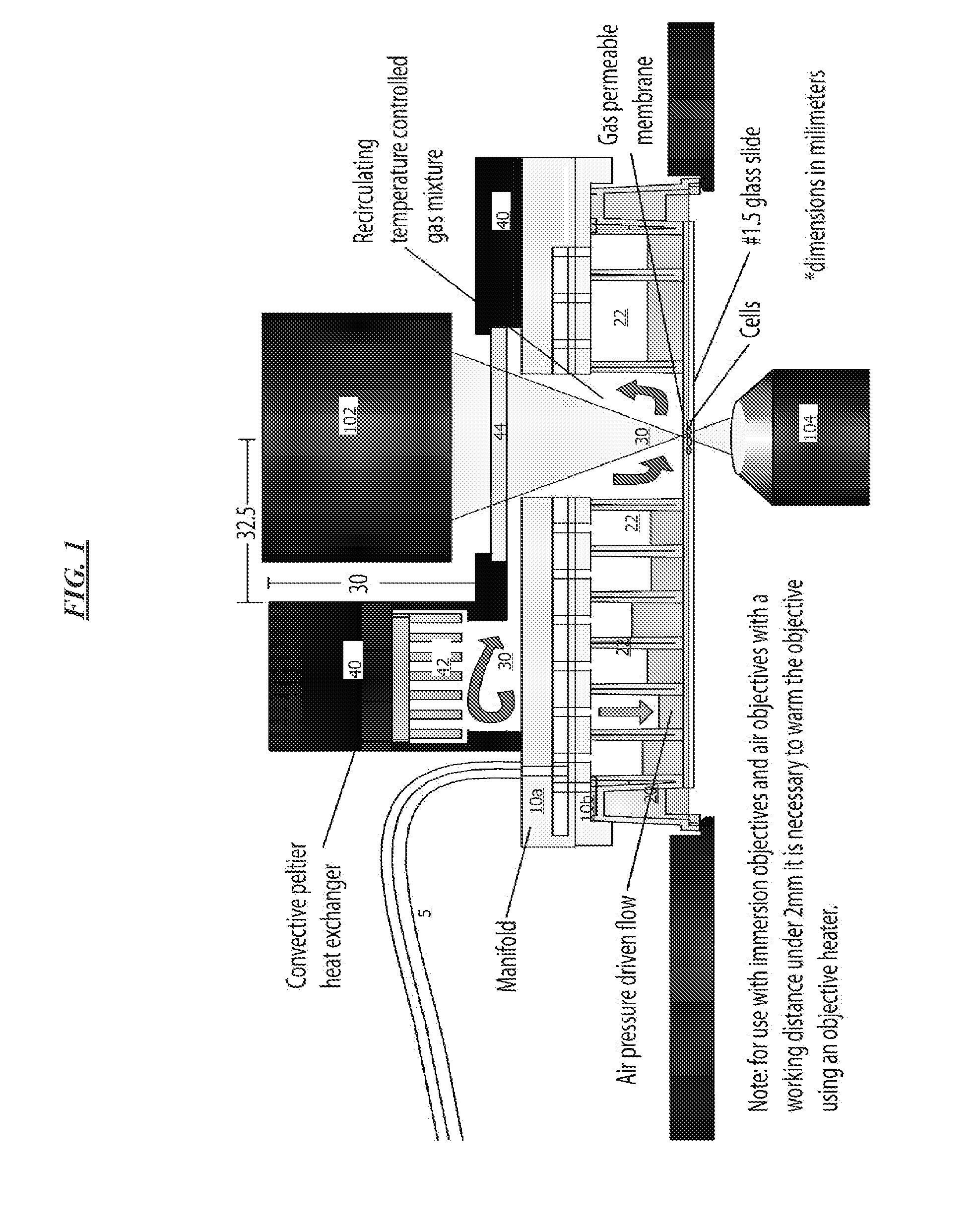
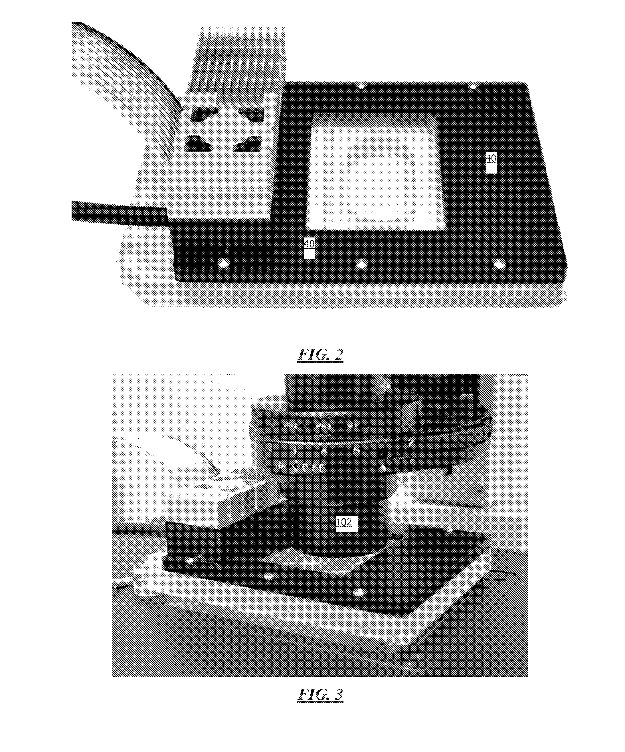
[0093]Cells were cultured using the micro-incubation system to control temperature and gas atmosphere. In one example, human cancer cells (HT-1080, MCF-7, MDA-MB-231) were cultured at 37 C and 5% CO2 to monitor cell division over 24 hours. Additional cell types, including yeast, bacteria, primary cells, neurons, etc. have been successfully cultured using the microincubation system. As an example, FIG. 13 shows NIH-3T3 mouse fibroblasts cultured using the microincubator system according to specific embodiments of the invention at t=0 (left) and after 15 hours (right) showing cell growth and viability. When no temperature or CO2 was controlled, the cells rapidly died within 2 hours.

Integrated Systems

[0094]Integrated systems for the collection and analysis of cellular and other data as well as for the compilation, storage and access of the databases of the invention, typically include a digital computer with software including an instruction set for sequence searching and / or ana...


PUM

PropertyMeasurementUnit
incubation volumeaaaaaaaaaa
volumeaaaaaaaaaa
depthaaaaaaaaaa

Abstract

A micro-incubator manifold for improved microfluidic configurations and systems and methods of manufacture and operation for a manifold and automated microfluidic systems.

Description

CROSS REFERENCE TO RELATED APPLICATIONS[0001]This application is a divisional of U.S. patent application Ser. No. 13 / 692,869, filed Dec. 3, 2012, which claims benefit of priority from U.S. Provisional Application 61 / 566,651, filed Dec. 3, 2011, which are hereby incorporated by reference for all purposes.[0002]This application incorporates each of the following provisional patent applications by reference:[0003]61 / 367,371 filed Jul. 23, 2010[0004]61 / 297,278 filed Jan. 21, 2010[0005]61 / 471,103 filed Apr. 4, 2011[0006]This application is related to material discussed in one or more of the following applications, each of which are incorporated herein by reference for all purposes: provisional patent application 61 / 037,297 filed Mar. 17, 2008, provisional patent application 61 / 018,882 filed Jan. 3, 2008, U.S. application Ser. No. 11 / 994,997, filed Aug. 11, 2008, which is a National Stage Entry of PCT / US06 / 26364, filed Jul. 6, 2006 and which claims priority from provisional patent applica...

Claims


Application Information

Patent Timeline
Patent Type & Authority Applications(United States)
IPC IPC(8): C12M1/00
CPCC12M29/24C12M23/16C12M23/40B01L3/502761B01L7/00C12M29/10C12M41/14C12M41/34C12M41/36B01L2200/0689B01L2200/147B01L2300/048B01L2300/1894B01L2400/0487C12M41/12C12Q1/02B01L3/563B01L3/502715B01L2200/143B01L2200/146B01L2300/0829B01L2300/14B01L2300/18C12M23/36C12M41/18C12M41/40C12M41/46C12M41/48G01N33/5005G01N33/574
Inventor LEE, PHILIP J.GAIGE, TERRY
Owner MILLIPORE CORP
Why Eureka
Try Eureka