Methods for Diagnosing Cancer by Characterization of Tumor Cells Associated with Pleural or Serous Fluids

a pleural or serous fluid and tumor cell technology, applied in the direction of material analysis, biochemistry apparatus and processes, instruments, etc., can solve the problem of inability to definitively cytological and symptomological diagnose a pleural effusion, abnormal collection of fluid, accumulation of fluid, etc., to achieve the effect of increasing diagnostic sensitivity and accuracy

Inactive Publication Date: 2014-10-02
JANSSEN DIAGNOSTICS LLC
View PDF5 Cites 27 Cited by
  • Summary
  • Abstract
  • Description
  • Claims
  • Application Information

AI Technical Summary

Benefits of technology

[0008]The methods described herein provide an advantageous alternative to the present methods of diagnosis of conditions related to excess serous fluids such as pleural fluids. These methods can identify malignant cells in pleural effusions and so increase the diagnostic sensitivity and accuracy of the current standard of care. It can also provide access to source material for individualized tumor in vitro growth and / or characterization using clinically relevant cellular and molecular markers

Problems solved by technology

For example, heart failure or cirrhosis can cause an imbalance between the pressure within blood vessels and the amount of protein in the blood, resulting in an accumulation of fluid, e.g., a transudate.
Injury or inflammation of the pleurae may cause abnormal collection of fluid, e.g., an exudate.
In a substantial number of cases, a definitive cytological and symptomological diagnosis cannot be made for a pleural effusion.
Generally immunocytochemical staining may not be performed due to undetectable numbers of tumor cells or missed cytological identification of tumor cells.
Each of these diagnostic options subjects the patients to the danger of undiagnosed, progressing disease and / or the disadvantages of the biopsy procedure.
While a pleural biopsy under direct visualization (thorascopy) has a sensitivity and accuracy of diagnosis of 95-100%., it is accompanied by disadvantages including the need for general anesthesia, some morbidity (i.e., infection and pain) and mortality risk, as well as high expense.

Method used


Image

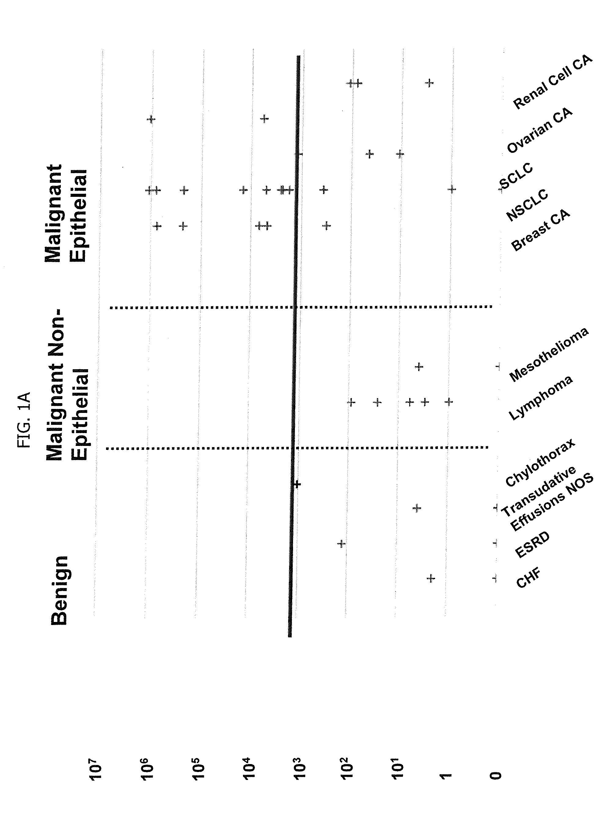
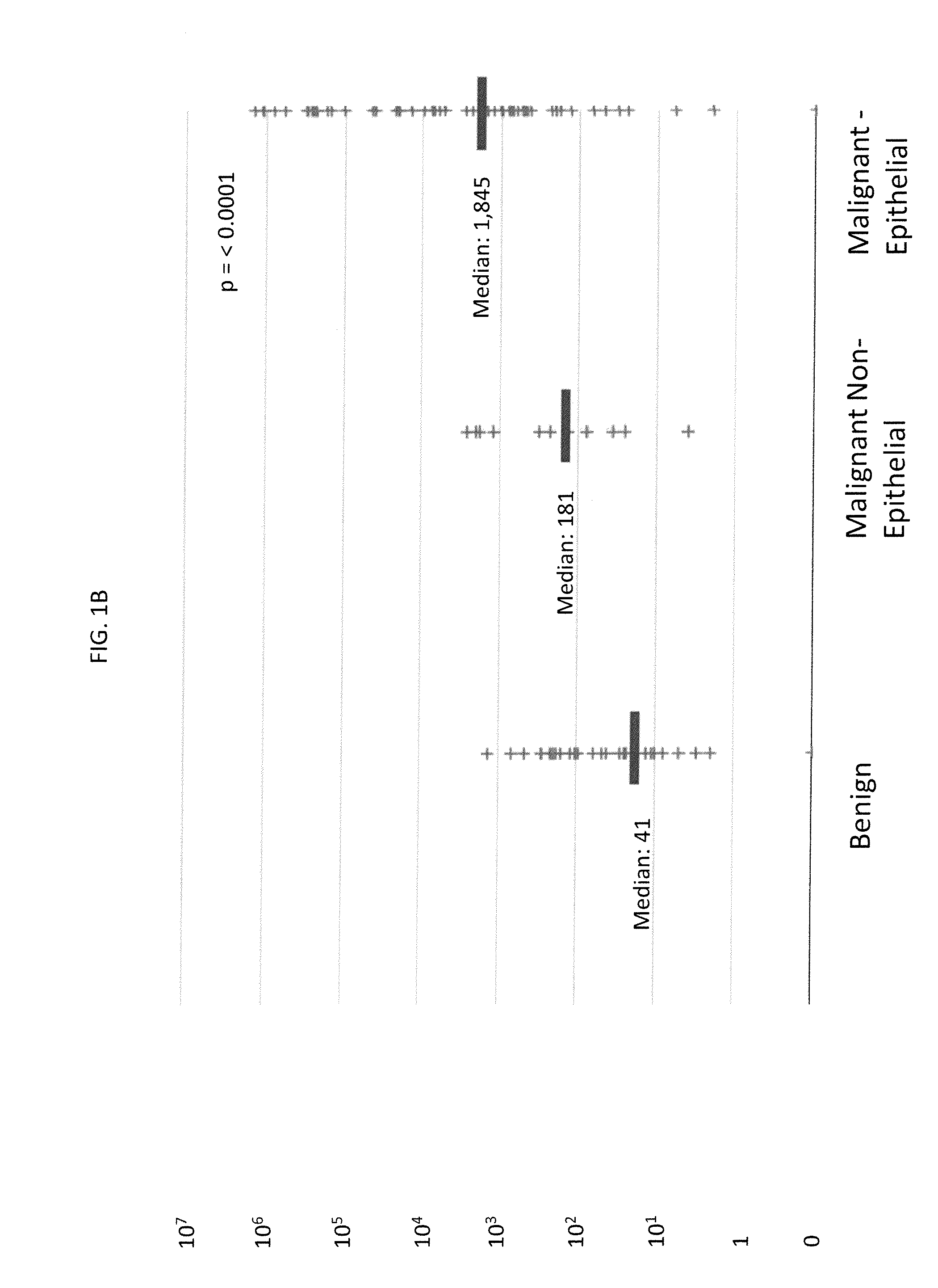
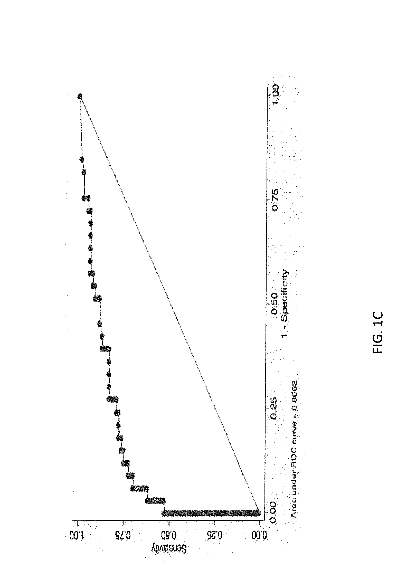
  • Methods for Diagnosing Cancer by Characterization of Tumor Cells Associated with Pleural or Serous Fluids
  • Methods for Diagnosing Cancer by Characterization of Tumor Cells Associated with Pleural or Serous Fluids
  • Methods for Diagnosing Cancer by Characterization of Tumor Cells Associated with Pleural or Serous Fluids

Examples


example 1

Pleural Fluid Sample

I. Collection

[0089]Pleural Fluid is usually collected in a sterile 50 cc syringe or sterile urine collection container for cytological examination and can be further treated as in steps II and III, if it is intended to be stored or shipped before being examined by cytological methods as in Example 2.

[0090]For the CellSearch assay of Example 3, unprocessed pleural fluid is withdrawn, as described above. The unprocessed fluid, optionally simply diluted 1:10 with sterile buffer, is transferred to a CellSave® tube.

[0091]The collected fluid in either instance is labeled with sample number and diagnosis, if any. Optimally, fluid is forwarded to the laboratory for processing within 30 minutes of being drawn. All procedures should be done sterilely in the biosafety hood. The pleural fluid samples are subject to the assay of Example 3 within 24 hours.

II. Optional Centrifugation Steps and Supernatant Harvesting for Cytological Examination or Storage Prior to the Method of ...

example 2

Cytologic Examination

I. Preparing the Samples

[0109]A. Centrifuge the remaining cell suspension from Example 1, step III, subparagraph I, at 1500 rpm for another 5 minutes.

[0110]B. Remove the supernatant and prepare to add freezing media.

[0111]C. Re-suspend the pellet in 4 cc of freezing media.

[0112]D. Labels should contain sample number, sample collection date, and an indication that the sample is pleural fluid.

[0113]E. Store in a Nalgene “Mr. Frosty” freezing container containing isopropanol overnight at −80° C. Transfer cryotubes to −140° C. at that time.

II. Cytological Analysis Using a CytoSpin® Apparatus

[0114]A. Transfer 2×10⁶ cells into 4 ml of PBS (with 0.5% FBS).

[0115]B. Pre-label the slides.

[0116]C. Prepare the slides mounted with the paper pad and the cuvette in the metal holder.

[0117]D. Load up to 200 μl of this suspension in each cuvette.

[0118]E. Spin at 750 rpm for 10 min.

[0119]F. Carefully detach the cuvette and the paper without damaging the fresh cytospin.

[0120]G. Mar...

example 3

Assay Using the CellSearch® System

[0123]The CellSearch® Circulating Tumor Cell system (Veridex LLC) is used to identify and enumerate the number of circulating tumor cells in a pleural fluid. The assay procedure is as described in Mayo Medical Laboratories Communique, Volume 36, No. 1 (Jan / February 2011), incorporated by reference herein, and summarized briefly below.

[0124]A. 7.5 ml of unprocessed and undiluted pleural fluid is placed in a CellSave tube (Veridex LLC).

[0125]B. The PBS is removed, and the cellular component is mixed with new buffer to a desired dilution, and then ferrofluid is added. The ferrofluid consists of nanoparticles with a magnetic core surrounded by a polymeric layer coated with antibodies targeting the Epithelial Cell Adhesion Molecule (EpCAM). The ferrofluid / antibody complex attaches specifically to epithelial cells in the cellular component.

[0126]C. The tube containing the ferrofluid / antibody complex is incubated in a magnetic cuvette, which attracts the f...


PUM

PropertyMeasurementUnit
temperatureaaaaaaaaaa
volumeaaaaaaaaaa
volumeaaaaaaaaaa

Abstract

A method for diagnosing or differentially diagnosing a cancer characterized by the presence of cancer cells in the pleural fluid of a mammalian subject, the method comprising contacting a sample of pleural fluid of the subject with colloidal magnetic particles coupled to a ligand which binds to a determinant on a cancer cell, but does not bind above a baseline threshold to other cellular and non-cellular components in pleural fluid; subjecting the pleural fluid-magnetic particle mixture to a magnetic field to produce a cell fraction enriched in ligand coupled-magnetic particle-bound cancer cells, if present in the pleural fluid; and analyzing the enriched fraction for the number of cancer cells in the pleural fluid. In certain aspects, this method involves preparing the pleural fluids for the above-noted method steps by, e.g., dilution of unprocessed pleural fluid. In certain aspect, the pleural fluid is subjected to the diagnostic method within 24 hours of withdrawal from the subject. This method has advantages to present diagnostic procedures for identifying malignant pleural effusions. The tumor cells present in pleural fluid can be characterized with cellular and molecular markers to determine prognostic and predictive factors.

Description

BACKGROUND OF THE INVENTION[0001]Normal pleural fluid is a thin film of serous fluid containing small numbers of white blood cells, a variety of proteins, and water, located between the visceral and parietal pleurae (i.e., the membranes lining the lungs and the chest cavity, or the pleural space). A pleural effusion is an abnormal accumulation of fluid in pleural space, which occurs in approximately 1.3 million people each year in the US. There are many causes of pleural effusions (see, Hooper, C et al, 2010 Thorax, Vol. 65 (Suppl 2):ii4-ii17). For example, heart failure or cirrhosis can cause an imbalance between the pressure within blood vessels and the amount of protein in the blood, resulting in an accumulation of fluid, e.g., a transudate. Injury or inflammation of the pleurae may cause abnormal collection of fluid, e.g., an exudate. Causes of exudates include infectious disease, bleeding disorders or trauma, inflammatory lung diseases (e.g., asbestosis), sarcoidosis or autoimm...

Claims


Application Information

Patent Timeline
Patent Type & Authority Applications(United States)
IPC IPC(8): G01N33/574C12Q1/68
CPCC12Q1/6841G01N33/574G01N33/57488G01N2333/705G01N1/40G01N1/4077
Inventor ALBELDA, STEVEN M.SCHWED, DANIELRAO, CHANDRA GALLACONNELLY, MARKFOULK, BRAD
Owner JANSSEN DIAGNOSTICS LLC
Why Eureka
Try Eureka