
Characterizing Melanoma

Active Publication Date: 2014-10-16
UNIV OF LOUISVILLE RES FOUND INC
View PDF4 Cites 5 Cited by
  • Summary
  • Abstract
  • Description
  • Claims
  • Application Information

AI Technical Summary

Benefits of technology

The patent describes a method for characterizing melanoma in a subject by isolating melanoma-derived microvesicles from a biological sample of the subject and measuring the presence or amount of microRNAs in the isolated microvesicles. The method can also involve determining the presence or amount of other biomarkers such as RNAs and polypeptides in the isolated microvesicles and comparing them to a reference to evaluate treatment efficacy and progression of the melanoma. The patent also describes a method for evaluating the presence or absence of melanoma biomarkers in a sample by isolating microvesicles from the sample and measuring the expression profile of biomarkers. The biological samples can include cell culture samples, milk, blood, serum, plasma, ascites, cyst fluid, pleural fluid, peritoneal fluid, cerebral spinal fluid, tears, urine, saliva, sputum, or combinations thereof.

Problems solved by technology

Despite considerable effort directed at early detection, few reliable and cost-effective screening tests have been developed that can diagnose cancer at an early stage.
Melanoma is characterized by both a rapidly-rising incidence and a growing lifetime risk.
Lack of a precise prognostic tool results in the imprecise application of adjuvant therapy by both under-treatment of those patients who are at high risk of recurrence and over-treatment of those who are actually at low risk.
There, however, still remains significant variability in overall risk assessment for individual patients.
Unfortunately, barriers exist in this research area.
One is that direct access to molecular information contained within the melanoma cells is, by definition, invasive in nature, i.e., occurs through biopsy or surgical removal of melanoma tissue, neither of which is amenable to frequent and longitudinal monitoring.
In addition, sampling of melanoma tissue relies on the assumption that the collected fragments are representative of the entire lesion, which is not always the case due to the regional tissue heterogeneity.

Method used


Image

  • Characterizing Melanoma
  • Characterizing Melanoma
  • Characterizing Melanoma

Examples


examples

Materials and Methods

[0108]Cell Lines and Culture Reagents.

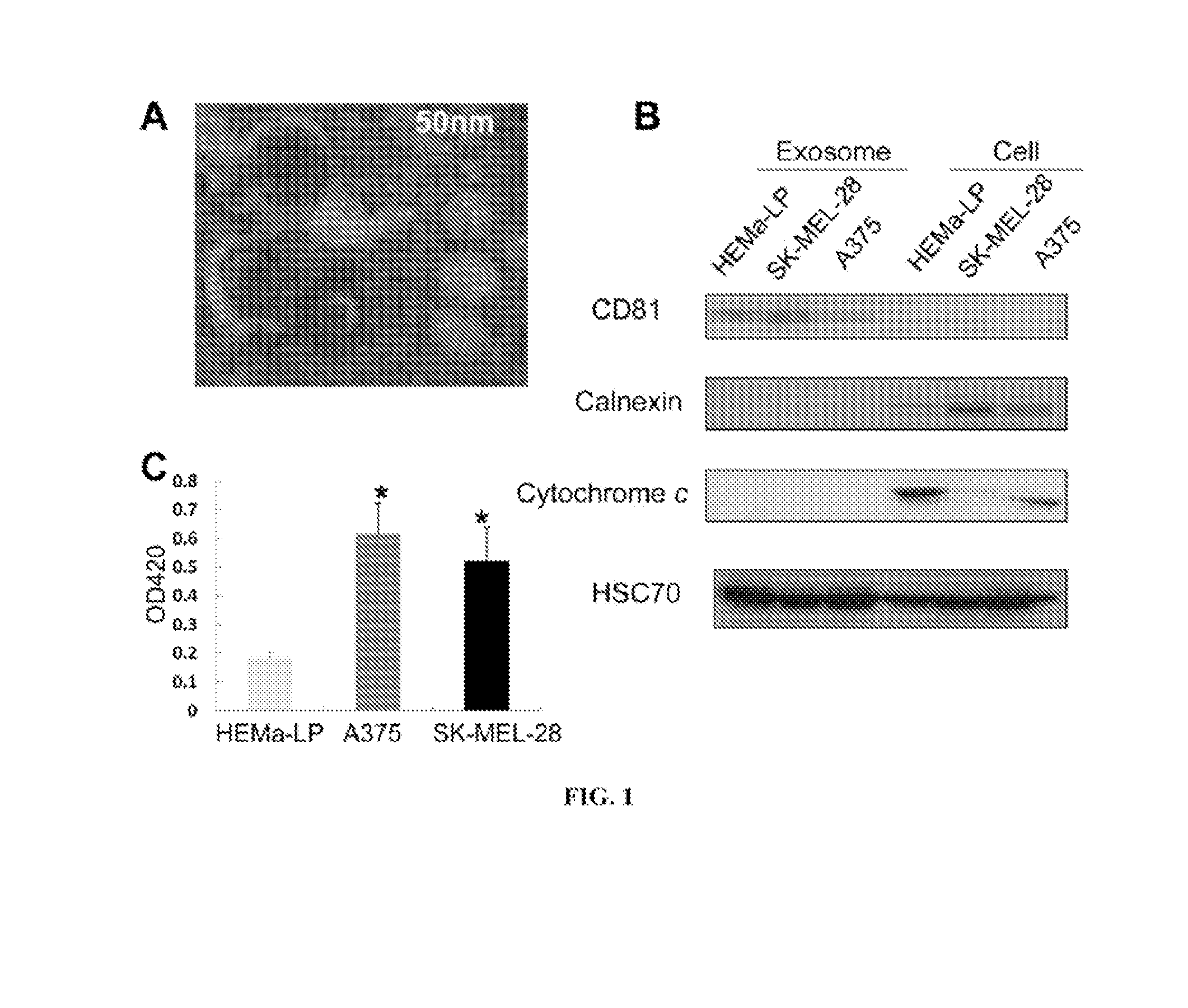
[0109]Two normal human epidermal melanocytes, HEMa-LP and NHEM-c cells, were purchased from Life Technologies (Carlsbad, Calif.) and PromoCell (Heidelberg, Germany), respectively. The human malignant melanoma cell lines A375 and SK-MEL-28 were purchased from American Type Culture Collection (Rockville, Md.). A375 cells and SK-MEL-28 cells were maintained in Dulbecco's Modified Eagle Medium (DMEM) and a minimal essential medium (α-MEM), respectively, supplemented with 10% exosome-depleted fetal bovine serum (FBS) and penicillin (100 U / mL) / streptomycin (100 μg / mL). FBS was depleted of exosomes by ultracentrifugation at 100,000×g for 16 h at 4° C. HEMa-LP cells and NHEM-c cells were cultured in Medium 254 supplemented with Human Melanocyte Growth Supplement-2 (HMGS-2) and Melanocyte Growth Medium M2 supplemented with Supplement Mix (PromoCell, Heidelberg, Germany), respectively, in a 5% CO2 incubator at 37° C. All other cell cu...


PUM

PropertyMeasurementUnit
Timeaaaaaaaaaa

Abstract

The presently disclosed subject matter provides methods of characterizing a melanoma in a subject by measuring amounts of one or more RNAs, miRNAs, and / or polypeptides present in cancer-derived microvesicles isolated from a biological sample from the subject.

Description

RELATED APPLICATIONS[0001]This application claims priority from U.S. Provisional Application Ser. No. 61 / 552,222 filed Oct. 27, 2011, the entire disclosure of which is incorporated herein by this reference.TECHNICAL FIELD[0002]The presently disclosed subject matter relates to methods for characterizing melanoma in a subject. In particular, the presently-disclosed subject matter relates to methods of characterizing a melanoma in a subject by measuring amounts of one or more RNAs and / or proteins present in melanoma-derived microvesicles isolated from a biological sample from the subject.INTRODUCTION[0003]The identification of cancer biomarkers suitable for the early detection and diagnosis of cancer holds great promise to improve the clinical outcome of subjects. It is especially important for subjects presenting with vague or no symptoms or with rapidly-progressing cancers. Despite considerable effort directed at early detection, few reliable and cost-effective screening tests have b...

Claims


Application Information

Patent Timeline
IPC IPC(8): C12Q1/68
CPCC12Q1/6886C12N2310/141C12Q2600/178C12Q2600/158
Inventor HAO, HONGYINGMCMASTERS, KELLY M.XIAO, DEYI
Owner UNIV OF LOUISVILLE RES FOUND INC
Learn More