Single shot high resolution conjunctival small vessel perfusion method for evaluating microvasculature in systemic and ocular vascular diseases

a small vessel, high-resolution technology, applied in the field of single shot high-resolution conjunctival small vessel perfusion method for evaluating microvasculature in systemic and ocular vascular diseases, can solve the problems of low sensitivity or specificity of methods, limited procedures, and registration of sequential images, so as to reduce anatomical boundaries and uniform diameter

Inactive Publication Date: 2014-12-04
UNIV OF MIAMI
View PDF0 Cites 13 Cited by
  • Summary
  • Abstract
  • Description
  • Claims
  • Application Information

AI Technical Summary

Problems solved by technology

However, these methods have not demonstrated high sensitivity or specificity due to, at least in part, a lack of high resolution microvasculature maps.
Other studies used orthogonal polarized spectral imaging to measure conjunctival capillary perfusion in a single vessel; however, this procedure was limited by the very small image field.
However, this method required a manual approach to the registration of sequential images, which was time consuming.
Improvements to this method include a semi-automated registration method was used on images of the human conjunctiva using a sequence of 62 images obtained with a Zeiss slit-lamp biomicroscope equipped with a digital charged couple device camera, but the image-to-geometry registration problem still requires the user to verify the correctness of the registration.
Therefore, user bias is still a limitation since certain key operations rely on the user to guide the registration.
When this method is applied to the conjunctiva, it is found that the resulting small vessel perfusion map is not as clear as the retinal perfusion map.
Because of the blurred images of the conjunctival perfusion maps obtained with RFI, it was more difficult to do image segmentation for quantitative analysis using commercial software like the Fractal Analysis Toolbox.

Method used


Image

  • Single shot high resolution conjunctival small vessel perfusion method for evaluating microvasculature in systemic and ocular vascular diseases
  • Single shot high resolution conjunctival small vessel perfusion method for evaluating microvasculature in systemic and ocular vascular diseases
  • Single shot high resolution conjunctival small vessel perfusion method for evaluating microvasculature in systemic and ocular vascular diseases

Examples


Embodiment Construction

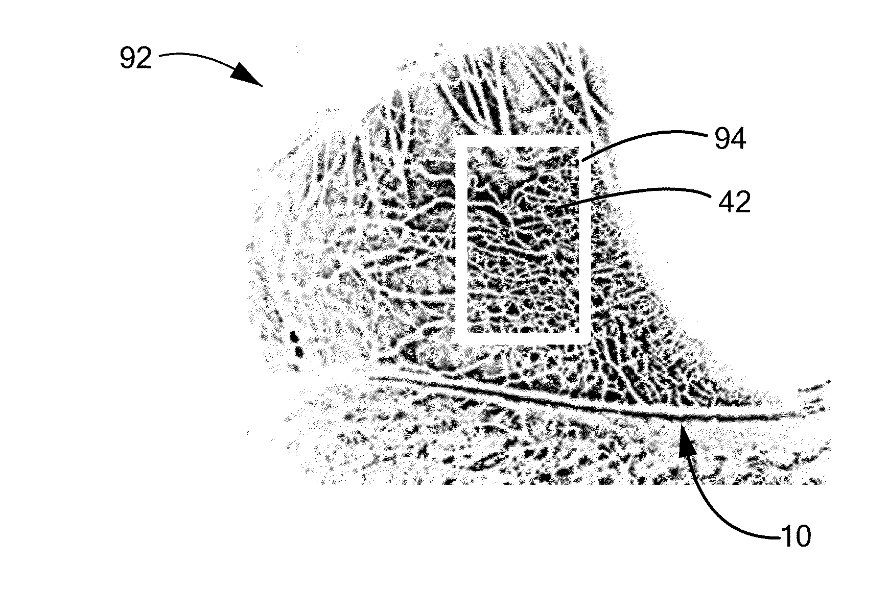
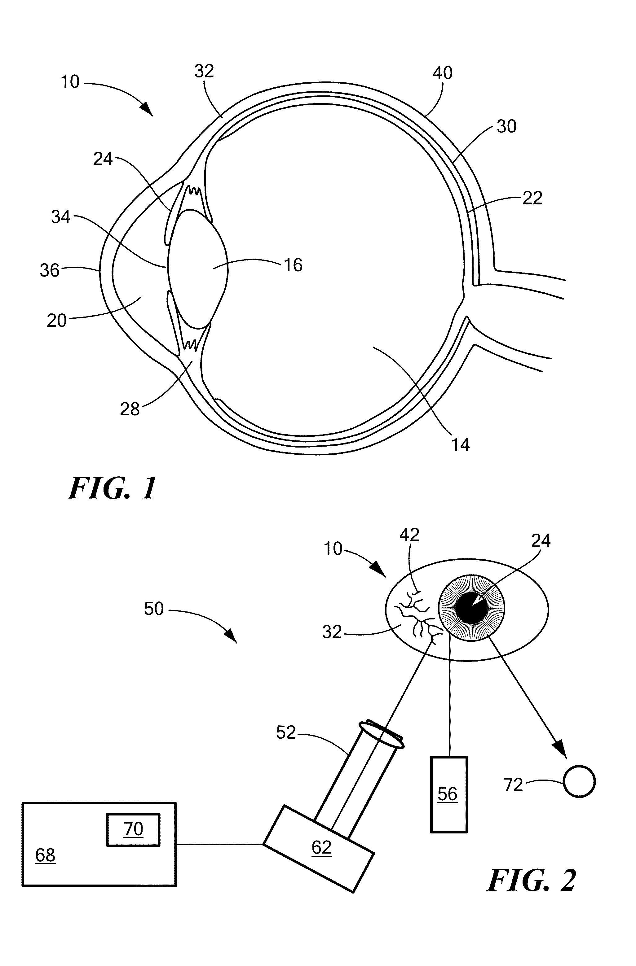
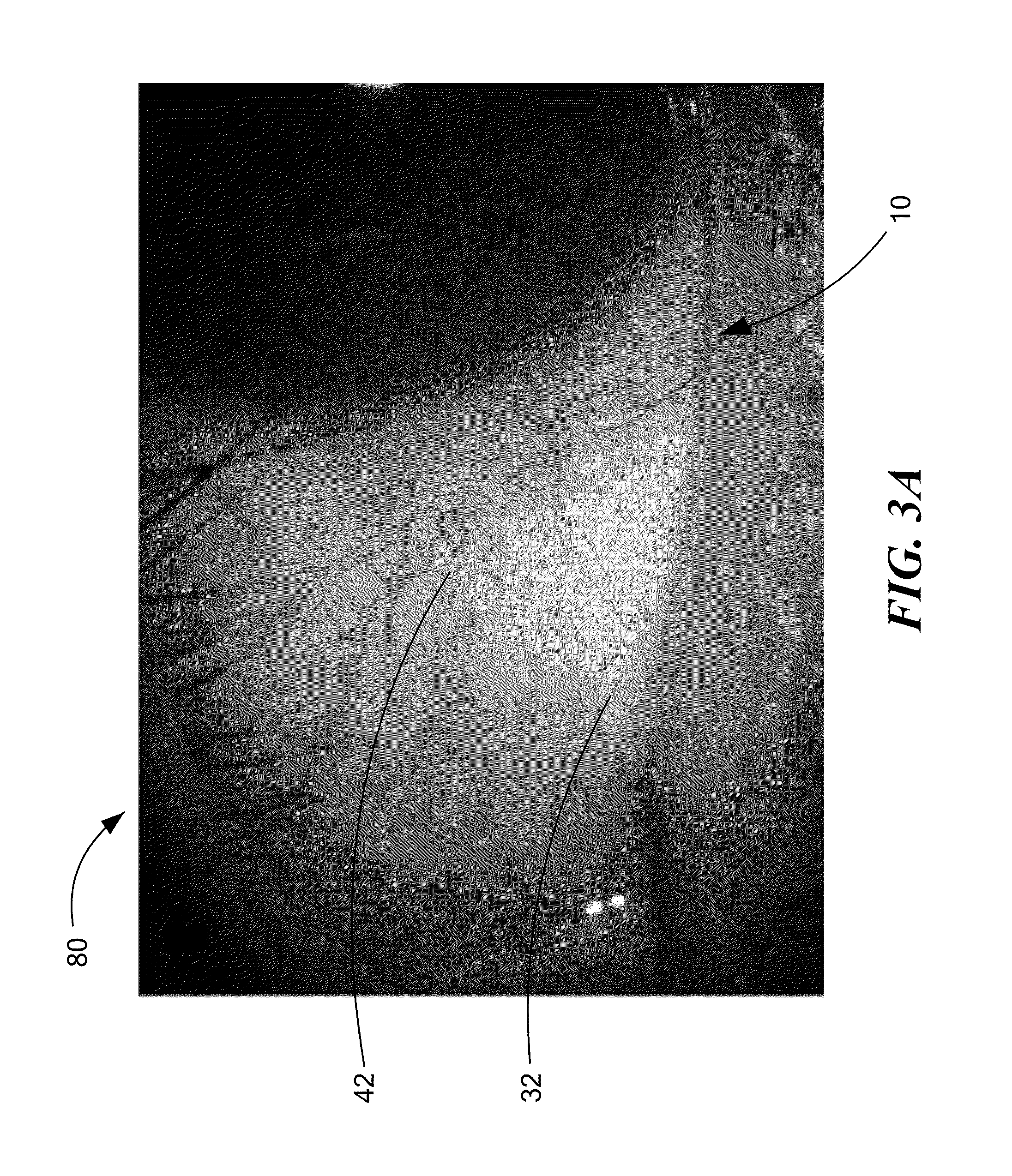
[0027]The present invention advantageously provides a method and system for generating high-resolution small vessel perfusion maps of the ocular or bulbar conjunctiva using a single raw image. Referring now to the drawing figures in which like reference designations refer to like elements, a cross-sectional view of a human eye 10 is shown in FIG. 1. The eye 10 generally includes a vitreous body 14, lens 16, aqueous humour 20, retina 22, iris 24, ciliary body 28, choroid 30, and the sclera 32. The aqueous humour 20, the iris 24, and the pupil 34 are covered by a transparent cornea 36, and the sclera 32, the white part of the eye, is covered by a conjunctiva 40. The conjunctiva 40 includes an epithelial layer, which contains, among other things, blood vessels 42 (which may be referred to herein as a “capillary bed”). The blood vessels 42 of the conjunctiva 40 may be visible against the white or whitish sclera 32 behind.

[0028]Referring now to FIG. 2, a non-limiting, exemplary system 50...


PUM


Abstract

A system and method for generating high-resolution, small vessel perfusion maps (nCPMs) of the ocular conjunctival microvasculature. Unlike current systems and methods, the present invention allows for the generation of nCPMs using only a single raw image obtained in a single image acquisition step. The method includes obtaining a single raw image of at least a portion of the microvasculature of the ocular conjunctiva. The raw image may be obtained using a digital camera and slit lamp. The red and blue color channels are removed from the raw image to create a color-adjusted image in which contrast between the microvasculature and the sclera is enhanced. The color-adjusted image is then inverted and saved as a grayscale inverted image. The grayscale inverted image may be skeletonized. The grayscale image and / or the skeletonized image may be quantitatively analyzed.

Description

CROSS-REFERENCE TO RELATED APPLICATION[0001]This application is related to and claims priority to U.S. Provisional Patent Application Ser. No. 61 / 828,388, filed May 29, 2013, entitled SINGLE SHOT HIGH RESOLUTION CONJUNCTIVAL SMALL VESSEL PERFUSION METHOD FOR EVALUATING MICROVASCULATURE IN SYSTEMIC AND OCULAR VASCULAR DISEASES, the entirety of which is incorporated herein by reference.STATEMENT REGARDING FEDERALLY SPONSORED RESEARCH OR DEVELOPMENT[0002]n / aFIELD OF THE INVENTION[0003]The present invention relates to a method and system for generating high-resolution small vessel perfusion maps of the ocular or bulbar conjunctiva using a single raw image.BACKGROUND OF THE INVENTION[0004]The conjunctiva, composed of non-keratinized, stratified columnar epithelium with interspersed goblet cells, is disposed on the inside of the eyelids and covers the sclera. The conjunctiva is involved in mucus and tear production as well as providing a protective barrier against pathogens. Microcirculat...

Claims


Application Information

Patent Timeline
Patent Type & Authority Applications(United States)
IPC IPC(8): G06T7/00A61B3/14
CPCG06T7/0012G06T2207/30041A61B3/145A61B3/12G06T5/003G06T5/30G06T2207/10024G06T2207/10056G06T2207/20044G06T2207/30104
Inventor JIANG, HONGDEBUC, DELIAWANG, JIANHUA
Owner UNIV OF MIAMI
Why Eureka
Try Eureka