
Lateral flow immunoassay for detecting vitamins

a technology of immunoassay and vitamin, applied in the field of vitamin and lateral flow immunoassay, can solve the problems of limiting the absorption of vitamin d, insufficient nutritional intake of vitamin d, and affecting the conversion ra

Inactive Publication Date: 2014-12-18
NANOSPEED DIAGNOSTICS
View PDF15 Cites 26 Cited by
  • Summary
  • Abstract
  • Description
  • Claims
  • Application Information

AI Technical Summary

Benefits of technology

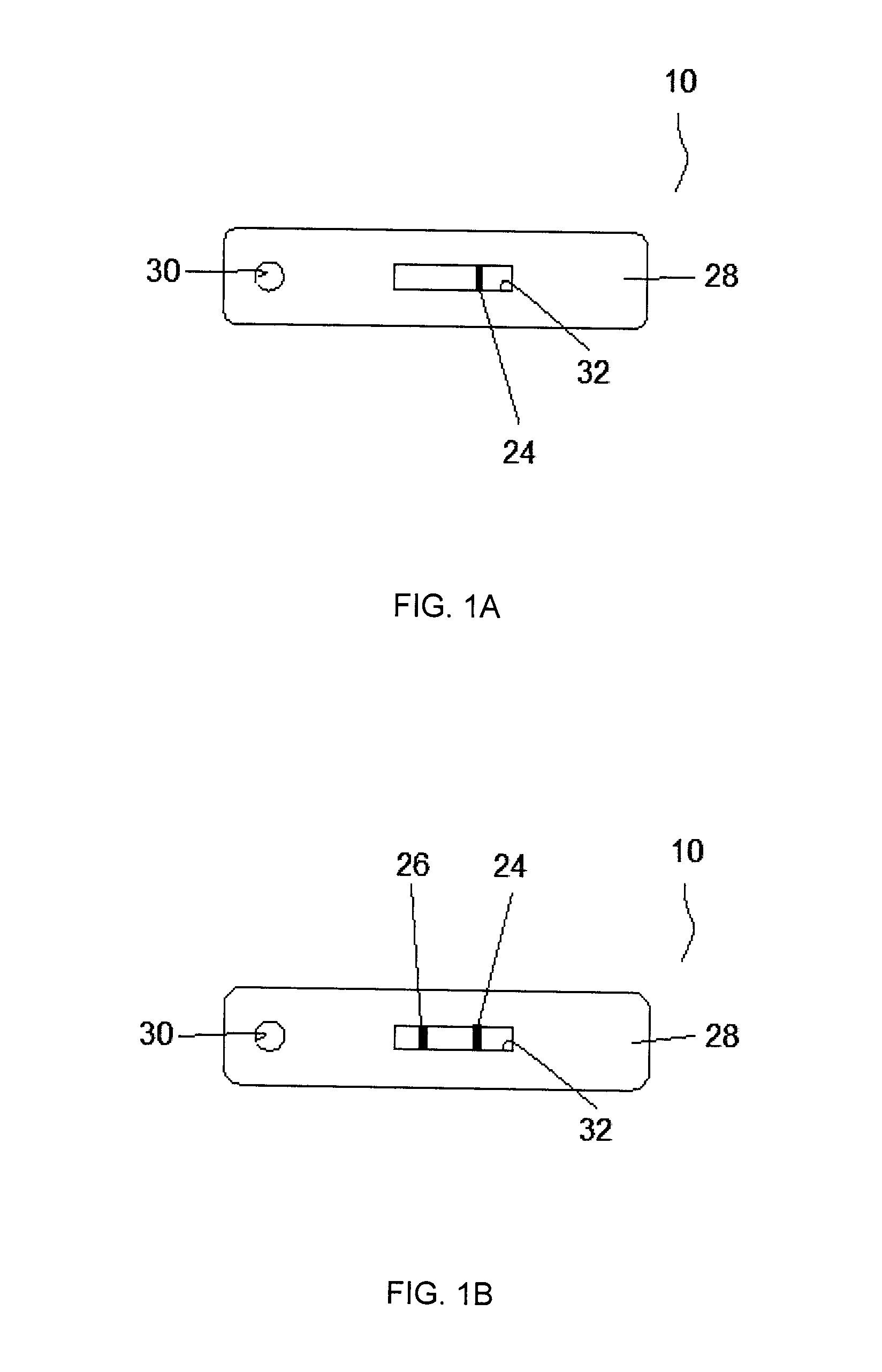
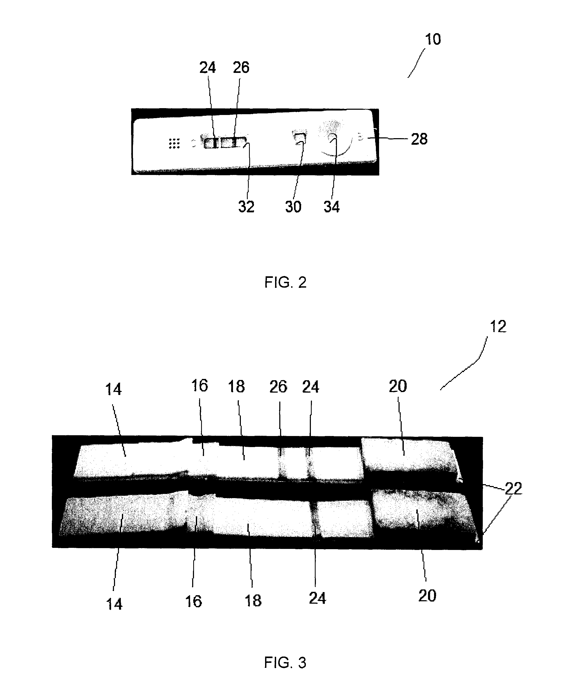
The present invention provides a method and kit for detecting vitamin D using a lateral flow immunoassay. The method involves applying a fluid sample to a test strip and allowing the sample to flow through a detection membrane. The test strip contains a conjugate pad with a labeled antibody against vitamin D and a capture reagent that binds to the labeled antibody but not the vitamin D. The sample can be a body fluid such as urine or blood, and the labeled antibody can be anti-25-hydroxy vitamin D3, anti-25-hydroxy vitamin D2, or anti-1,25-dihydroxy vitamin D3. The detection membrane can also contain a control band for a second capture reagent specific to an antibody or antigen. The test results can be observed in about ten minutes or less. The kit includes the test strip, a sample collector, and a diluent.

Problems solved by technology

The deficiency can also result from inadequate nutritional intake of vitamin D, disorders which limit vitamin D absorption, conditions which impair the conversion of vitamin D into active metabolites, resistance to the effects of vitamin D, and use of certain medicines including phenytoin, phenobarbital and rifampin.
Vitamin D deficiency results in impaired bone mineralization, rickets in children, and osteomalacia and osteoporosis in adults.
Although sunshine is available in most of the populated regions in world, reports suggest that a minimum of 25% of adolescents and more than 50% of undernourished communities in developed countries are vitamin D3 deficient, leading to severe osteomalacia and osteoporosis in adults and rickets in infants.
The situation is worse among women during pre-partum and post-partum periods due to indoor stays, reduced exposure to sunshine, poor dietary calcium intake and no vitamin supplements.
This situation adversely affects the active trans-placental transport of calcium and vitamin supplements to the developing fetus, leading to fetal hypovitaminosis D, and neonatal / infantile rickets in infants.
Using such methods, it may take at least one to two weeks to obtain the test results.
Further, current analytical instruments tend to be time-consuming and complex, requiring sophisticated experimental set-ups, specialized training, and considerable expense.

Method used


Image

  • Lateral flow immunoassay for detecting vitamins
  • Lateral flow immunoassay for detecting vitamins
  • Lateral flow immunoassay for detecting vitamins

Examples


example 1

Detection of Vitamin D in a Fluid Sample

[0075]A lateral flow immunoassay was used to detect vitamin D3 in a fluid sample. Within the test strip, the detection membrane comprised nitrocellulose membrane immobilized with 25-hydroxy vitamin D3 conjugated to bovine serum albumin (BSA) at the test band. Anti-mouse IgG antibodies were immobilized at the control band. The particulate conjugate comprised monoclonal antibodies against 25-hydroxy vitamin D3 labelled with colloidal gold (40 nm).

[0076]Anti-vitamin D antibody was raised in mice by immunizing animals with purified antigen conjugated to keyhole limpet hemocyanin. Different clones were obtained after fusion and their activity was checked against 25-hydroxy vitamin D3. A clone specific for 25-hydroxy vitamin D3 was selected and further re-cloning was performed. The clone that showed the highest titer in the ELISA assay using 25-hydroxy vitamin D3 coated plate was selected. Cells were grown in cell culture medium and supernatant rich...

example 2

Assessment of the Analytical Performance of the Immunoassay

[0079]The immunoassay was evaluated by using 25-hydroxy vitamin D3 conjugated to bovine serum albumin (D-BSA) of known vitamin D concentration. 20 μL of known concentrations of D-BSA were applied on the test strip followed by 7 drops of chase buffer and the appearance of test and control lines was checked after 10 minutes. The assay was repeated to confirm the reproducibility of the results (Table 1).

TABLE 1Test Results1st Set2nd SetD-BSA SampleTestNo Test ControlTestNo TestControl(ng / ml)LineLineLineLineLineLine188 ✓✓✓✓125 ✓✓✓✓62✓✓✓✓4.7✓✓✓✓0✓✓✓✓

[0080]The test line appeared up to a vitamin D concentration of 4.7 ng / ml. There were no test lines at vitamin D concentrations of 62 ng / ml or greater. The results were consistent in both assays. The control line appeared in all the test strips, confirming the validity and proper performance of the assays. These results demonstrate that variations in vitamin D concentrations and the c...

example 3

Comparison of immunoassay to conventional assay

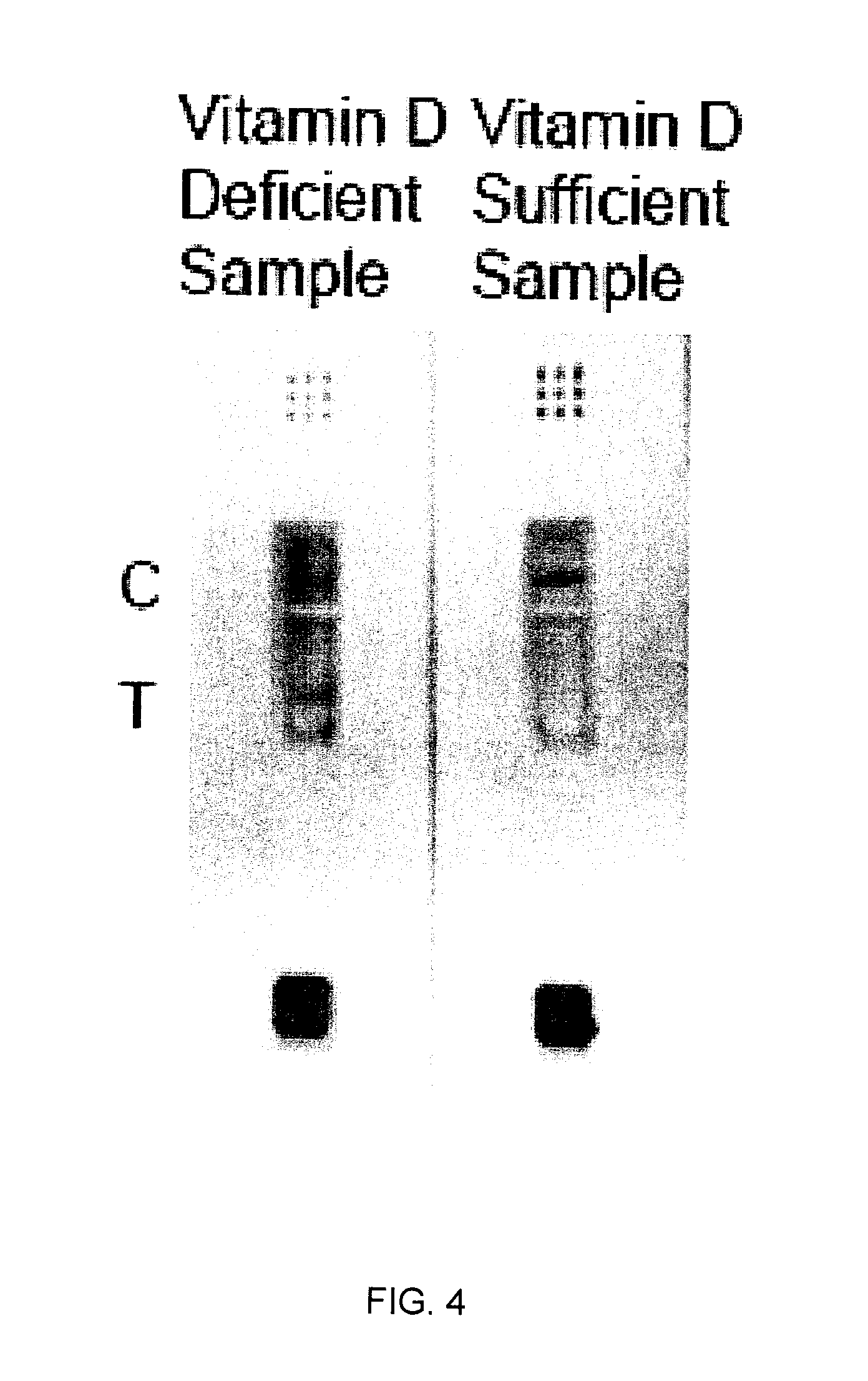
[0083]The immunoassay of the present invention yields qualitative results which are consistent with results obtained using a conventional quantitative 25-hydroxy vitamin D3 assay (LIAISON™ 25 OH Vitamin D TOTAL Assay, DiaSorin Canada Inc., Mississauga, ON, Canada). Table 3 compares the results obtained for the same samples using the two different assays.

TABLE 3Comparison of immunoassay to conventional vitamin D testQuantitativeTest results(Liaison ™,Qualitative Test results ofDiaSorin)immunoassay25(OH)test strip (Yes / No)Patient #Vit D nmol / LResultStrip ResultStrip Comment1125SufficientSufficientNo Test Line2113SufficientSufficientNo Test Line892SufficientSufficientNo Test Line980SufficientSufficientNo Test Line1074DeficientDeficientTest Line1158DeficientDeficientTest Line1235DeficientDeficientTest Line

[0084]FIG. 4 shows actual test results from the immunoassay of the present invention comparing vitamin D deficient and sufficient blood s...


PUM

PropertyMeasurementUnit
diameter sizeaaaaaaaaaa
concentrationsaaaaaaaaaa

Abstract

The invention is directed to a method for detecting analytes, particularly vitamins, using a lateral flow immunoassay. The method may be used to detect vitamin D, particularly 25-hydroxy vitamin D3 or 1,25-dihydroxy vitamin D3. The method involves obtaining a fluid sample from a subject; applying the sample to a lateral flow test strip comprising a conjugate pad capable of releasing a labelled antibody against vitamin D, and a detection membrane comprising a first capture reagent specific to the antibody and immobilized on a test band; and detecting the presence or absence of vitamin D in the sample. The presence of a detectable signal in the test band is indicative of vitamin D deficiency.

Description

FIELD OF THE INVENTION[0001]The present invention is directed to a method for detecting or quantifying analytes, particularly a vitamin, using a lateral flow immunoassay.BACKGROUND OF THE INVENTION[0002]Vitamin D is a group of fat-soluble prohormones, of which the two major forms are vitamin D2 (or ergocalciferol) and vitamin D3 (or cholecalciferol). Vitamin D3 is produced in skin exposed to sunlight, and is obtained through consumption of fish liver oils, salt water fish, mushrooms, fortified milk or other foods, and dietary supplements. Vitamin D3 is biologically inert and must undergo two hydroxylations in the body for activation. First, the liver converts vitamin D3 to 25-hydroxy vitamin D (25(OH)D or calcidiol), which is then converted by the kidneys to physiologically active 1,25-dihydroxy vitamin D (1,25(OH)2D or calcitriol).[0003]Vitamin D promotes bone formation; muscle contraction; nerve conduction; absorption of calcium and phosphate in the gut; calcium reabsorption by th...

Claims


Application Information

Patent Timeline
Patent Type & Authority Applications(United States)
IPC IPC(8): G01N33/82
CPCG01N2800/02G01N33/82G01N33/558G01N33/54388
Inventor GUPTA, RAJANGUPTA, SEEMA
Owner NANOSPEED DIAGNOSTICS
Learn More