Medical image processor and storage medium

a technology of medical image and storage medium, applied in image data processing, image enhancement, instruments, etc., can solve the problems of poor prognosis of cases where her2 is positive, low accuracy, and high accuracy, and achieve the effect of facilitating prognostic expectation and determining the future treatment plan

Active Publication Date: 2015-02-19
KONICA MINOLTA INC
View PDF16 Cites 16 Cited by
  • Summary
  • Abstract
  • Description
  • Claims
  • Application Information

AI Technical Summary

Benefits of technology

[0032]According to the present invention, a physician is able to quantitatively understand an expression amount of a specifi

Problems solved by technology

HER2 protein is considered to be a prognostic factor of breast cancer, and it is known that especially in a case where lymph node metastasis is positive, the prognosis of cases where HER2 is positive is significantly poor

Method used


Image

  • Medical image processor and storage medium
  • Medical image processor and storage medium
  • Medical image processor and storage medium

Examples


first embodiment

Configuration of Pathological Diagnosis Assistance System 100

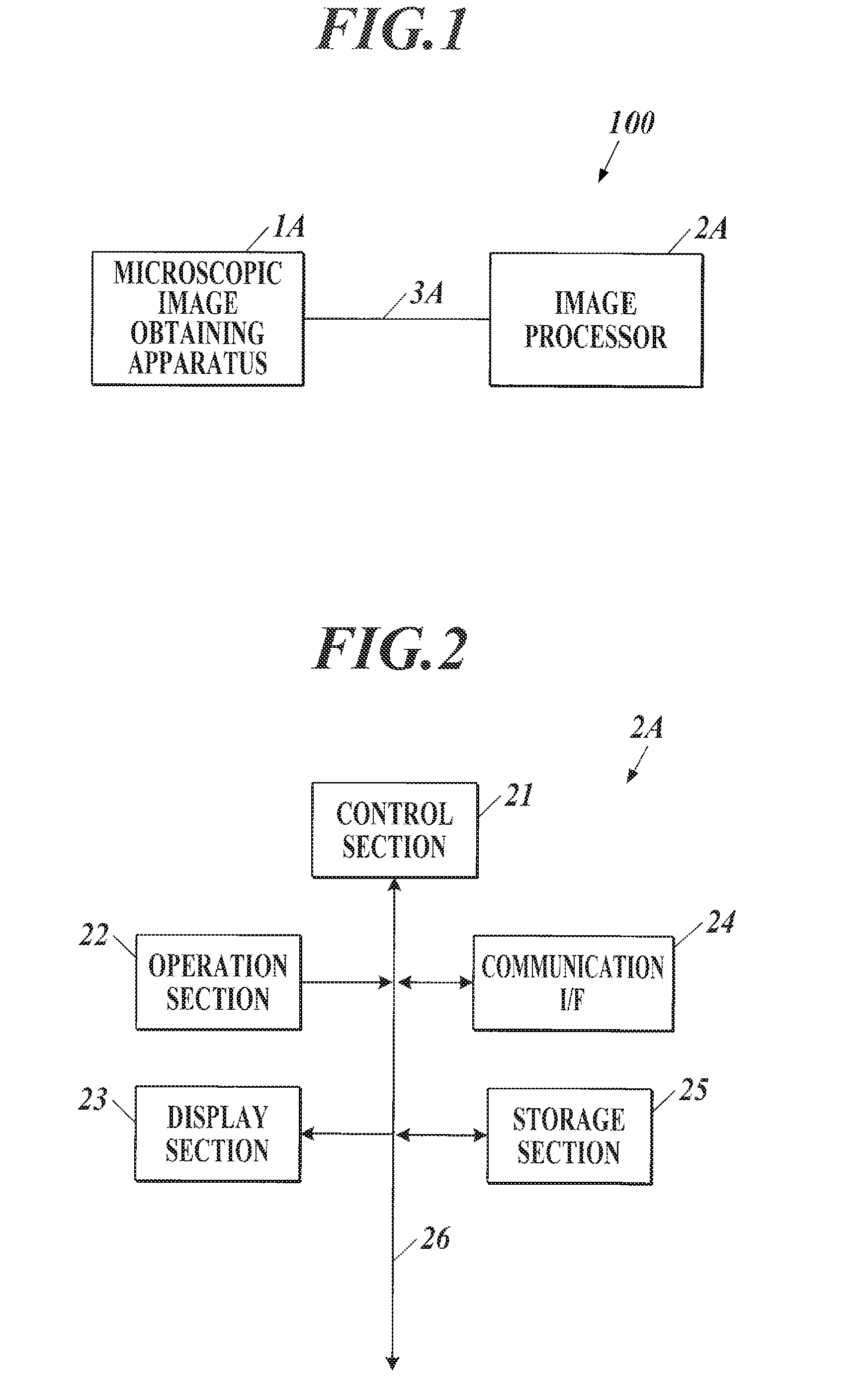
[0059]FIG. 1 shows an example of an entire configuration of a pathological diagnosis assistance system 100 of the first embodiment. The pathological diagnostic assistance system 100 obtains a microscopic image of a tissue slice of a human body stained with a predetermined staining reagent, and outputs a feature amount quantitatively expressing a specific biological substance in the tissue slice of the observation target by analyzing the obtained microscopic image.

[0060]As shown in FIG. 1, the pathological diagnosis assistance system 100 includes a microscopic image obtaining apparatus 1A and an image processor 2A connected to each other through an interface such as a cable 3A so as to be able to transmit and receive data. The method of connecting the microscopic image obtaining apparatus 1A and the image processor 2A is not limited. For example, the microscopic image obtaining apparatus 1A and the image processor 2A can be...

synthesis example 1

Fluorescent Organic Dye Included Silica: Synthesis of Cy5 Included Silica Nanoparticle

[0113]The Cy5 included silica nanoparticle (nanoparticle 1) is made by the method including the following steps of (1) to (5).

[0114]step (1): 1 mg (0.00126 mmol) of N-hydroxysuccinimideester derivative of Cy5 (GE healthcare) and 400 μL (1.796 mmol) of tetraethoxysilane are mixed.

[0115]step (2): 40 mL of ethanol and 10 mL of 14% aqueous ammonium are mixed.

[0116]step (3): The mixed liquid prepared in step (1) is added while stirring the mixed liquid made in step (2) at room temperature. The stirring continues for 12 hours from when the adding starts.

[0117]step (4): Centrifugal separation is performed on the reacted mixture at 10000 G for 60 minutes and the supernatant is removed.

[0118]step (5): Ethanol is added, precipitate is dispersed and centrifugal separation is performed again. Cleaning is performed using ethanol and pure water, one time each with the similar process.

[0119]The obtained nanoparti...

synthesis example 2

Quantum Dot Included Silica: Synthesis of CdSe / ZnS Included Silica Nanoparticle with Emission Wavelength of 655 Nm

[0120]The CdSe / ZnS included silica nanoparticle (hereinafter referred to as nanoparticle 2) is made by the method including the following steps (1) to (5).

[0121]step (1): 10 μL of CdSe / ZnS decane dispersion liquid (Invitrogen, Qdot 655) and 40 μL of tetraethoxysilane are mixed.

[0122]step (2): 4 mL of ethanol and 1 mL of 14% aqueous ammonium are mixed.

[0123]step (3): The mixed liquid made in step (1) is added while stirring the mixed liquid made in step (2) at room temperature. The stirring continues for 12 hours from when the adding starts.

[0124]step (4): Centrifugal separation is performed on the reacted mixture at 10000 G for 60 minutes and the supernatant is removed.

[0125]step (5): Ethanol is added, precipitate is dispersed and centrifugal separation is performed again. Cleaning is performed using ethanol and pure water, one time each with the similar process.

[0126]Th...


PUM


Abstract

A medical image processor and storage medium are shown. According to one implementation, a medical image processor includes an input unit, an operation unit, a cell nucleus extracting unit, a fluorescent bright point extracting unit, a feature amount calculating unit, and an output unit. The input unit is used to input a cell shape image showing a shape of a cell and a fluorescent image showing expression of a specific protein as a fluorescent bright point. The operation unit is used to specify an analysis target region. The cell nucleus extracting unit extracts a region of a cell nucleus. The fluorescent bright point extracting unit extracts a fluorescent bright point. The feature amount calculating unit calculates a feature amount showing an expression amount of the specific protein. The output unit outputs the calculated feature amount.

Description

TECHNICAL FIELD[0001]The present invention relates to a medical image processor and a program.BACKGROUND ART[0002]In pathological diagnosis, first, after processing of dehydrating the obtained tissue so as to be fixed and blocking with paraffin, the above is cut into thin pieces with a thickness of 2 to 8 μm, the paraffin is removed, and the above is stained to observe with a microscope. The pathologist performs diagnosis based on morphologic information such as change in size and shape of a nucleus of a cell, change in pattern as a tissue, etc., and stain information in the microscopic image. Methods known as the tissue staining method used in pathological diagnosis include a conventional chromatic staining method (for example, Hematoxylin and Eosin staining; hereinafter referred to as HE staining) and a chromatic staining method using enzyme (for example, DAB (diaminobenzidine) staining).[0003]In pathological diagnosis, specifying the protein overexpressing in the tissue slice and...

Claims


Application Information

Patent Timeline
IPC IPC(8): G06K9/00G06T7/00G06K9/46
CPCG06K9/0014G06K9/4661G06T7/0012G06T2207/30024G06T2207/20021G06T2207/30096G06T2207/10064G01N21/6456G01N21/6458G06T2207/20104G06T2207/20221G06V20/695
Inventor TSUNOMORI, AKINORIOKADA, HISATAKEGONDA, KOHSUKEOHUCHI, NORIAKIWATANABE, MIKA
Owner KONICA MINOLTA INC
Why Eureka
Try Eureka