

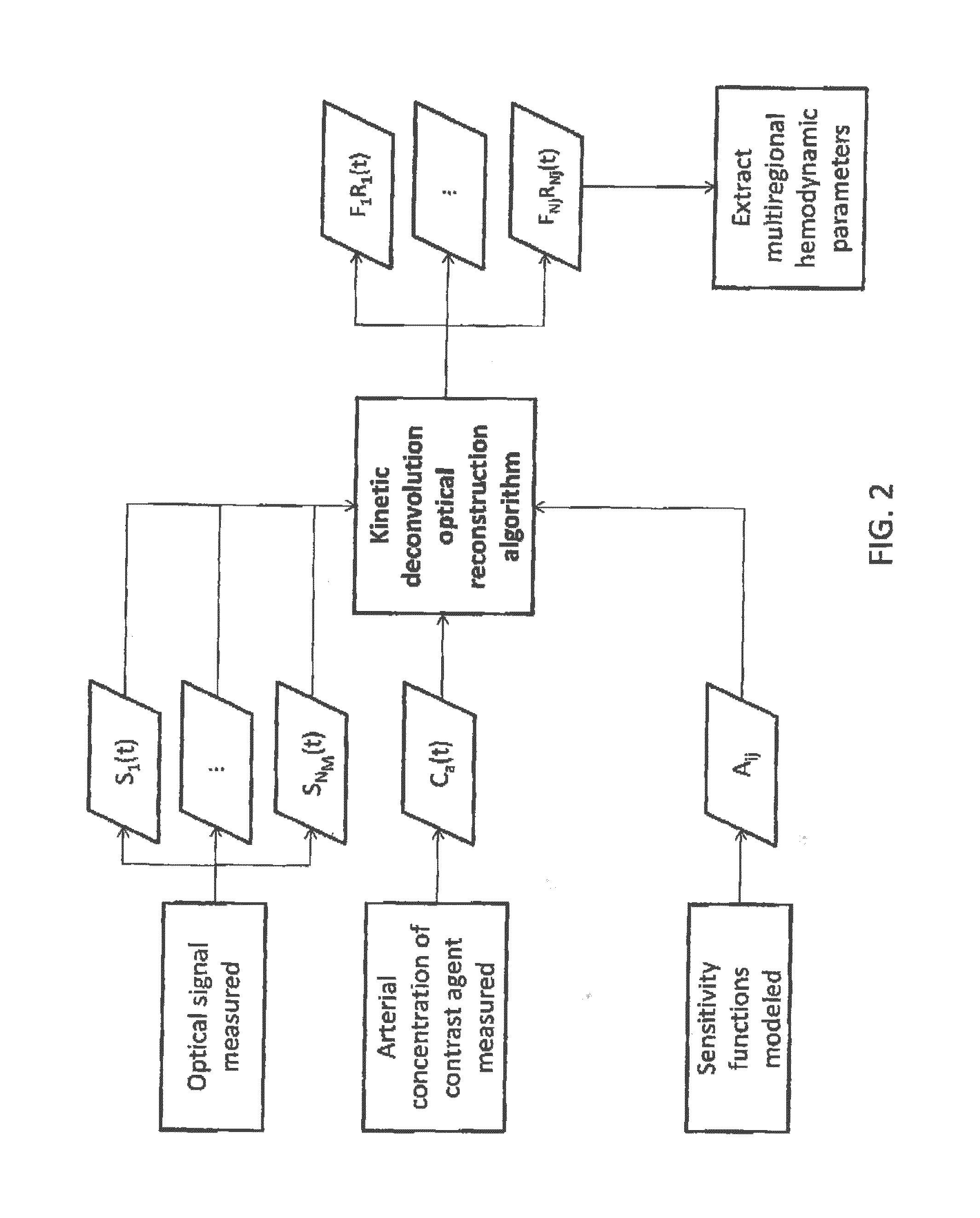
Kinetic deconvolution optical reconstruction method
- Summary
- Abstract
- Description
- Claims
- Application Information
AI Technical Summary
Benefits of technology
Problems solved by technology
Method used
Image

Examples
example 1
[0070]In a three-sub-region or three-layer medium, continuous-wave diffuse reflectance measurements were simulated for source-detector positions of 10, 20, 30 and 40 mm, as shown in FIG. 3A. Layer-specific sensitivity functions (i.e., mean partial path lengths) for each source-detector pair were simulated using Monte Carlo simulations [2]. The time-dependent tracer concentration was generated by convolving a simulated Ca(t) with layer-specific FR(t) functions generated using a gamma variant model [7]. Input F values of 10, 75 and 45 mL / min / 100 g and input MT values of 12, 4, and 4.2 sec were used for layers 1, 2 and 3, respectively. Dynamic changes in μa for layers 1 and 2 were determined from the generated FR(t) functions assuming that the contrast agent was indocyanine green (FIG. 3C). Gaussian noise levels of 5% were added to the optical signal, Ca(t), and the sensitivity functions before two analytical methods were used to extract FR(t). The recovered values of F, BV, and MTT we...
example 2
[0071]Simulations were performed using NIRFAST (Dartmouth College, NH) with fan-beam geometry (five detectors directly across from the source, spaced 22.5° apart) [5]. The contrast agent was chosen to be fluorophore. The cylindrical medium used for Example 2 comprised three hemodynamic regions and is shown in FIG. 4A. The source is illustrated as arrow AA and the detectors are illustrated as arrows BB. Fluorescence and transmission signals were simulated for sixteen (16) equally spaced projections, while the concentration of the contrast agent fluorophore was varied. Specifically, input F values of 10, 75 and 45 mL / min / 100 g were used for sub-regions or layers 1, 2 and 3 (shown in FIGS. 4B), respectively. In the TS approach, fluorophore concentration maps were reconstructed independently for each time-point using the Levenberg-Marquardt minimization algorithm (the time-averaged input concentration of each pixel was used as an initial guess). Regions-of-interest were the same ones us...
example 3
[0072]Numerical experiments were conducted to compare the accuracy and precision of the KDOR and TS methods. Hemodynamic input parameters used to generate the forward data were held constant for all iterations. The input parameters were blood flow (BF), blood volume (BV) and mean transit time (MTT). For the extracerebral layer (ECL), BF=5 mL / min / 100 g, BV=1 mL / 100 g, and MTT=12 s. For the brain, BF=50 mL / min / 100 g, BV=4 mL / 100 g and MTT=5 s. Reconstruction was repeated 100 times on the forward data to compare the precision of the KDOR and TS methods.
[0073]Brain specific absorption curves obtained from the TS and KDOR methods are shown in FIGS. 5A and 5B. The input curve is shown in FIGS. 5A and 5B and is identified by reference numeral 500. The change in absorption coefficient recovered with the KDOR method is shown in FIG. 5A. The KDOR absorption curve was generated by convolving the recovered FR(t) with the original arterial input function. The change in absorption coefficient rec...
PUM
 Abstract
Description
Claims
Application Information