Lipid-Based Drug Carriers for Rapid Penetration Through Mucus Linings

Active Publication Date: 2015-03-26
THE JOHN HOPKINS UNIV SCHOOL OF MEDICINE
View PDF7 Cites 74 Cited by
  • Summary
  • Abstract
  • Description
  • Claims
  • Application Information

AI Technical Summary

Benefits of technology

[0009]Mucus-penetrating liposomal nanoparticles and methods of making and using thereof are described herein. The nanoparticles contain one or more lipids, one or more PEG-conjugated lipids, and optionally one or more additional materials that physically and/or chemically stabilize the particles. The concentration of the PEG-conjugated lipid is from about 8 mole percent to about 30 mole percent. The liposomes can be prepared by any means known in the art. The nanoparticle have an average diameter of about 100 nm to about 300 nm, preferably from about 100 nm to about 250 nm, more preferably from about 100 nm to about 200 nm. The particles are mobile in mucus. “Mobile” as used herein means particles which travel a distance of at least over 10 times its radius (about 100 nm) in a 20 second movie, which corresponds to abo

Problems solved by technology

However, it remains challenging to encapsulate water soluble agents (e.g, proteins such as antibodies or growth factors) in hydrophobic polymeric particles, especially for those with small sizes at sub-micron scale such as MPP.
However, these systems are generally

Method used


Image

  • Lipid-Based Drug Carriers for Rapid Penetration Through Mucus Linings
  • Lipid-Based Drug Carriers for Rapid Penetration Through Mucus Linings
  • Lipid-Based Drug Carriers for Rapid Penetration Through Mucus Linings

Examples


example 1

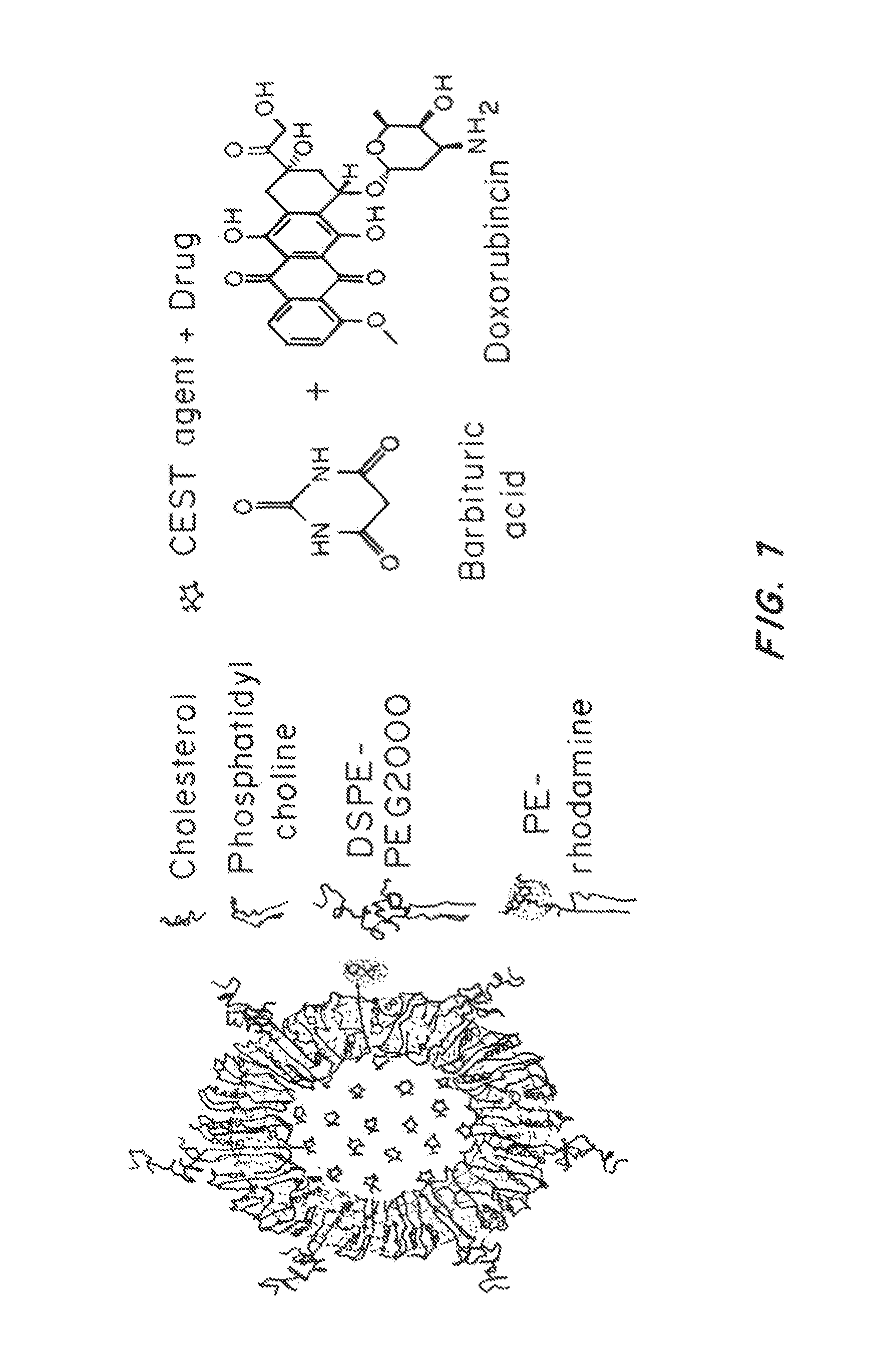
Preparation of Mucus-Penetrating Liposomal Particles

[0203]Liposomal particles were prepared using a conventional thin film hydration and extrusion method. 12 mg of a mixture of phosphatidylcholine (PC, e.g., egg PC or hydrogenated soy PC), PEG-phosphatidylethanolamine (PEG-PE) and cholesterol (CHOL) were dissolved in chloroform at pre-determined molar ratios. A small proportion of Rhodamine labeled PE (Rho-PE) was added to the mixture to enable visualization of the liposomal particles via fluorescence microscopy. The mixture was placed in a rotavap with reduced atmosphere pressure to evaporate the organic solvent. The resulting lipid film was hydrated with phosphate buffered saline (PBS) while agitated using a water bath sonicator to form multi-lamellar vehicles (MLV). The suspension was subsequently extruded through polycarbonate filters with pore sizes of 400 nm and 200 nm to generate unilamellar vehicles (LUV, i.e., liposomes with single bilayer membrane).

[0204]To load CEST agent...

example 2

Characterization of Liposomal Particles

[0207]The size and zeta-potential of liposomal particles were measured using dynamic light scattering and laser Doppler electrophoresis, respectively. Data are shown in Table 2.

TABLE 2Compositions of liposomal particles and their basic characterizationSizeMolar Ratio (%)Sample(Z-ave)PolydispersityZeta Potential(PC:CHOL:Rho-PE:PEG-PE)Abbreviation (d. nm)Index(mV)0.28:0.72:0.002:0.00 0% PEG2220.41−2.50.26:0.68:0.002:0.05 5% PEG1600.16−2.00.25:0.65:0.002:0.1010% PEG1750.06−2.20.22:0.58:0.002:0.2020% PEG1320.15−1.6

[0208]The size of the liposomal particles of all tested compositions ranged from 130 to 230 nm, well below the mesh dimension of human CVM (estimated to be >500 nm). Higher ratio of PEG-conjugated lipids reduced the size of the particles, likely due to the formation of liposomal micelles. A high proportion of cholesterol was used to enhance the stability of the liposomal particles.

example 3

Stability of Mucus-Penetrating Liposomal Particles

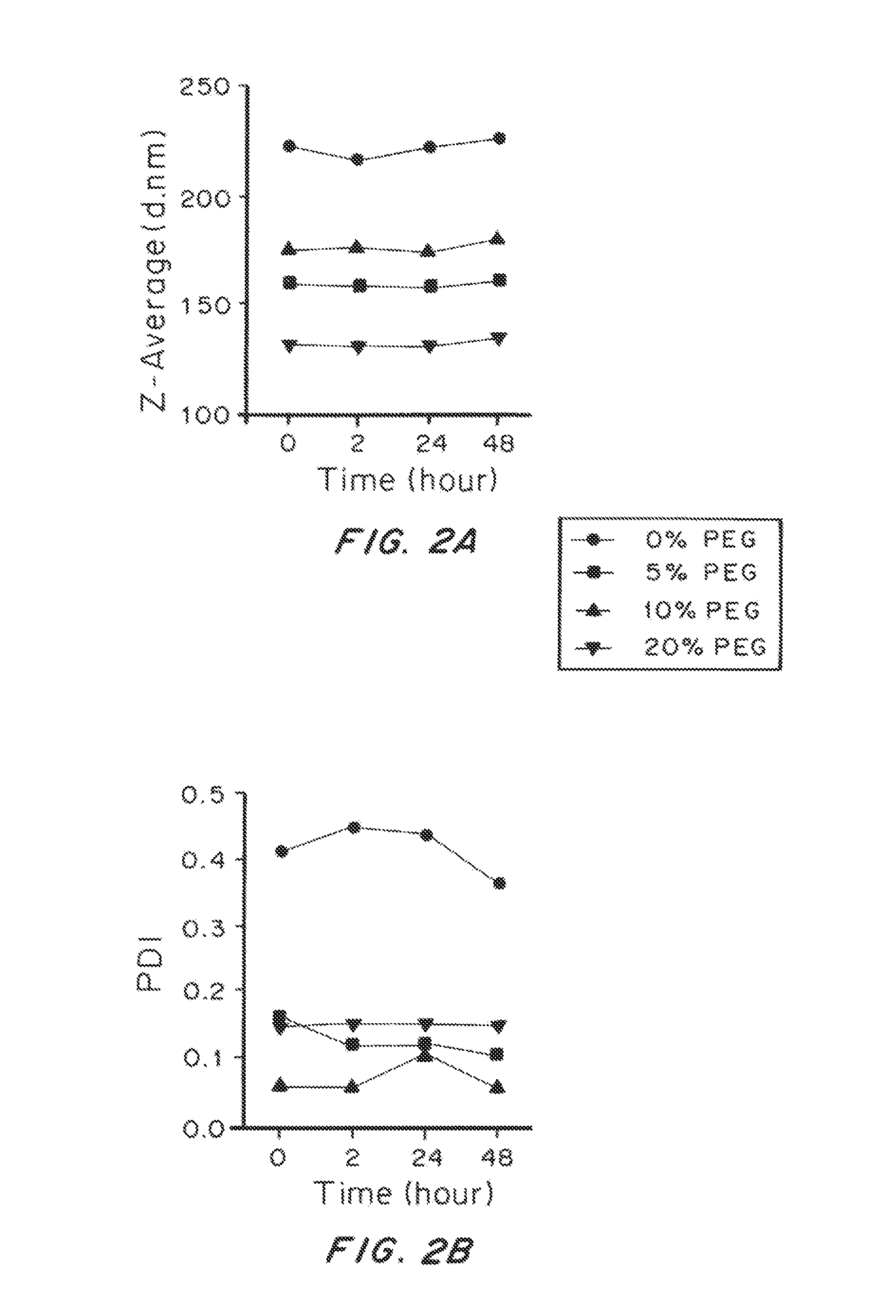
[0209]To test the stability of the liposomal particles of different compositions, we incubated the particles in phosphate buffered saline (PBS) at 37° C. and monitored the change of size and polydispersity over time, as shown in FIG. 2. The size and polydispersity index (PDI) of the particles were steady for all the experimental groups for over 48 hrs. However, the PDI of liposomal particles with no PEG was consistently higher than other experimental group, indicating their poorer quality and weaker stability as compared to the PEGylated ones.


PUM

PropertyMeasurementUnit
Fractionaaaaaaaaaa
Fractionaaaaaaaaaa
Timeaaaaaaaaaa

Abstract

Mucus-penetrating liposomal nanoparticles and methods of making and using thereof are described herein. The nanoparticles contain one or more lipids, one or more PEG-conjugated lipids, and optionally one or more additional materials that physically and/or chemically stabilize the particles. The nanoparticle have an average diameter of about 100 nm to about 300 nm, preferably from about 100 nm to about 250 nm, more preferably from about 100 nm to about 200 nm. The particles are mobile in mucus. The liposomes can further contain one or more therapeutic, prophylactic, and/or diagnostic agent to be delivered to a mucosal surface, such as the CV tract, the colon, the nose, the lungs, and/or the eyes. The liposomes can further contain one or more CEST agents to allow real time imaging of the particles in a live animal. The particles may also further contain an imaging agent, such as a fluorescent label.

Description

CROSS-REFERENCE TO RELATED APPLICATIONS[0001]This application claims priority to U.S. Ser. No. 61 / 642,776 entitled “Lipid-Based Drug Carriers for Rapid Penetration Through Mucus Linings”, filed on May 4, 2012. The contents of this application are incorporated herein by reference in their entirety.STATEMENT REGARDING FEDERALLY SPONSORED RESEARCH OR DEVELOPMENT[0002]This invention was made with Government Support under Agreement R01EB015031 awarded to Michael McMahon by the National Institutes of Health. The Government has certain rights in the invention.FIELD OF THE INVENTION[0003]This invention is in the field of liposomal nanoparticles for drug delivery, particularly mucus-penetrating liposomal nanoparticles.BACKGROUND OF THE INVENTION[0004]Mucus is a viscous and adhesive substance that efficiently traps and removes pathogens, hazard particles, and conventional drug or gene carriers from mucosal surfaces, including those that cover the female reproductive tract, GI tract, lung, air...

Claims


Application Information

Patent Timeline
IPC IPC(8): A61K47/48A61K49/18A61K31/704
CPCA61K47/48815A61K49/1812A61K31/704A61K9/1271A61K47/6911A61P25/04A61P25/08A61P35/00A61P43/00Y10S977/773Y10S977/906Y10S977/907
Inventor HANES, JUSTINCHAN, KANNIE WAI YANMCMAHON, MICHAEL T.YANG, MINGYU, TAO
Owner THE JOHN HOPKINS UNIV SCHOOL OF MEDICINE
Why Eureka
Try Eureka