

Monoclonal antibody against duck tembusu virus, hybridoma cell line and application thereof
- Summary
- Abstract
- Description
- Claims
- Application Information
AI Technical Summary
Benefits of technology
Problems solved by technology
Method used
Image

Examples
example 1
Preparation and Identification of a Monoclonal Antibody against Duck Tembusu Virus
1 Materials and Methods
1.1 Viruses, Cells and Experimental Animals
[0026]Duck Tembusu virus (DTMUV) Fengxian strain (FX2010 strain) was isolated and preserved by the laboratory; the duck embryo fibroblasts (DEF cells) and SP2 / 0 cells were provided by the laboratory; the clean grade BALB / c mice were purchased from Shanghai Laccas Experimental Animals Co., Ltd. The PCAGGS-DTMUV-E recombinant eukaryotic expression plasmids were constructed and preserved by our laboratory.
1.2 The Main Materials and Test Serum
[0027]DMEM was purchased from GBICO; 8-azaguanine, PEG1450, HAT, HT, Freund's complete adjuvant, Freund's incomplete adjuvant and HRP labeled goat-anti-mouse IgG were purchased from Sigma. DTMUV positive and negative sera, positive sera of avian influenza virus (AIV), Newcastle disease virus (NDV), reticuloendotheliosis virus (REV), type-I duck hepatitis virus (DHV-1), reovirus (RV) and avian leukemia v...
example 2
Identification of a Monoclonal Antibody against Duck Tembusu Virus E Protein
[0035]1. Identification of a Monoclonal Antibody again Duck Tembusu Virus E Protein
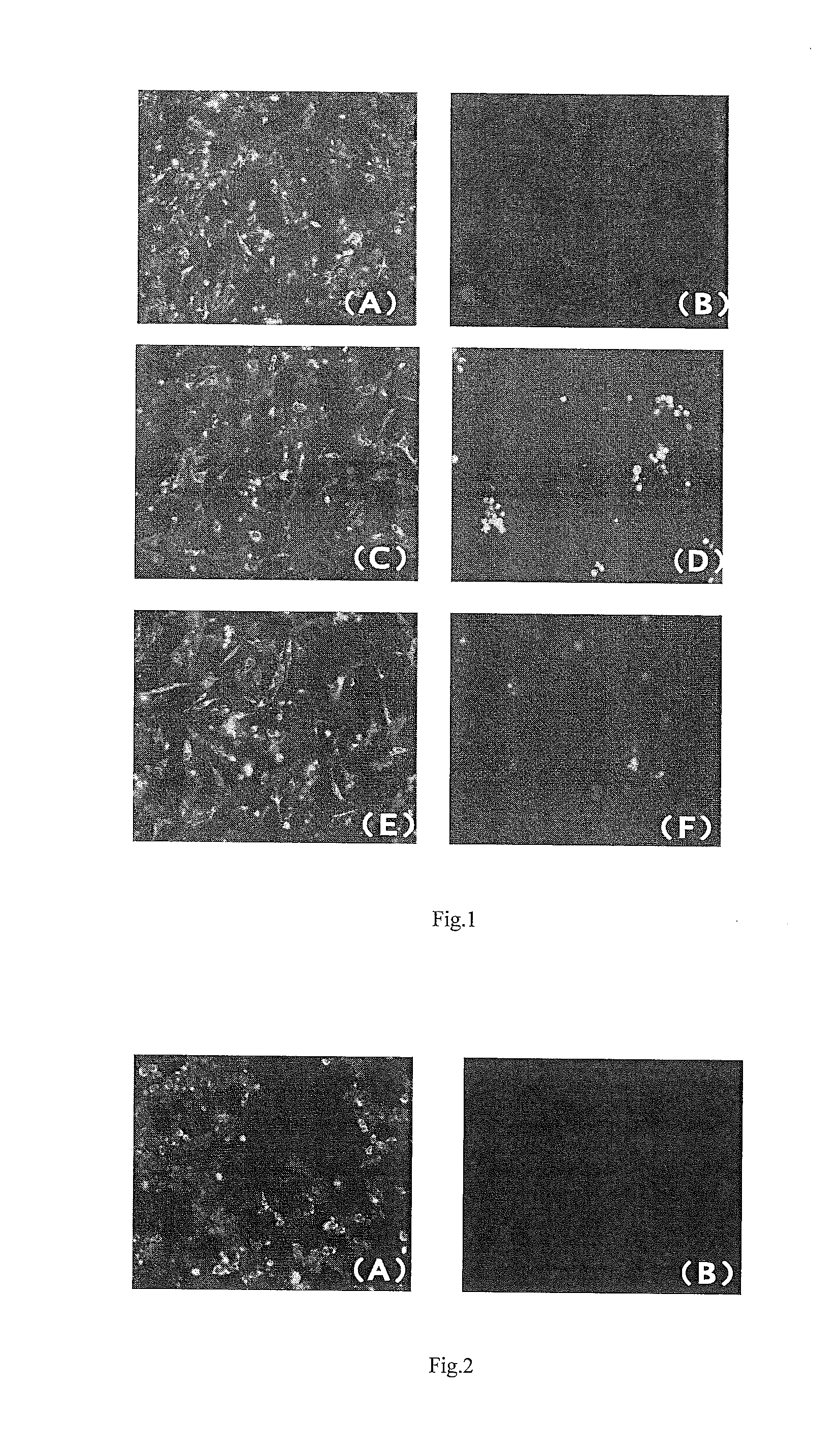
[0036]293 cells were cultured in 6-well plates, when the cell monolayers grown, 293 cells were transfected by eukaryotic expression recombinant plasmid pCAGGS-DTMUV-E, meanwhile, negative control holes were set. 24 hours after transfection, the supernatant was discarded, and the cells were fixed with 4% paraformaldehyde. After washed once by PBST, hybridoma cultural supernatant was added, and incubated for 1 h at 37° C., washed three times using PBST, added with FITC-goat anti-mouse IgG antibody, continued to incubate for 1 hour at 37° C., washed three times using PBST, and finally observed under a fluorescence microscope. Those with green fluorescence were judged as positive, and those without fluorescence were judged as negative.
[0037]Results: Through IFA detection of 293 T cells transfected by PCAGGS-DTMUV-E eukaryotic plas...
example 3
Establishment of Block ELISA Method (Blockinge ELISA, B-ELISA) for Detecting DTMUV Antibody
1. Preparation of Negative Serum
[0041]6-week-old SPF ducks were used, to collect blood from heart, and then the serum was separated, and dispensed in 0.2 mLEp tubes, kept at −20° C. for future use.
2. Preparation of Positive Serum
[0042]6-week-old SPF ducks were infected nasally by 103.5ELD50DTMUV FX2010 isolates. 3 weeks later, blood was collected from heart, and then the serum was separated, and dispensed in 0.2 mLEp tubes, kept at −20° C. for future use.
3. The Procedures for DTMUV Antibody Block ELISA
[0043]The purified DTMUV was diluted to 0.03 mg / mL using 0.05 mol / L carbonate buffer solution (pH 9.6), coated 96-well microtiter plate, 100 μL each well, overnight at 4° C., and closed for 1 h at 37° C. using PBS (0.01 mol / L, pH 7.4) solution containing 5% skim milk powder, and then washed three times with PBS (PBST) containing 0.5 mL / L TWEEN 20. The DTMUV -positive and negative sera diluted by ...
PUM
| Property | Measurement | Unit |
|---|---|---|
| Fraction | aaaaa | aaaaa |
| Inhibition | aaaaa | aaaaa |
| Inhibition | aaaaa | aaaaa |
Abstract
Description
Claims
Application Information