Method for detection of microorganism and kit for detection of microorganism

a microorganism and kit technology, applied in the field of microorganism detection and kit for microorganism detection, can solve the problems of foodtuffs actually subjected to pasteurization treatment, methods that cannot be said to be sufficient as methods for distinguishing live and dead states, and methods that require about two days to obtain, etc., to achieve high detection sensitivity

Inactive Publication Date: 2015-03-26
MORINAGA MILK IND CO LTD
View PDF3 Cites 1 Cited by
  • Summary
  • Abstract
  • Description
  • Claims
  • Application Information

AI Technical Summary

Problems solved by technology

However, the plate culture method requires time of about two days to obtain a result.
However, if a bacterial DNA is targeted, the background originated from dead cells originally contained in the test sample is also detected, and therefore a positive result obtained by PCR does not necessarily suggest the presence of live bacteria.
Therefore, the current situation in the fields of food sanitation and clinical test is that PCR is not used widely, although it is a highly sensitive and quick technique.
Therefore, this method cannot be said to be sufficient as a method for distinguishing the live and dead states.
However, the following problems remain in these methods for distinguishing live and dead states of microorganisms such as bacteria using the PCR method.
However, there are not foodstuffs actually subjected to such pasteurization treatments, especially the treatment of the latter type.
Moreover, there are not supposed detection of only live microorganisms subjected to the currently major pasteurization method in the food industry, low temperature long time pasteurization (LILT pasteurization), high temperature short time pasteurization (HTST pasteurization), or ultra high temperature pasteurization (UHT pasteurization), and detection of only live specific pathogenic bacteria in clinical specimens of infectious disease patients administered with antibiotics.
Moreover, in the case of a test sample of a foodstuff or clinical specimen containing dead cell background at a concentration of 104 cfu / ml or higher, the amounts of the final PCR amplification products derived from dead cells exceed the detection limit of the technique of Patent document 1, and therefore it is impossible to determine whether a positive response of a test sample obtained by PCR is derived from live cells or dead cells.
That is, the above procedure requires to perform troublesome extraction of total RNA and uses two steps of reverse transcription reaction and PCR.
Therefore, this technique is inferior to usual PCR targeting DNA in quantification performance and quickness.
Therefore, the technique lacks stability.
Furthermore, in a foodstuff or clinical specimen containing dead cells at a high concentration, only live cells of 1 / 10 of that concentration can be detected by this technique.
Therefore, it is difficult to apply this technique in the fields of food sanitation inspection and clinical test, which require quickness, high sensitivity and accuracy.Patent document 1: International Patent Application Unexamined Publication in Japan No. 2000-530118Patent document 2: International Patent Publication WO2002 / 052034 pamphlet

Method used


Image

  • Method for detection of microorganism and kit for detection of microorganism
  • Method for detection of microorganism and kit for detection of microorganism
  • Method for detection of microorganism and kit for detection of microorganism

Examples


example 1

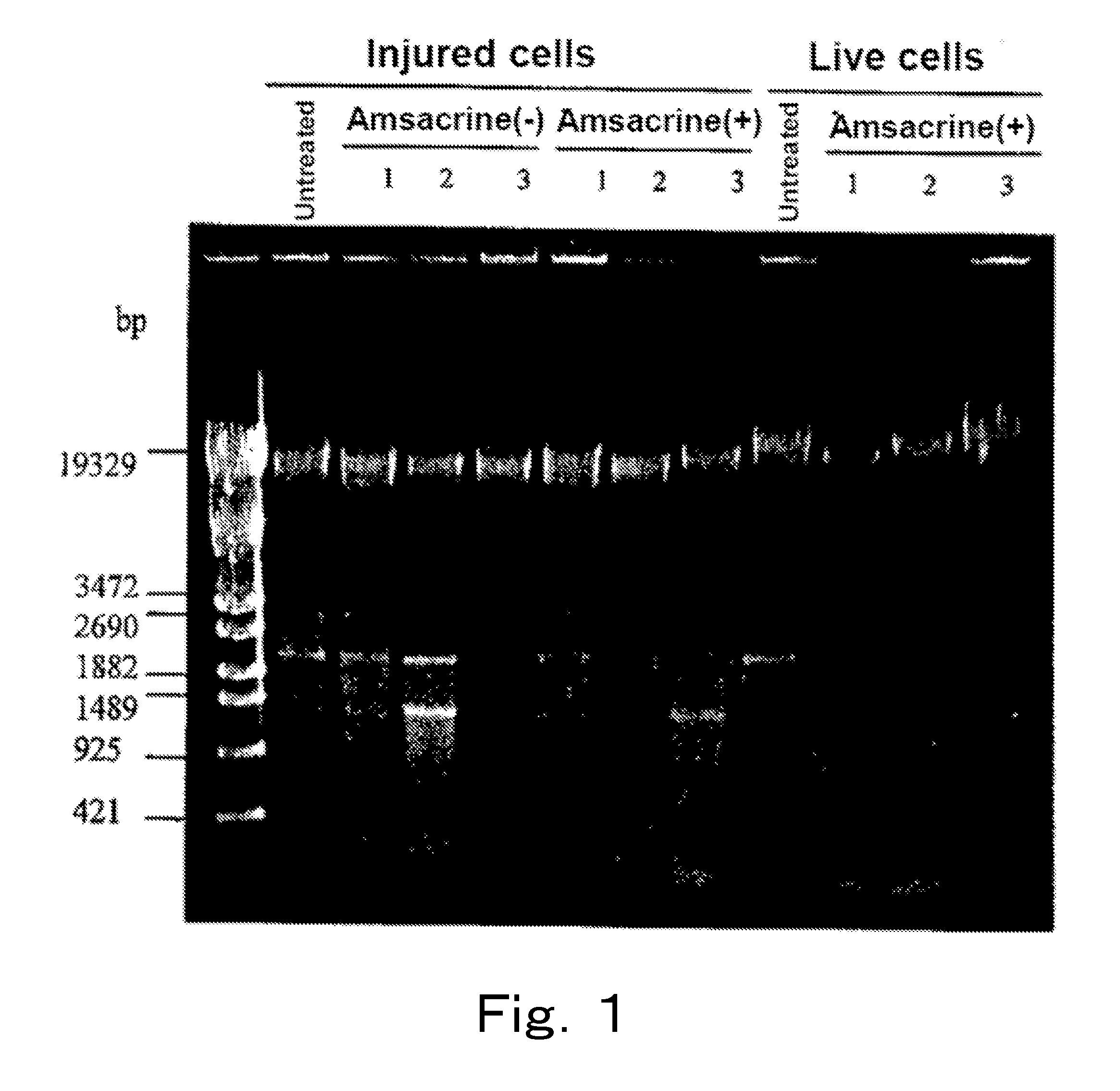
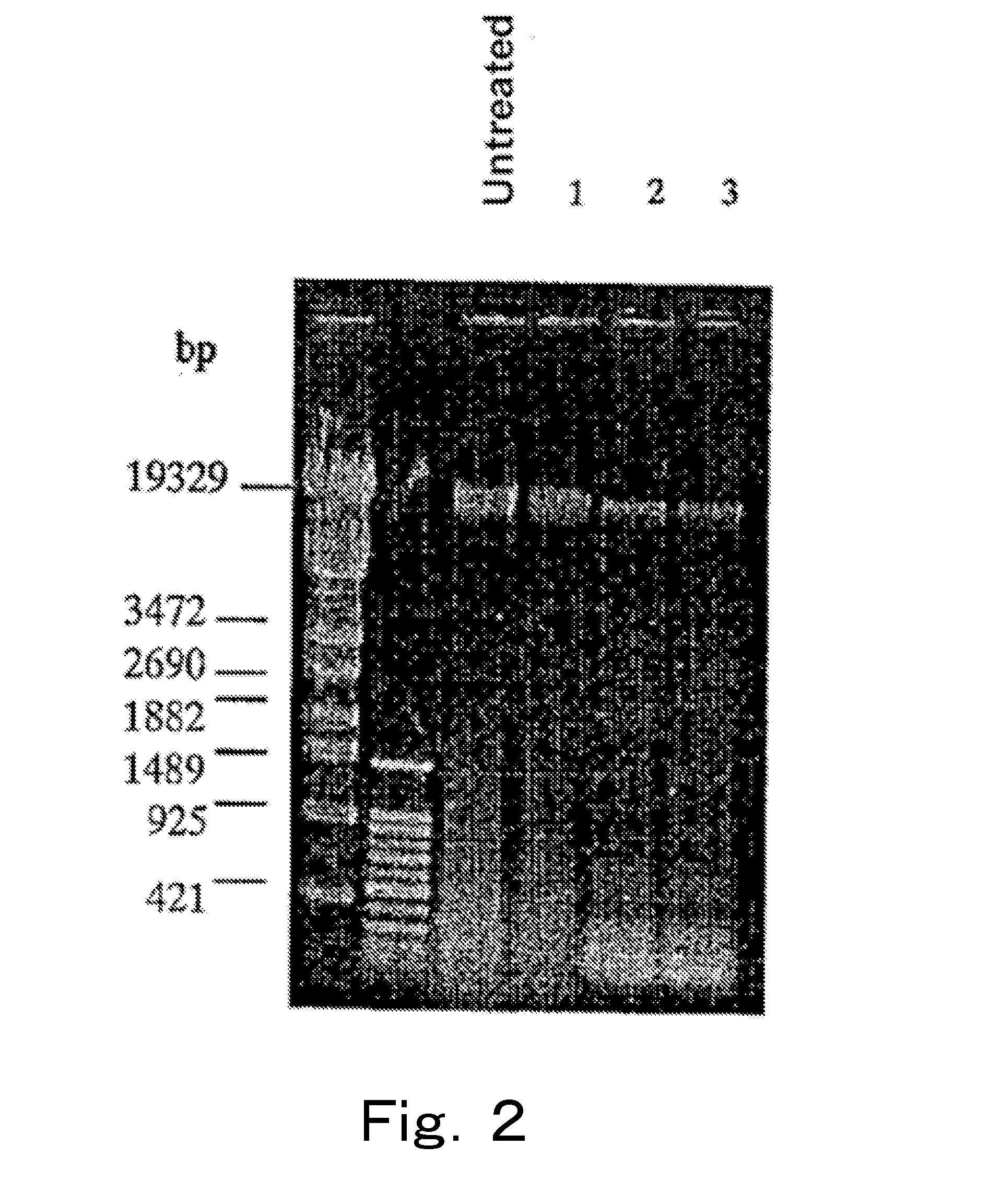
[0103]Live cells and injured cells of a microorganism were treated with a topoisomerase poison, respectively, and cleavage degree of each chromosomal DNA was examined.

1. Preparation of Samples

1-1) Preparation of Live Cell and Injured Cell Suspensions

[0104]Listeria monocytogenes (Listeria monocytogenes JCM 2873, henceforth also referred to as “Listeria”), which is a gram-positive bacterium, was cultured at 30° C. by using the BHI broth, 40 ml of the culture medium in which the cells were at the logarithmic phase was subjected to refrigerated centrifugation at 4° C. and 8,000×g for 15 minutes, and the supernatant was removed. 40 ml of physiological saline was added to the cells, and the mixture was sufficiently stirred, and subjected to similar refrigerated centrifugation, the supernatant was removed, and then 10 ml of physiological saline was added to the cells to prepare a live cell suspension. When live cell count of this live cell suspension was measured on a standard agar plate m...

example 2

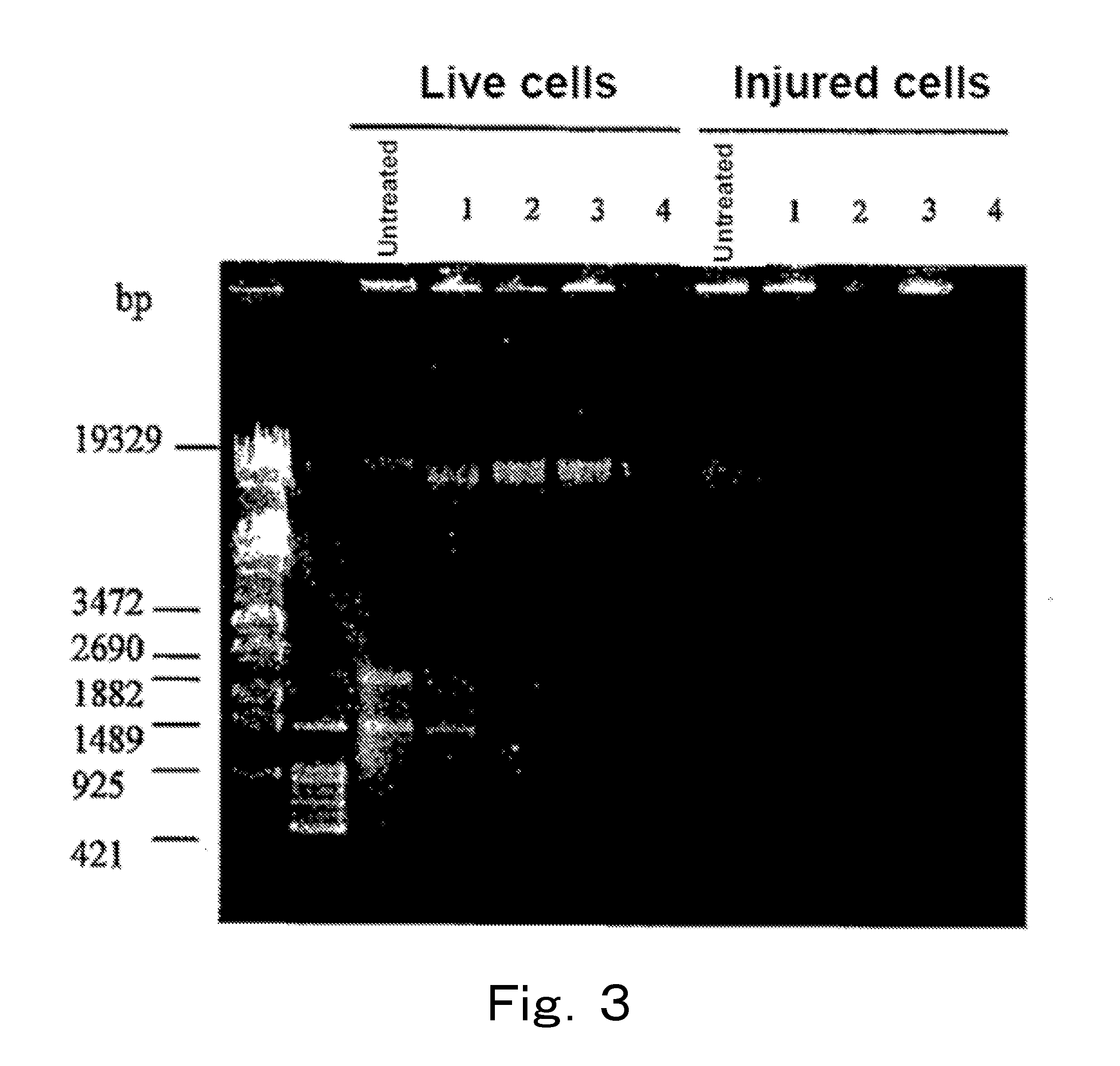
[0117]Analysis was performed by PCR targeting the 23S rRNA gene using chromosomal DNAs of live cells and injured cells of microorganisms treated with a DNA gyrase poison.

1. Preparation of Samples

1-1) Preparation of Live Cell and Injured Cell Suspensions

[0118]Enterobacter sakazakii (Enterobacter sakazakii ATCC 51329 strain, henceforth also referred to as “Enterobacter”), which is a gram-negative bacterium, was cultured at 37° C. by using the BHI broth, 40 ml of the culture medium in which the cells were at the logarithmic phase was subjected to refrigerated centrifugation at 4° C. and 8,000×g for 15 minutes, and the supernatant was removed. 40 ml of physiological saline was added to the cells, and the mixture was sufficiently stirred, and subjected to similar refrigerated centrifugation, the supernatant was removed, and then 10 ml of physiological saline was added to the cells to prepare a live cell suspension. When live cell count of this live cell suspension was measured on a stand...

example 3

[0153]Analysis was performed by PCR targeting the 16S rRNA gene or the 23S rRNA gene using chromosomal DNAs of live cells and injured cells of microorganisms treated with EMA.

1. Preparation of Samples

1-1) Preparation of Gram-Negative Bacterium (Live Cell and Injured Cell) Suspensions

[0154]Escherichia coli DH5α (henceforth also referred to as “Escherichia coli”), Citrobacter koseri (Citrobacter koseri JCM 1658, henceforth also referred to as “Citrobacter”), Salmonella enteritidis (Salmonella enteritidis IID 604, henceforth also referred to as “Salmonella”), and Klebsiella oxytoca (Klebsiella oxytoca JCM 1665, henceforth also referred to as “Klebsiella”) were each cultured at 37° C. by using the BHI broth, 40 ml of the culture medium in which the cells were at the logarithmic phase was subjected to refrigerated centrifugation at 4° C. and 8,000×g for 15 minutes, and the supernatant was removed. Then, 40 ml of physiological saline was added to the cells, and the mixture was sufficientl...


PUM

PropertyMeasurementUnit
timeaaaaaaaaaa
temperatureaaaaaaaaaa

Abstract

Live cells of a microorganism in a test sample are detected by the following steps:a) the step of treating the test sample with a topoisomerase poison and / or a DNA gyrase poison,b) the step of extracting DNA from the test sample, and amplifying a target region of the extracted DNA by PCR, andc) the step of analyzing an amplification product.

Description

RELATED APPLICATIONS[0001]This application is a continuation of U.S. application Ser. No. 11 / 994,823, filed Jan. 4, 2008 which is the U.S. National Phase under 35 U.S.C. §371 of International Application PCT / JP2006 / 302893, filed Feb. 17, 2006, which was published in a non-English language.TECHNICAL FIELD[0002]The present invention relates to a method for detecting a microorganism contained in foodstuffs or clinical samples, and a kit for detecting a microorganism. More precisely, the present invention relates to a method and kit for detection of a microorganism that enable selective detection of live cells of a microorganism contained in foodstuffs or clinical samples.BACKGROUND ART[0003]The plate culture method has been conventionally used for measurement of general live bacterial counts in foodstuffs, clinical samples or environments. However, the plate culture method requires time of about two days to obtain a result.[0004]Because of the improvements of sterilization techniques a...

Claims


Application Information

Patent Timeline
Patent Type & Authority Applications(United States)
IPC IPC(8): C12Q1/68C12N15/11C12Q1/04
CPCC12Q1/6806C12Q2600/158C12Q1/689C12Q1/686C12Q1/6888C12Q2521/519C12Q1/04C12Q2561/113
Inventor YOSHIDA, SHINICHISOEJIMA, TAKASHI
Owner MORINAGA MILK IND CO LTD
Why Eureka
Try Eureka