Method for producing ciliary marginal zone-like structure
a ciliary marginal and structure technology, applied in the field of ciliary marginal structure production, can solve the problems of high efficiency of stem cells and no known method for producing such structure from pluripotent stem cells, and achieve the effect of high efficiency
- Summary
- Abstract
- Description
- Claims
- Application Information
AI Technical Summary
Benefits of technology
Problems solved by technology
Method used
Image
Examples
example 1
Production Example of Cell Aggregate Containing Retinal Tissue Using Human ES Cell—1
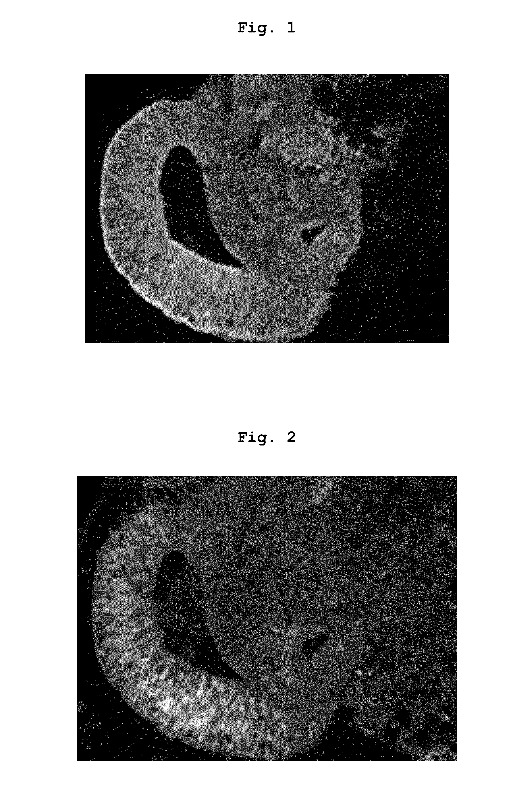
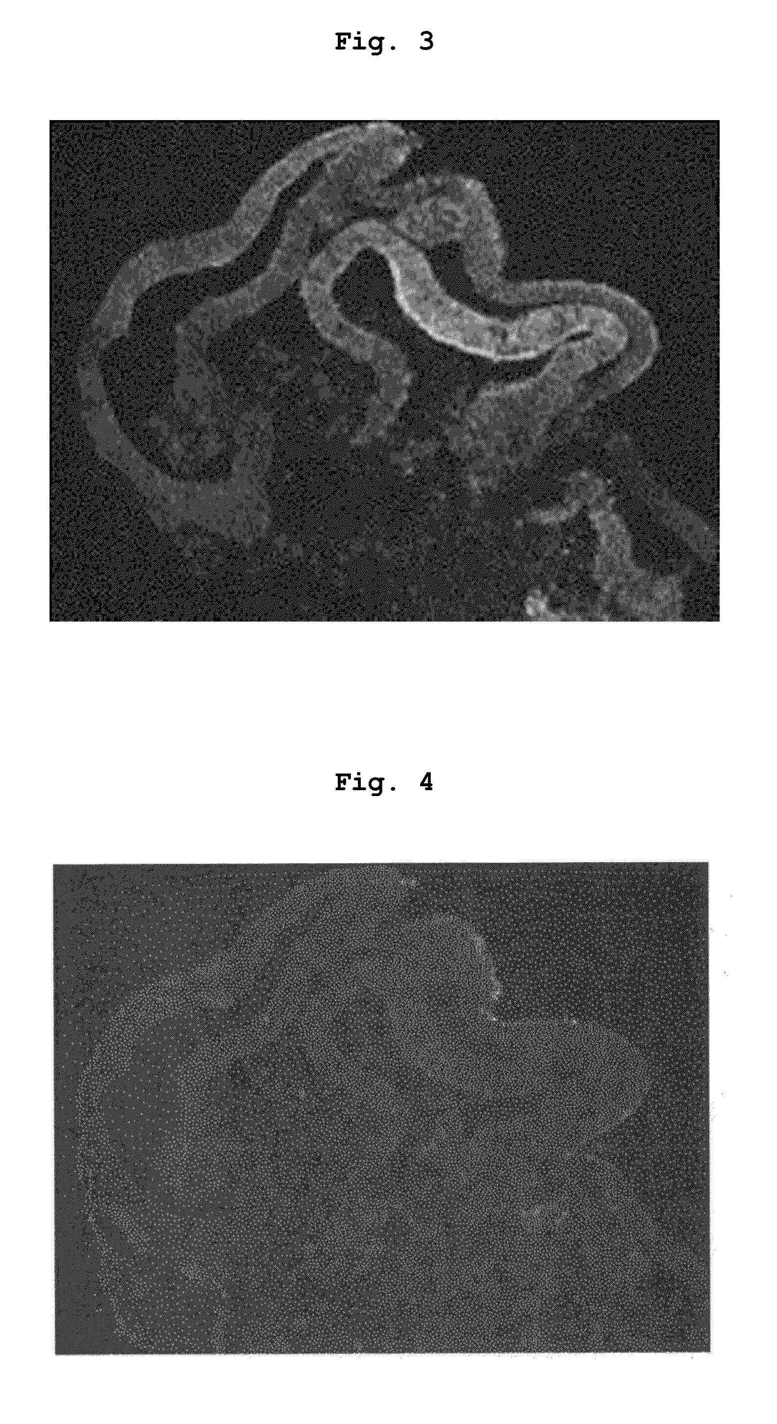
[0093]RAX::GFP knock-in human ES cells (derived from KhES-1; Nakano, T. et al. Cell Stem Cell 2012, 10(6), 771-785) were cultured according to the methods described in “Ueno, M. et al PNAS 2006, 103(25), 9554-9559” and “Watanabe, K. et al. Nat Biotech 2007, 25, 681-686”. As the medium, DMEM / F12 medium (Invitrogen) added with 20% KSR (Knockout Serum Replacement; Invitrogen), 0.1 mM 2-mercaptoethanol, 1 mM pyruvic acid and 5 to 10 ng / ml bFGF was used. The aforementioned cultured ES cells were singly dispersed in 0.25% trypsin-EDTA (Invitrogen), and the singly dispersed ES cells were floated in a 100 μl serum-free medium to 9×10³ cells per well of a non-cell adhesive 96-well culture plate (SUMILON spheroid plate, SUMITOMO BAKELITE CO., LTD.), and suspension-cultured at 37° C., 5% CO2. The serum-free medium used then was a serum-free medium obtained by adding 20% KSR, 0.1 mM 2-mercaptoethanol, 1 mM pyruv...
example 2
Production Example of Cell Aggregate Containing Retinal Tissue Using Human ES Cell—2
[0095]By a method similar to that in Example 1, suspension culture was performed for a longer time. As a result, on day 25 from the start of the suspension culture, a cell aggregate containing a retinal tissue in which Chx10 positive cells were found in about 80% or about 90% of the tissue was also obtained.
example 3
Culture of Cell Aggregate Containing Retinal Tissue in Serum-Free Medium Containing Substance Acting on Wnt Signal Pathway
[0096]A cell aggregate containing a retinal tissue on day 18 from the start of the suspension culture, which was produced by the method described in Example 1, was suspension-cultured in a serum-free medium containing a substance acting on the Wnt signal pathway (3 μM CHIR99021) for 3 days.
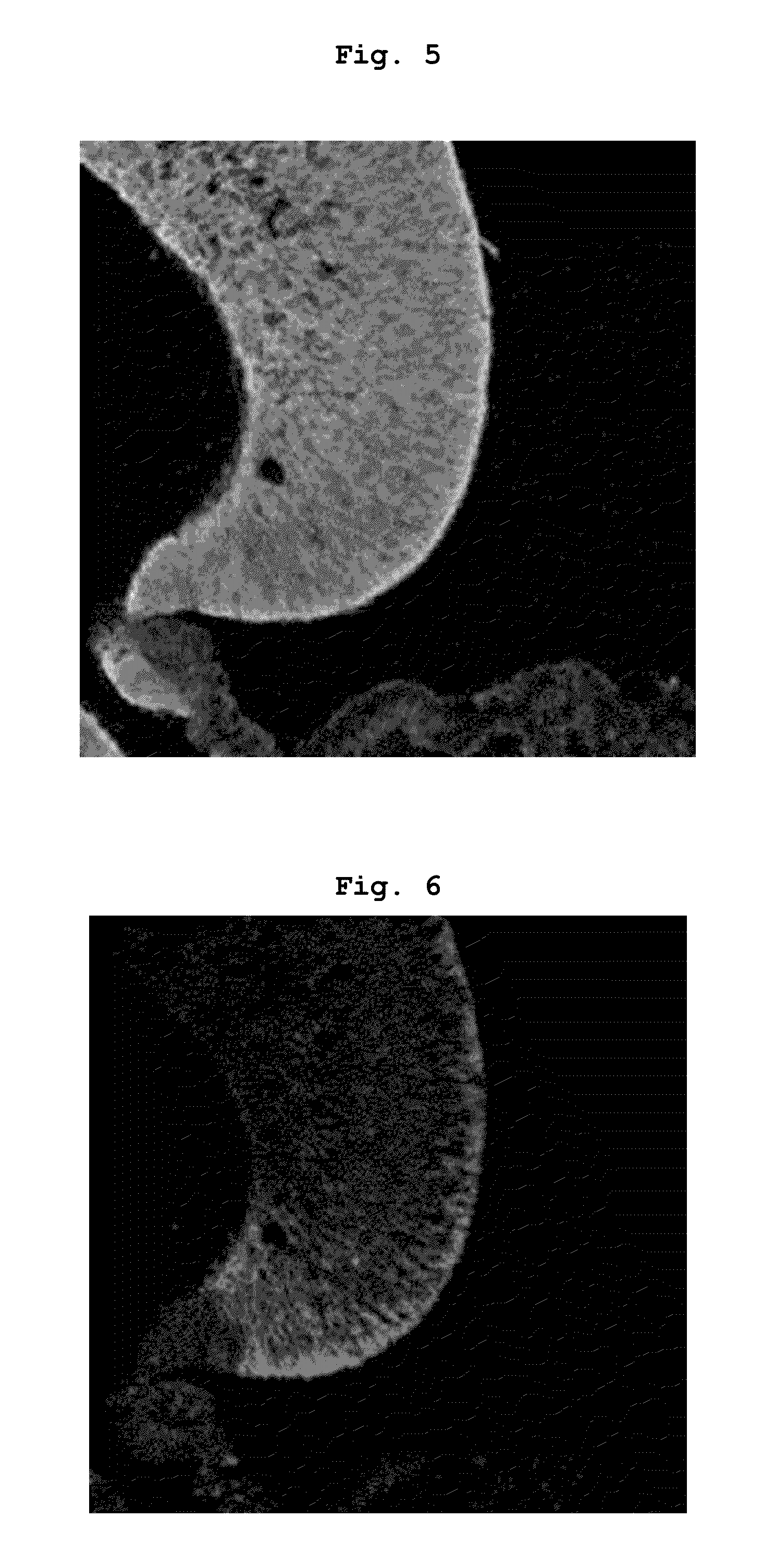
[0097]The obtained cell aggregate was fixed with 4% para-formaldehyde to prepare a cryosection. The prepared cryosection was subjected to fluorescence microscopic observation of a GFP fluorescence image (FIG. 3) and immunostaining with Chx10 which is one of the neural retinal progenitor cell markers (FIG. 4). In the retinal tissue contained in the aforementioned cell aggregate, which had been suspension-cultured in a serum-free medium containing a substance acting on the Wnt signal pathway for 3 days, Chx10 positive cells were found in only about 3% of the tissue and a clear de...
PUM
 Abstract
Description
Claims
Application Information
 - R&D
- Intellectual Property
- Life Sciences
- Materials
- Tech Scout
- Unparalleled Data Quality
- Higher Quality Content
- 60% Fewer Hallucinations
Browse by: Latest US Patents, China's latest patents, Technical Efficacy Thesaurus, Application Domain, Technology Topic, Popular Technical Reports.
© 2025 PatSnap. All rights reserved.Legal|Privacy policy|Modern Slavery Act Transparency Statement|Sitemap|About US| Contact US: help@patsnap.com


