Controlling extracellular matrix protein microstructure with ultrasound
- Summary
- Abstract
- Description
- Claims
- Application Information
AI Technical Summary
Benefits of technology
Problems solved by technology
Method used
Image
Examples
example 1
Ultrasound Exposure Effects Collagen Fiber Structure
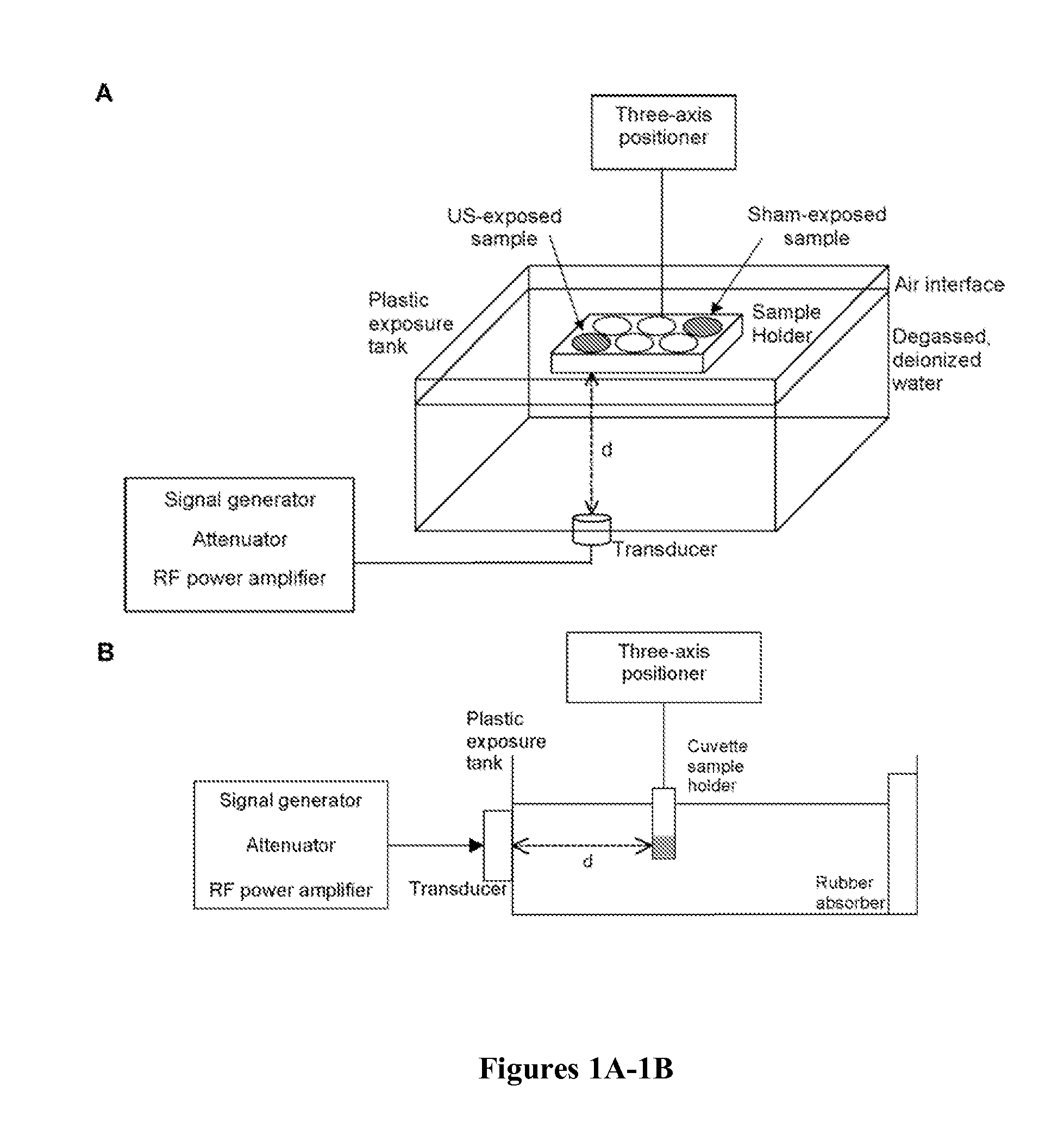
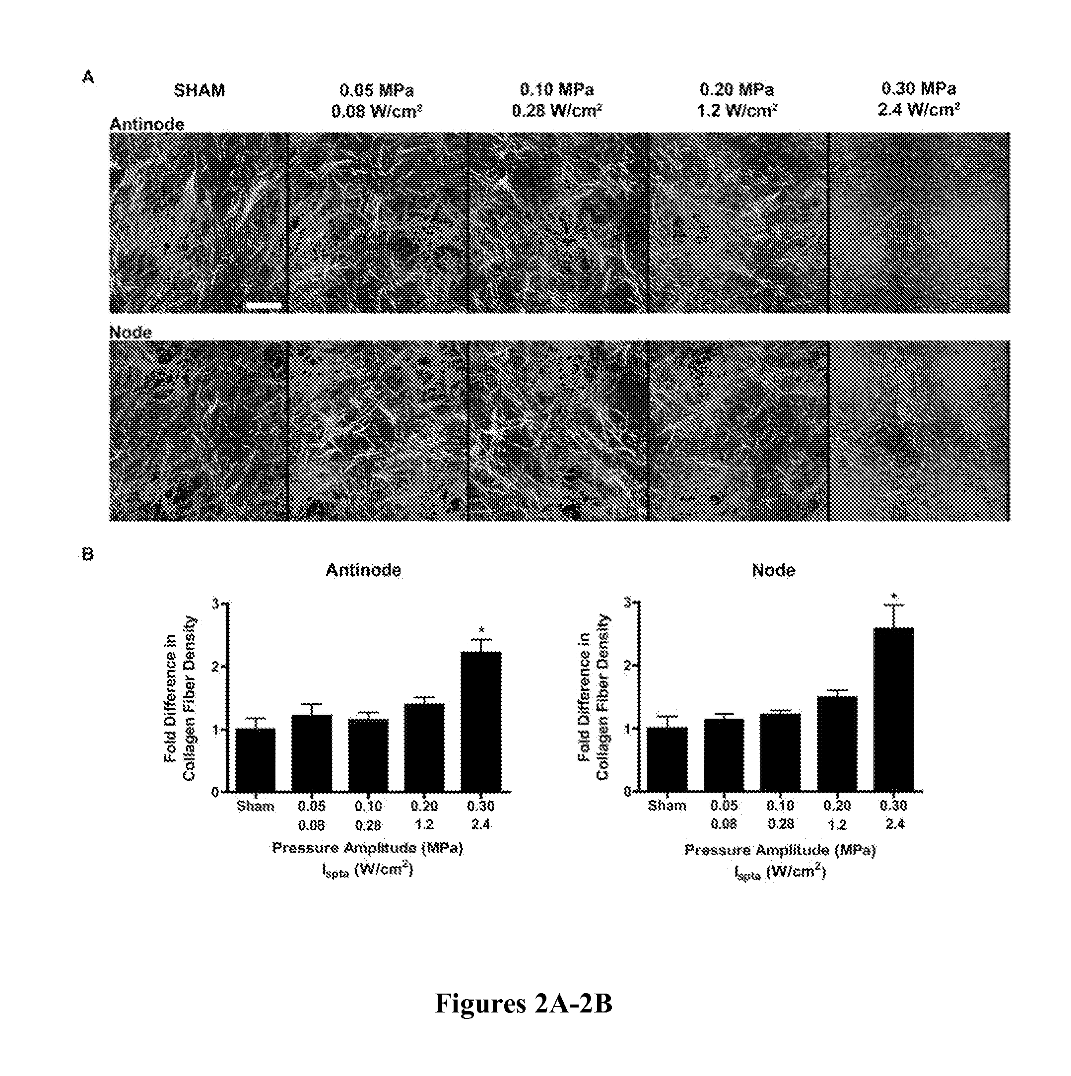
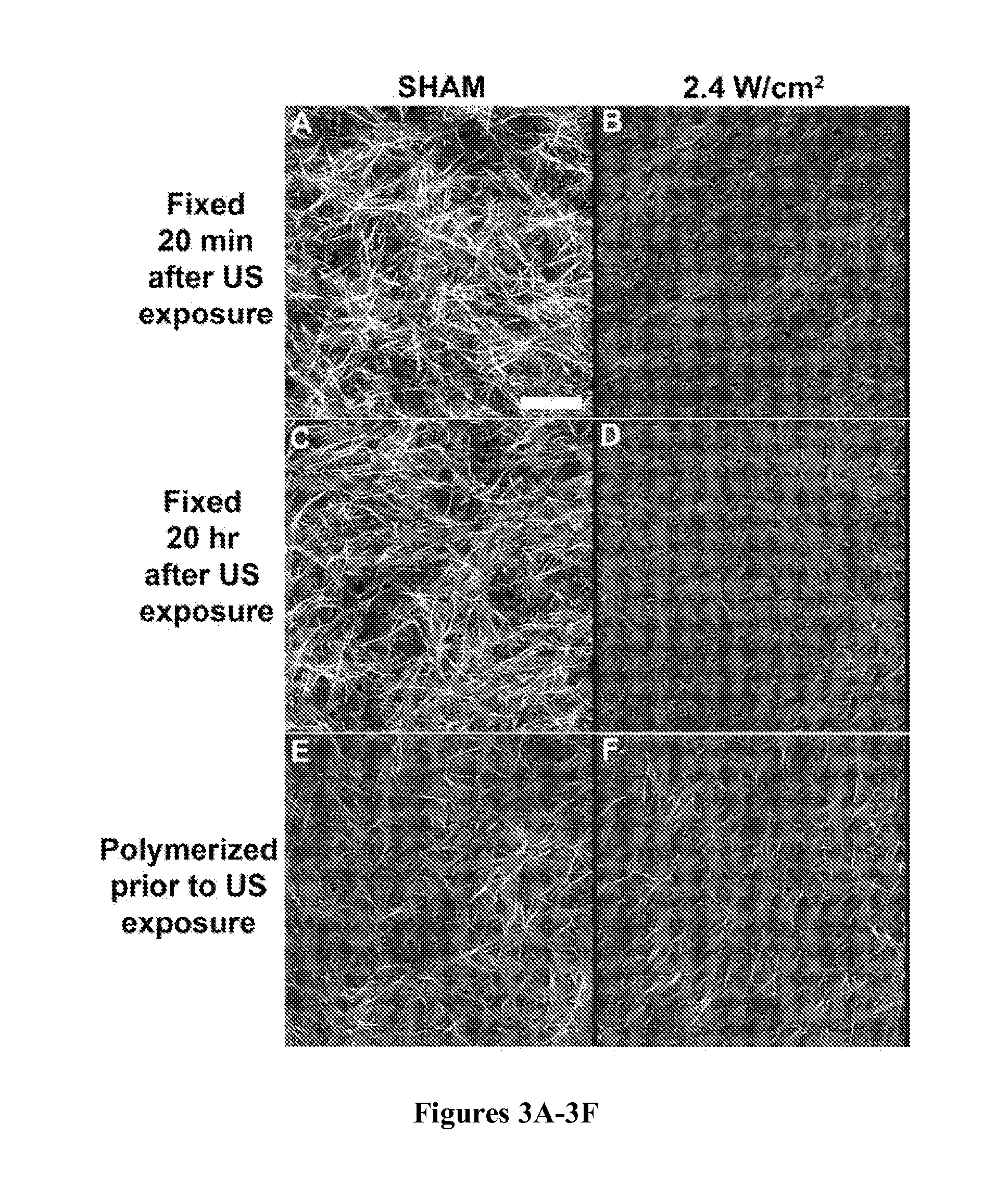
[0086]To investigate effects of ultrasound on collagen fiber microstructure, unpolymerized solutions of type I collagen were exposed to 1 MHz, CW ultrasound of various peak positive pressures and intensities using the apparatus diagramed in FIG. 1A. This exposure geometry produces an acoustic standing wave field throughout the collagen sample volume (Garvin et al., “Controlling the Spatial Organization of Cells and Extracellular Proteins in Engineered Tissues Using Ultrasound Standing Wave Fields,”Ultrasound Med. Biol. 36(11):1919-32 (2010), which is hereby incorporated by reference in its entirety). Therefore, collagen fiber morphology of polymerized gels was assessed at imaging depths corresponding to either pressure antinodes or nodes using second-harmonic generation microscopy imaging. Sham-exposed collagen gels were characterized by long, thick, loosely packed collagen fibers (FIG. 2A). In contrast, as the ultrasound exposure ...
example 2
Ultrasound-Mediated Changes in Collagen Fiber Structure Depend on Ispta
[0088]Other studies have shown that collagen fiber microstructure is affected by polymerization temperature (Roeder et al., “Tensile Mechanical Properties of Three-Dimensional Type I Collagen Extracellular Matrices With Varied Microstructure,”J. Biomech. Eng. 124:214-222 (2002); Sander & Barocas, “Biomimetic Collagen Tissues: Collagenous Tissue Engineering and Other Applications” in P. Fratzl, ed., Collagen Structure and Mechanics, Springer, New York, N.Y., pp. 475-504 (2008), which are hereby incorporated by reference in their entirety). Thus, experiments were conducted to investigate the role of ultrasound-induced heating in the observed effects on collagen fiber microstructure. Two investigations were performed to independently assess the roles of acoustic pressure and Ispta. To examine the role of Ispta, solutions of type I collagen were exposed to 1-MHz ultrasound standing wave fields of various Ispta but c...
example 3
Temperature Measurements and Thermal Effects of Ultrasound
[0090]Temperatures of collagen gels were measured during ultrasound exposure. Sham-exposed gels reached a steady-state temperature of 18.6±0.3° C. (FIG. 5A; ‘Sham, RT water bath’). In contrast, the steady-state temperature within collagen gels exposed at 2.4 W / cm2 was 26.3±1.7° C. (FIG. 5A). This temperature rise was simulated in sham-exposed samples by bulk heating using a water bath heated to 28.5° C. (FIG. 5A). The morphology of collagen fibrils of gels polymerized in the 28.5° C. water bath was similar to that of collagen gels exposed to ultrasound at 2.4 W / cm2 (FIG. 5B). In these gels, fibrils appeared thinner, shorter and more densely packed than those of sham-exposed collagen gels polymerized at room temperature (FIG. 5B). Quantitative analysis of images showed a 2.5-fold increase in fiber density for samples polymerized in the 28.5° C. water tank and ultrasound-exposed samples at 2.4 W / cm2, as compared to sham collage...
PUM
| Property | Measurement | Unit |
|---|---|---|
| Mechanical strength | aaaaa | aaaaa |
| Microstructure | aaaaa | aaaaa |
Abstract
Description
Claims
Application Information
 - R&D
- Intellectual Property
- Life Sciences
- Materials
- Tech Scout
- Unparalleled Data Quality
- Higher Quality Content
- 60% Fewer Hallucinations
Browse by: Latest US Patents, China's latest patents, Technical Efficacy Thesaurus, Application Domain, Technology Topic, Popular Technical Reports.
© 2025 PatSnap. All rights reserved.Legal|Privacy policy|Modern Slavery Act Transparency Statement|Sitemap|About US| Contact US: help@patsnap.com


