Method and apparatus for measuring and displaying dental plaque, gingiva, and alveolar bone

a technology of dental plaque and apparatus, applied in the field of apparatus and to a method for measuring and displaying dental plaque, gingiva, and/or alveolar bone, can solve the problems of difficult recognition of the attachment of dental plaque to the surface of the tooth, failure to provide details of the state of dental plaque attachment, and strong discomfort of patients, so as to encourage more reliable dental practice, high reproducibility and reliability, and excel in reproducibility

Active Publication Date: 2015-07-02
NAT CENT FOR GERIATRICS & GERONTOLOGY
View PDF9 Cites 8 Cited by
  • Summary
  • Abstract
  • Description
  • Claims
  • Application Information

AI Technical Summary

Benefits of technology

[0038]The methods and apparatus according to the present invention have the following features and provide an advantageous effect of encouraging more reliable dental practice.[Objectivity and Universality]
[0039]In dental clinical practice and dental examination of related art, visual inspection, palpation, and other subjective inspection methods are primarily executed and results of the methods differ from each other. In contrast, the present invention can provide an evaluation method that objectively quantifies dental plaque, gingiva, and alveolar bone. The method and apparatus according to the present invention that automatically extract a dental plaque region in an image with no involvement of human evaluation by using a computer can acquire consistent data irrespective of inspection location and inspection operator, whereby the method and apparatus excel in reproducibility and can be used in a universal manner.[Image Generation and Digitization]
[0040]The method and apparatus according to the present invention can evaluate an entire layer of dental plaque, which cannot be detected with a dental image diagnosis apparatus based, for example, on X-rays, in the form of a two-dimensional image. In particular, the method and apparatus according to the present invention allow evaluation of a tomographic image and hence allow evaluation in the depth direction, whereby even dental plaque under a gingival cuff, which cannot be detected by visual inspection, can be detected. Further, dental plaque attachment can be evaluated based on a three-dimensional image of the dental plaque, whereby the thickness, length, cross-sectional area, surface area, and volume, of the dental plaque can be expressed in the form of an image and digitized. Even gingiva and alveolar bone, quantification of which has not been even attempted, can be quantified in the same manner.[Quantification and Creation of Database]
[0041]The methods and apparatus according to the present invention, which allow quantitative measurement, provide high reproducibility and reliability. Further, the methods and apparatus according to the present invention allow measurement and evaluation in the time course. The capability of image generation and digitization allows the methods and apparatus according to the present invention to be used in dental examination, and results from the methods and apparatus can be provided in the form of a numerical database.[Safety]
[0042]The methods and apparatus according to the present invention, which use near infrared light as observation light, allow inspection without medical exposure, which is inevitable in a dental x-ray method of related art. Further, since the methods and apparatus according to the present invention are based on a noncontact, noninvasive inspection method, inspection can be performed without damage to a structure of an attachment associated with periodontal tissue and without dissemination of bacteria into surrounding periodontal pockets.[Reliability, High Sensitivity, and Specificity]
[0043]The near infrared light used in the methods and apparatus according to the present invention can pass through mutan, glucan, and other substances that form dental plaque and reach and visualize dentin. The methods according to the present invention, which can generate an image of an entire layer of dental plaque and distinguish specifically the dental plaque from the dentin, are high-sensitivity, high-specificity, highly reliable inspection methods.[Effective in Informed Consent]

Problems solved by technology

Since the color of dental plaque is white or translucent white, which is the same color as that of the surface of a tooth, it is difficult to recognize the attachment of the dental plaque on the surface of the tooth.
The method is, however, a two-level evaluation method that simply evaluates presence or absence and hence fails to provide details of the state of dental plaque attachment.
Further, the dental plaque coloring operation itself gives a patient strong discomfort, and removal of the coloring solution after the coloring operation is cumbersome.
Moreover, since portions other than dental plaque are also colored, the inspection method undesirably has low specificity and many other disadvantages.
The method does not rely on coloring and cannot therefore clearly show the boundary among dental plaque, a tooth, and a periodontal tissue.
It cannot, however, be said that the method is a quantitative evaluation method because there are large differences among the four levels.
The method, however, has problems of poor reproducibility and lack of true objectivity, that is, multiple inspection operators who execute the method cannot provide consistent values.
The method, however, has problems of bitterness of the coloring agent and low stability of the coloring agent when the coloring agent is stored.
Fluorescent dyes, such as chlorophyll and fluorescein, cannot also sufficiently dye dental plaque.
The method, however, has a problem of inability to detect dental plaque formed in an initial stage.
The method, however, has difficulty in distinguishing the dental plaque from gingiva.
In reality, no image processing software based on a typical optical photograph or an optical photograph using a specific wavelength has been brought into practice, and it can hardly be said that development of the image processing software is underway.
The collection of impressions twice, i.e., before and after the removal of the dental plaque is, however, cumbersome, and practicability of the method is therefore very low in the clinical field.
Further, the dental plaque measurement and evaluation based on the plaster cast is not realistic.

Method used


Image

  • Method and apparatus for measuring and displaying dental plaque, gingiva, and alveolar bone
  • Method and apparatus for measuring and displaying dental plaque, gingiva, and alveolar bone
  • Method and apparatus for measuring and displaying dental plaque, gingiva, and alveolar bone

Examples


first embodiment

Method and Apparatus for Measuring and Displaying Dental Plaque

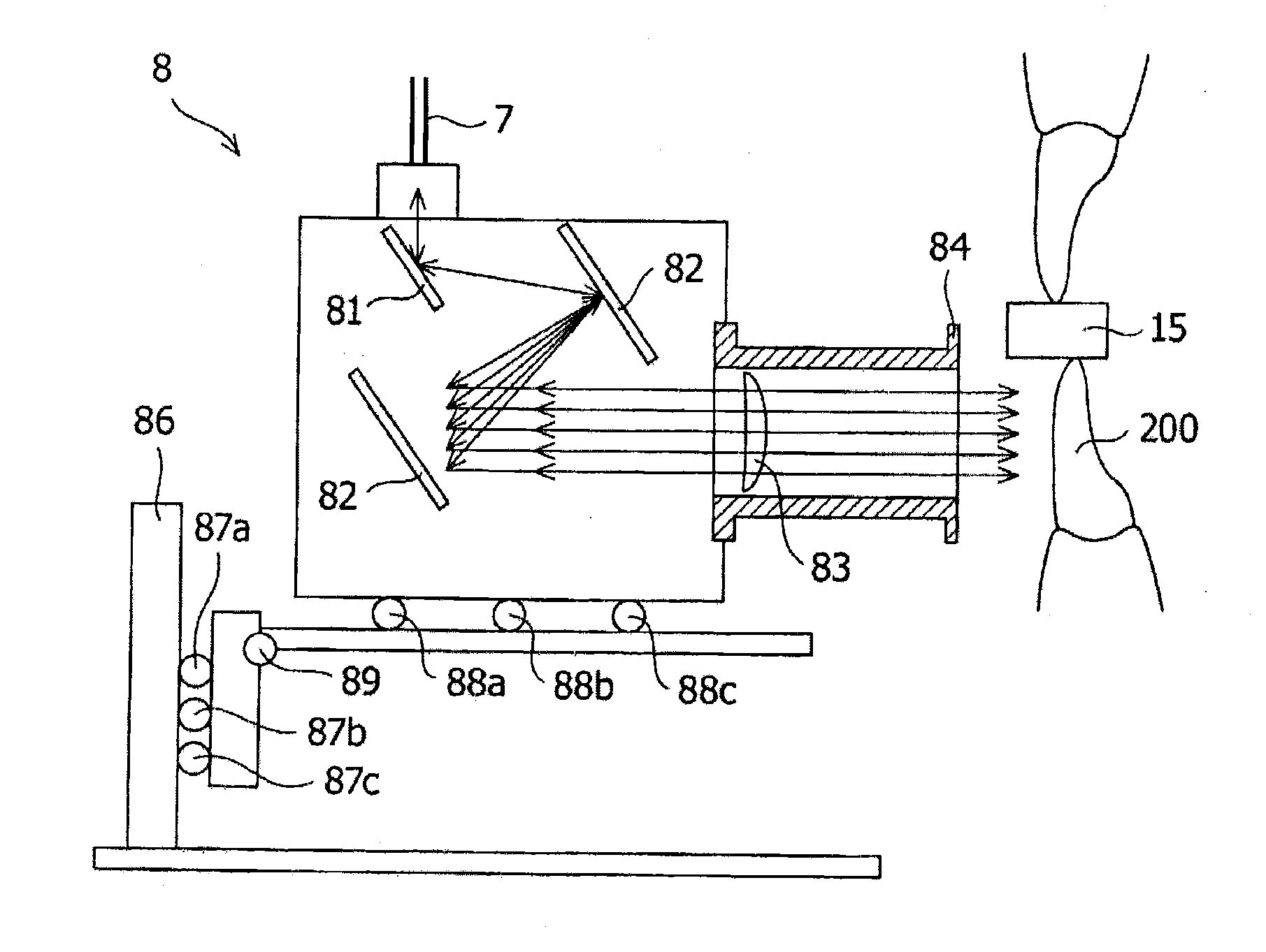
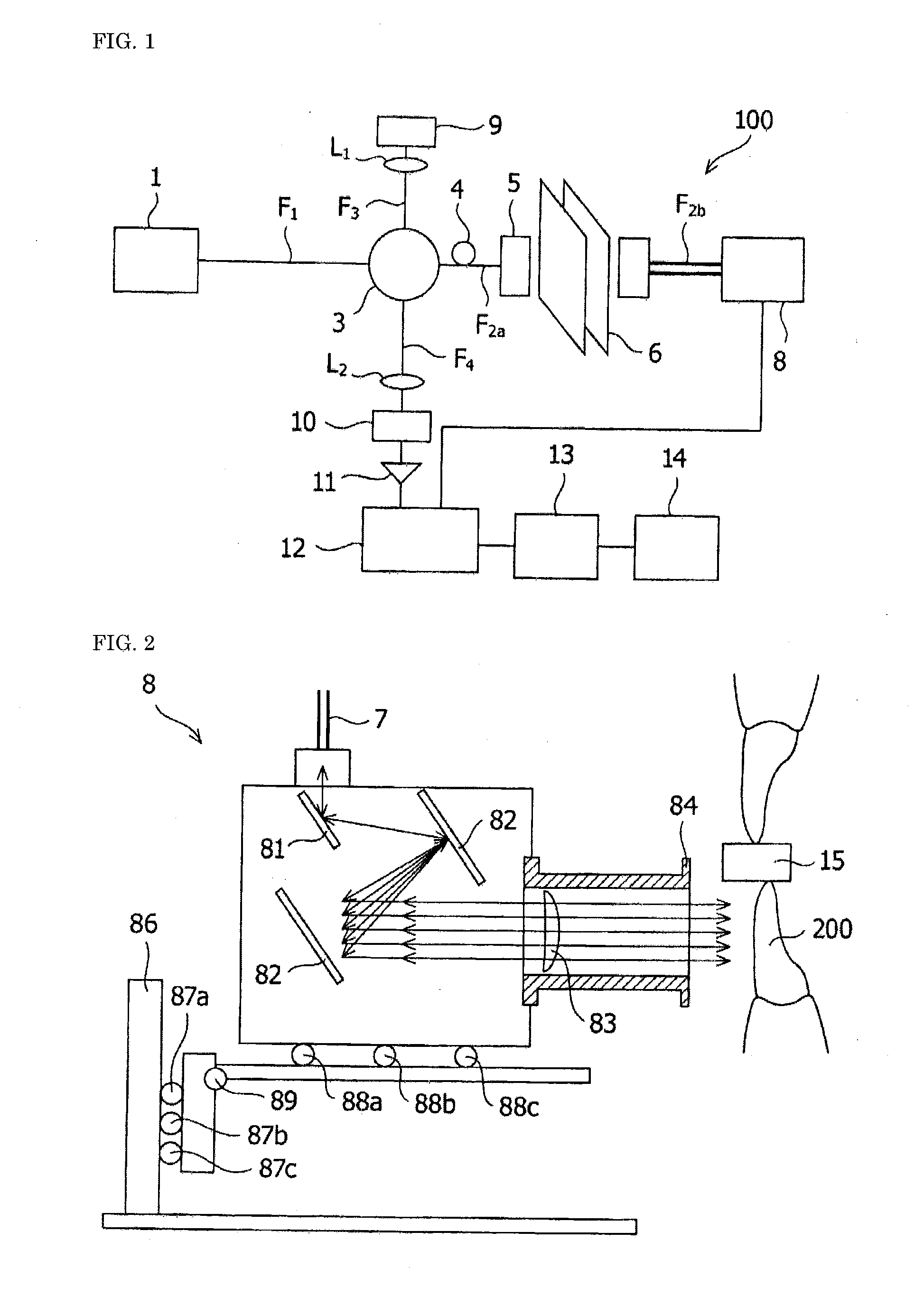
[0068]An embodiment of the present invention relates to an apparatus for measuring and displaying dental plaque. The apparatus for measuring and displaying dental plaque according to the present embodiment uses an OCT (optical coherent tomography) apparatus to selectively measure dental plaque, in particular. The OCT apparatus is capable of measuring an intravital tissue with very high microscopic resolution. Further, the OCT apparatus, which uses a light source that emits near infrared light, which can reach a site under a body surface, can perform measurement not only in a surface portion of a subject but also a deep portion under the body surface of the subject. Near infrared light, which is electromagnetic radiation having no detrimental effect on a living body, unlike roentgen-rays (X-rays), allows noninvasive inspection of a subject in an exact sense. The OCT apparatus in the present invention is, in particular, pr...

second embodiment

Method and Apparatus for Measuring and Displaying Gingiva and / or Alveolar Bone

[0135]Another embodiment of the present invention relates to a method for measuring and displaying gingiva and / or alveolar bone. In the method for measuring and displaying gingiva and / or alveolar bone, the same OCT apparatus as that described in the first embodiment can be used. Infrared light is so applied to a tooth and periodontal tissue by using the OCT apparatus that scattering intensity values of interference light from the gingiva and the alveolar bone are provided, and a two-dimensional optical coherence tomographic image and / or a three-dimensional optical coherence tomographic image can then be generated.

[0136]To quantitatively evaluate swelling of gingiva and damage to alveolar bone, which lead to periodontitis, in particular, the present embodiment is characterized in that a gingiva region and an alveolar bone region are evaluated based on a two-dimensional optical coherence tomographic image an...

example 1

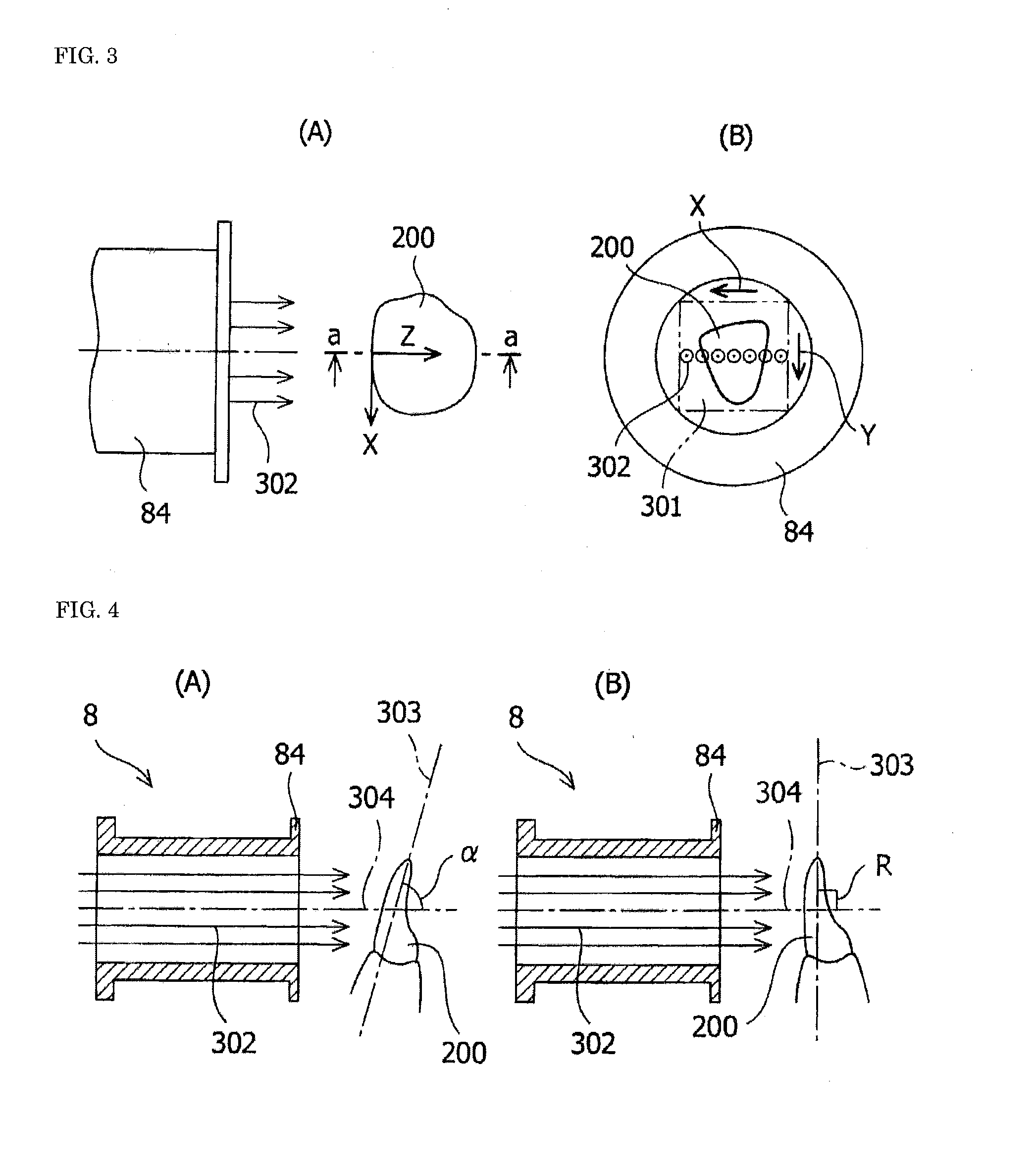
Measurement of Gray Level Values in Dental Plaque Region and Enamel Region (Two-Dimensional Tomographic Image: 50 Slices)

[0150]A light source that emits near infrared light, which is harmless to living bodies, was used as the light source and the dental plaque measuring probe shown in FIG. 2 was used to capture an image of a front tooth of a patient as the subject. Image software photoshop (manufactured by Adobe Systems Software Ireland Ltd) was used to measure gray level values in 150 sites in a dental plaque region and an enamel region in a two-dimensional tomographic image generated through conversion of scattering intensity values obtained from the subject into gray level values. As a result, it was ascertained that the gray level values in the enamel region and the gray level values in the dental plaque region differed from each other.

[0151]Dental plaque region: 169.4 in average (maximum of 207 to minimum of 140)

Enamel region: 95.9 in average (maximum of 119 to minimum of 63)

(S...


PUM


Abstract

A method and an apparatus for measuring and displaying dental plaque are provided, and the method includes the steps of dividing near infrared light output from a light source into measurement light and reference light, applying the measurement light toward a tooth in an oral cavity and scanning the tooth with the measurement light, producing interference light from reflected light and back-scattered light from the tooth and the reference light, generating an optical coherence tomographic image based on a scattering intensity value of the interference light, extracting a dental plaque region having a specific scattering intensity value from the optical coherence tomographic image, and quantifying the dental plaque. A method and an apparatus for measuring and displaying gingiva and / or alveolar bone are further provided. A method and an apparatus for quantifying dental plaque, digitizing the dental plaque, and generating an image of the dental plaque are further provided.

Description

TECHNICAL FIELD[0001]The present invention relates to an apparatus and to a method for measuring and displaying dental plaque, gingiva, and / or alveolar bone. The present invention relates particularly not only to a dental plaque measurement / display apparatus and a dental plaque measurement / display method capable of displaying dental plaque present on a tooth surface including a surface of a tooth facing an adjacent tooth and an occlusal surface of a tooth, in an interdental space, in a gingival sulcus, and in a periodontal pocket in the form of two-dimensional and three-dimensional images and instantly calculating quantified values, such as the thickness, length, cross-sectional area, volume, and / or surface area of the dental plaque but also to a gingiva and / or alveolar bone measurement / display method and a gingiva and / or alveolar bone measurement / display apparatus capable of quantifying gingiva and / or alveolar bone to provide the amount of change in swelling of the gingiva and / or t...

Claims


Application Information

Patent Timeline
Patent Type & Authority Applications(United States)
IPC IPC(8): A61B5/00A61C19/04
CPCA61B5/0088A61B5/0066A61C19/04A61B5/0086A61B5/4552A61B2576/02
Inventor SUMI, YASUNORIOZAWA, NOBUYOSHIGONDA, YOHEI
Owner NAT CENT FOR GERIATRICS & GERONTOLOGY
Why Eureka
Try Eureka