Transcranial photoacoustic/thermoacoustic tomography brain imaging informed by adjunct image data

a technology of image data and brain imaging, applied in the field of system and method of brain imaging using adjunct image data, can solve the problems of inconvenient use of patients, high-resolution human brain imaging modalities such as x-ray computed tomography (ct) and magnetic resonance imaging (mri), and high cost of high-resolution human brain imaging modalities such as ct and mri

Active Publication Date: 2015-09-03
CALIFORNIA INST OF TECH
View PDF1 Cites 36 Cited by
  • Summary
  • Abstract
  • Description
  • Claims
  • Application Information

AI Technical Summary

Benefits of technology

[0011]In one aspect, a method of imaging a brain situated within a skull of a subject is provided. The method includes obtaining an adjunct image dataset corresponding to a region of interest comprising the skull of the subject. analyzing the adjunct image dataset to obtain a spatial model of one or more acoustic properties of the region of interest, obtaining one or more photoacoustic imaging signals corresponding to the brain of the subject through the skull of the subject at a skull spatial position, registering the spatial model with the skull spatial position to establish an imaging model, and reconstructing a photoacoustic image of the subject's brain by implementing an image construction method using the imaging model and the one or more photoacoustic signals.
[0012]In this aspect, the adjunct image dataset may be obtained using a device chosen from an X-ray CT scanning device and an MRI device. The adjunct image dataset may include a spatial map of porosity and the one or more acou

Problems solved by technology

Existing high-resolution human brain imaging modalities such as X-ray computed tomography (CT) and magnetic resonance imaging (MRI) are expensive and employ bulky and generally non-portable imaging equipment.
Moreover, X-ray CT employs ionizing radiation and is therefore undesirable for use with patients who require frequent monitoring of brain diseases or injuries.
Alternatively, ultrasonography is an established portable pediatric brain imaging modality, but its image quality degrades severely when employed after the closure of the fontanels and therefore is not effective for imaging adults.
Flat pressure transducers, which are used typically for pressure measurements associated with macroscopic regions of interest, generally function over a relatively small field-of-view

Method used


Image

  • Transcranial photoacoustic/thermoacoustic tomography brain imaging informed by adjunct image data
  • Transcranial photoacoustic/thermoacoustic tomography brain imaging informed by adjunct image data
  • Transcranial photoacoustic/thermoacoustic tomography brain imaging informed by adjunct image data

Examples


example 1

Reconstruction of Biological Phantoms

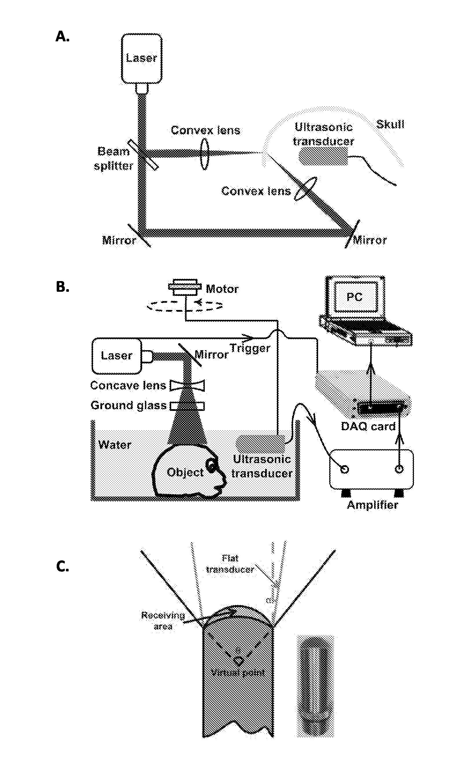
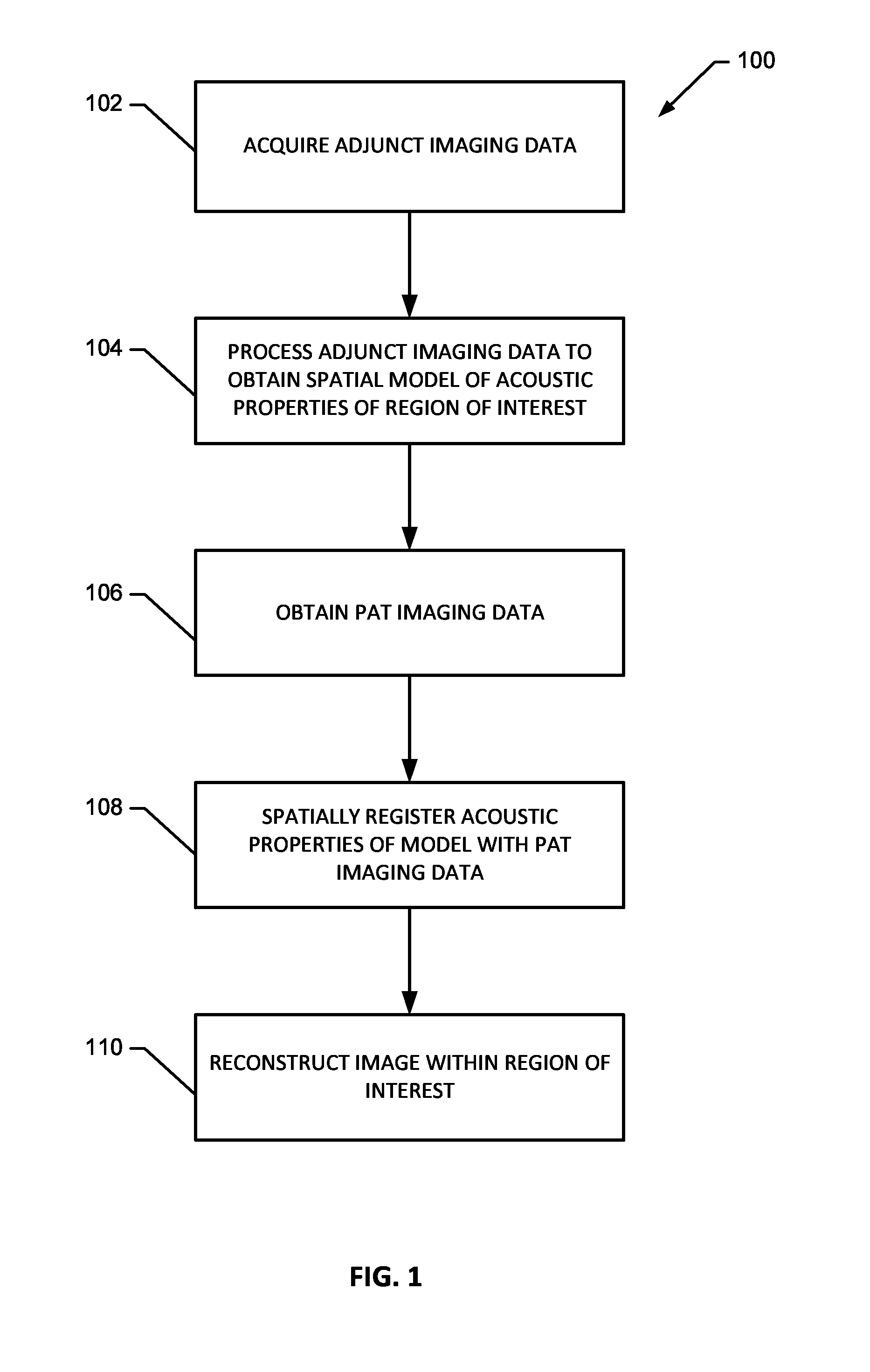
[0128]The reconstruction methodology is comprised of two primary steps. First, the spatially varying SOS and density distributions of the to-be-imaged subject's skull are determined by use of adjunct X-ray CT data. These distributions are subsequently employed with the time-reversal image reconstruction method described above for estimation of p0(r) within the brain tissue from knowledge of the measured data {{circumflex over (p)}(rm, t)}m=1M.

[0129]Two biological phantoms that employed a monkey skull were employed in the experimental studies to demonstrate the effectiveness of the present methodology. The first phantom was the head of an 8 month old rhesus monkey that was obtained from the Wisconsin National Primate Research Center. The hair and scalp were removed from the skull. A second, more simple, phantom was constructed by removing the brain of the monkey and replacing it by a pair of iron needles of diameter 1 mm that were embedded in agar...

example 2

Images of Biological Phantoms

[0131]To accommodate the 2-D PAT imaging system and 2-D image reconstruction method, imaging phantoms were moved to the image plane, approximately 2 cm below the top of the skull. Note that this was not the plane in which the cortical vessels are normally found; the primate brain was moved to align the cortical vessels with the imaging plane. For in-vivo transcranial PAT applications in which the cortical structure is of interest, the geometry of the skull necessitates a full 3-D treatment of the image reconstruction problem. Additionally, accurate measurement of the transducer's electrical impulse response (EIR) and subsequent deconvolution of its effect on the measured data may further improve image reconstruction accuracy. Alternative reconstruction methods to the time-reversal image reconstruction method employed in this study may yield additional improvements in image quality.

[0132]Virtual point detectors were developed and applied for transcranial ...

example 3

Images of Needle Phantom

[0135]The reconstructed images corresponding to the head phantom containing the needles are displayed in FIG. 6. FIG. 6A displays the control image of the needles, without the skull present, reconstructed by use of a back-projection method. FIGS. 6B and 6C display reconstructed images of the phantom when the skull was present, corresponding to use of back-projection and the time-reversal reconstruction method utilized to estimate the SOS and density maps, respectively. All images were normalized to their maximum pixel value, and were displayed in the same grey-scale window. Due to the skull-induced attenuation of the high-frequency components of the PA signals, which was not compensated for in the reconstruction process, the spatial resolution of the control image in FIG. 6A appears higher than the images in FIGS. 6B and 6C. However, the image reconstructed by use of the time-reversal method in FIG. 6C contains lower artifact levels and has an appearance clos...


PUM


Abstract

Systems and methods of reconstructing photoacoustic imaging data corresponding to a brain of a subject through a skull of a subject utilizing a reconstruction method that incorporates a spatial model of one or more acoustic properties of the brain and skull of the subject derived from an adjunct imaging dataset.

Description

RELATED APPLICATIONS[0001]This application claims priority to U.S. Provisional Application 61 / 762,415 filed on Feb. 8, 2013 and to U.S. Provisional Application 61 / 715,671 filed on Oct. 18, 2012, of which all contents are hereby incorporated herein in their entirety.GOVERNMENTAL RIGHTS IN THE INVENTION[0002]This invention was made with government support under Grant Nos. EB010049, EB009715, CA134539, EB000712, CA136398, and EB008085 awarded by the National Institutes of Health. The government has certain rights in the invention.FIELD OF THE INVENTION[0003]The invention generally relates to systems and methods for transcranial photoacoustic imaging of the brain using adjunct image data.BACKGROUND OF THE INVENTION[0004]Existing high-resolution human brain imaging modalities such as X-ray computed tomography (CT) and magnetic resonance imaging (MRI) are expensive and employ bulky and generally non-portable imaging equipment. Moreover, X-ray CT employs ionizing radiation and is therefore...

Claims


Application Information

Patent Timeline
IPC IPC(8): A61B5/00A61B5/055A61M31/00A61B6/03
CPCA61B5/0095A61B5/0042A61B6/032A61B5/0035A61B5/055A61B2576/026A61B5/4848A61B5/7282A61M31/005A61B5/4839A61B5/725A61B6/501G01N29/2418
Inventor WANG, LIHONG V.NIE, LIMINGCAI, XINMASLOV, KONSTANTINANASTASIO, MARK A.HUANG, CHAOSCHOONOVER, ROBERT W.
Owner CALIFORNIA INST OF TECH
Why Eureka
Try Eureka