Compositions and Methods for Reducing Neointima Formation

Active Publication Date: 2016-03-03
UNIV OF GEORGIA RES FOUND INC
View PDF1 Cites 4 Cited by
  • Summary
  • Abstract
  • Description
  • Claims
  • Application Information

AI Technical Summary

Benefits of technology

[0013]The composition can include a targeting signal for enhancing delivery of the CTPS1 inhibitor to vascular smooth muscle cells. The targeting signal can facilitate binding of the composition to smooth muscle cells by targeting a cell surface ligand such as Tissue Factor or αvβ3 integrin; or the targeting signal can target the compositions to the vicinity of vascular injuries by binding to a marker of clots or thrombosis such as fibrin, gpIIb/IIIa, tissue factor/VIIA complex, activated clotting factor Xa, activated clotting factor IXa, or the fibrin condensation product d-dimer. The targeting signal can be operably linked to the CTPS1 inhibitor or to the delivery vehicle.
[0014]Medical devices that are coated with or otherwise incorporate a composition including a CTPS1 inhibitor are also disc

Problems solved by technology

However, currently available anti-neointimal drugs indiscriminately block the proliferation of both SMCs

Method used


Image

  • Compositions and Methods for Reducing Neointima Formation
  • Compositions and Methods for Reducing Neointima Formation
  • Compositions and Methods for Reducing Neointima Formation

Examples


Example

Example 1

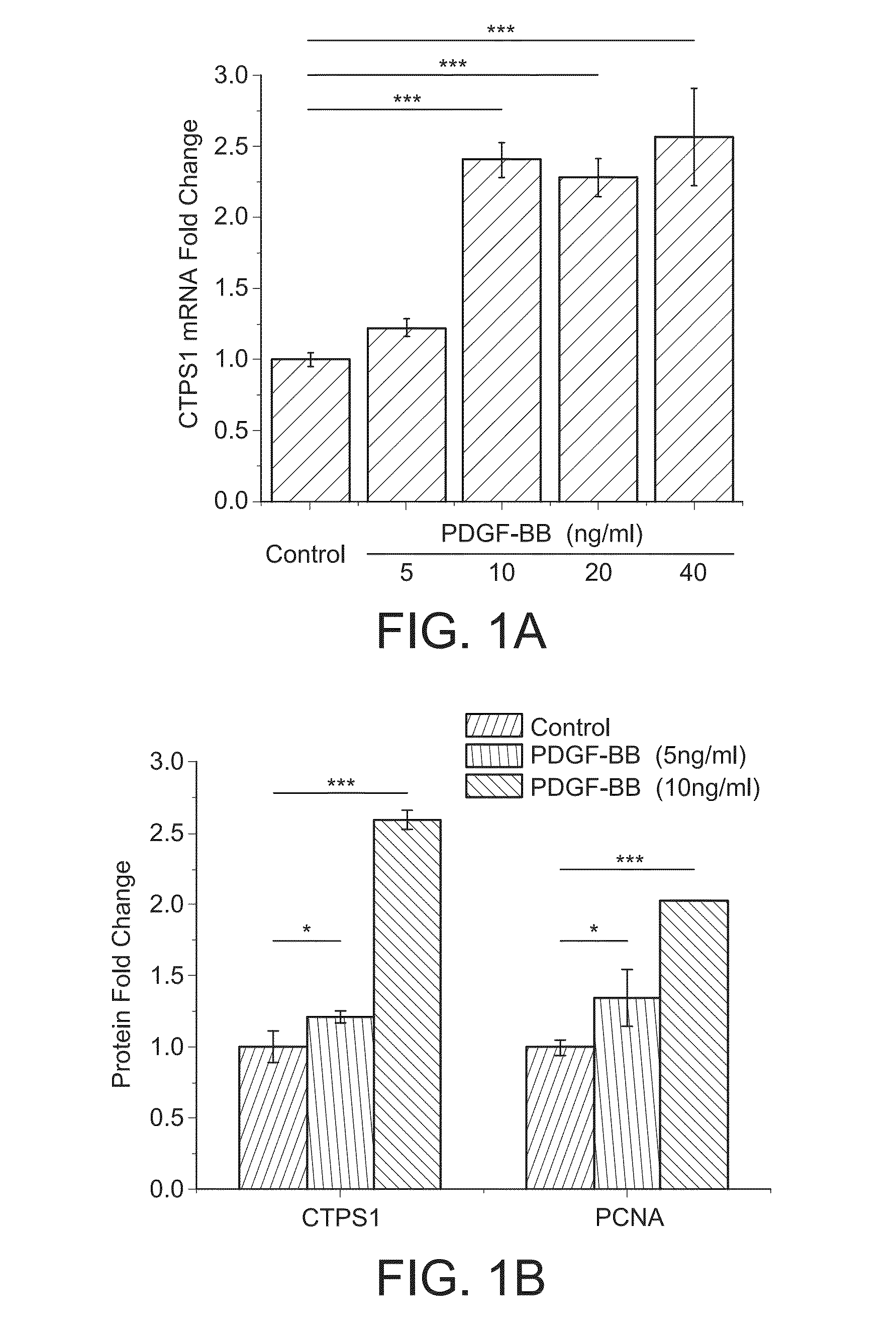
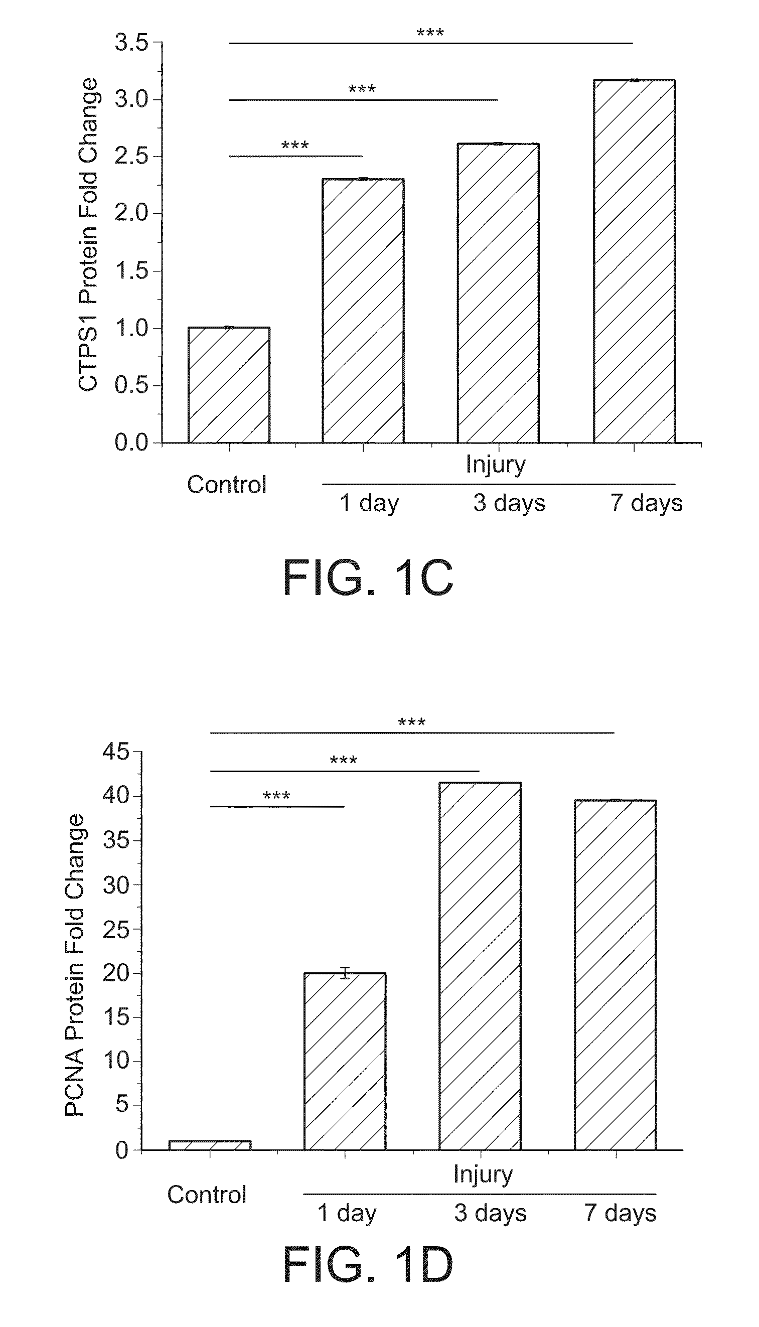
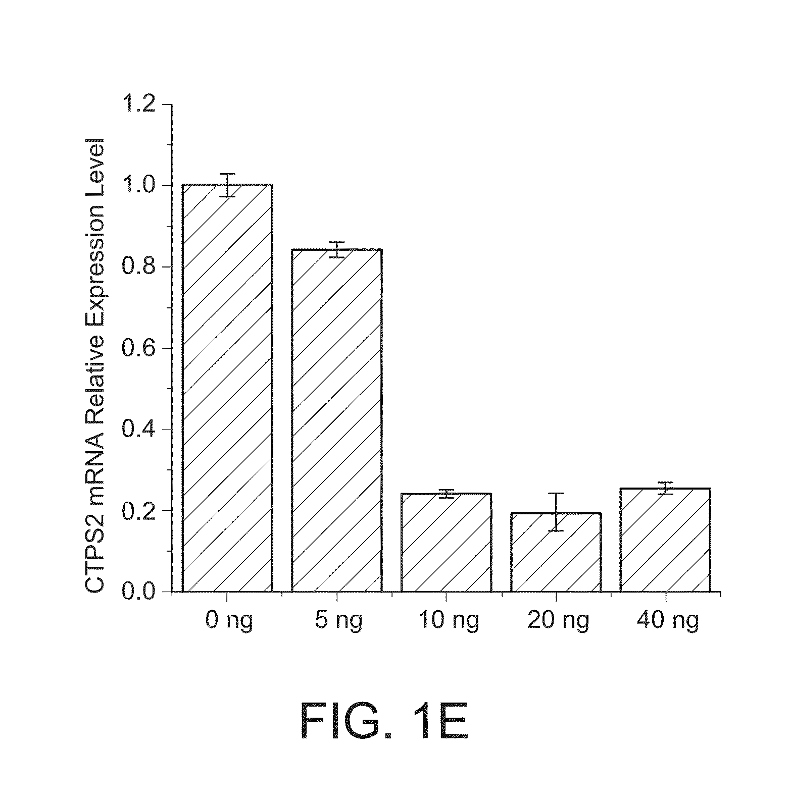
CTPS1 is Up-Regulated in Cultured SMCs in Vitro and Neointimal SMCs in Vivo

Materials and Methods

Reagents and Cell Culture

[0202]Rat aortic smooth muscle cells (SMCs) were cultured by enzyme-digestion method from rat thoracic aorta as described previously (Hofer, et al., Proc Natl Acad Sci U S A., 98:6412-6416 (2001), Marquez, et al., Biochem Pharmacol., 41:1821-1829 (1991), (Berg, et al., Eur. J. Biochem., 216:161-167 (1993)). SMC phenotype of the cultured cells was confirmed by the expression of smooth muscle alpha-actin and SM22-alpha.

[0203]Endothelial cell C166 was purchased from ATCC and grown at 37° C. in a humidified atmosphere of 5% CO2 in DMEM (Invitrogen) supplemented with 10% FBS.

[0204]CTPS1 (sc-131474), PCNA (sc-56), CDK1 (sc-137034) and phospho-CDK1 (T161) (sc-101654) antibodies were from Santa Cruz Biotechnology (Santa Cruz, Calif.). NME1 (#3345S) and NME2 (SAB1400187) antibodies were purchased from Cell Signaling (Danvers, Mass.), and Sigma-Aldrich (St. Louis, ...

Example

Example 2

Blocking CTPS Activity Inhibits SMC Proliferation and Migration

Materials and Methods

Reagents

[0211]CPEC (compound 375575) was obtained from the Open Chemical Repository of National Cancer Institute Developmental Therapeutics Program (DTP).

Construction of Adenoviral Vectors

[0212]NME1 and NME2 cDNA were individually subcloned into the XhoI site of pShuttele-IREShrGFP-1 (Agilent) and was confirmed by sequencing. Adenoviral vectors expressing CTPS1 and NME2 short hairpin RNA (shRNA) (shCTPS1 and shNME2) were constructed and the viruses were purified as described previously (Shi, et al. Arterioscler. Thromb. Vasc. Biol., 31:e19-e26 (2011)). The shRNA sequences were as follows: shCTPS1 top strand: 5′-CGC GTC GCG CTA GAG CAC TCT GCA TTG GCC ATT AAT TCA AGA GAT TAA TGG CCA ATG CAG AGT GCT CTA GCG CTT TTT TCC AAA-3′ (SEQ ID NO:12); shCTPS1 bottom strand: 5′-AGC TTT TGG AAA AAA GCG CTA GAG CAC TCT GCA TTG GCC ATT AAT CTC TTG AAT TAA TGG CCA ATG CAG AGT GCT CTA GCG CGA-3′ (SEQ ID NO:13...

Example

Example 3

[0217]Blockade of CTPS1 Activity or Expression does not Induce SMC Apoptosis but Impairs Cell Cycle Progression

Materials and Methods

TUNEL Assay

[0218]In vivo cell apoptosis was evaluated by detecting DNA fragmentation using the terminal deoxynucleotidyl transferase (TdT)-mediated dUTP-digoxigenin nick end-labeling method (TUNEL kit, Roche, USA). Apoptotic cells were observed under a fluorescent microscope. In vitro cell apoptosis was measured by Flow Cytometry. Cells were stained with both Annexin V-FITC (BD Biosciences) and propidium iodide (PI) and analyzed on a FACSCALIBUR™ (Becton Dickinson). The percentages of positive-stained cells were quantified using CELLQUEST™ software (Becton Dickinson). Non-stained cells served as controls.

Cell Cycle Flow Cytometry Analysis

[0219]1×10⁶ cells were harvested and resuspended in 500 μl of reaction buffer containing 1 μl of Nuclear-IDTM Red dye (Nuclear-IDTM Red Cell Cycle Analysis Kit, Enzo Life Sciences, USA). After mixing, cells wer...


PUM

PropertyMeasurementUnit
Cell proliferation rateaaaaaaaaaa

Abstract

Compositions, devices, grafts and methods for reducing or preventing anti-neointima following cardiovascular injuries and interventions are disclosed. The compositions, devices, and grafts typically include an effective amount of a CTP synthase 1 inhibitor to reduce proliferation of vascular smooth muscle cells, without substantial reducing the proliferation of endothelial cells. Methods of reducing neointima formation, accelerating re-endothelialization, and reducing restenosis in a subject using the compositions, devices, and grafts are also disclosed.

Description

STATEMENT REGARDING FEDERALLY SPONSORED RESEARCH OR DEVELOPMENT[0001]This invention was made with Government Support under Agreement HL107526 awarded by the National Institutes of Health. The Government has certain rights in the invention.CROSS-REFERENCE TO RELATED APPLICATIONS[0002]This application is a continuation-in-part of International Application No. PCT / U.S. 2014 / 037955 filed May 14, 2014, which claims benefit of U.S. Provisional Application No. 61 / 823,163, filed May 14, 2013, and the contents of each of which are incorporated by reference herein in its entireties.REFERENCE TO SEQUENCE LISTING[0003]The Sequence Listing submitted as a text file named “UGA—2021_PCT_ST25.txt,” created on May 13, 2014, and having a size of 14,231 bytes is hereby incorporated by reference pursuant to 37 C.F.R. § 1.52(e)(5).FIELD OF THE INVENTION[0004]The field of the invention is generally related to compositions, devices, and methods for enhancing recovery from vascular injury or surgery, and re...

Claims


Application Information

Patent Timeline
IPC IPC(8): A61L31/16A61K31/4412A61L31/08
CPCA61L31/16A61L31/08A61K31/4412A61K35/74A61L2300/416A61L2300/434C12N2310/11C12N2310/12C12N2310/14C12N2310/141C12N2310/16C12N2310/531C12N15/1137C12Y603/04002A61L27/54A61K45/06A61K31/00A61K31/198A61K31/42A61K31/7068A61K31/7072A61L27/507A61K2300/00A61P9/10
Inventor CHEN, SHIYOUTANG, RUI
Owner UNIV OF GEORGIA RES FOUND INC
Why Eureka
Try Eureka