
Method for detecting cells

a cell identification and cell technology, applied in the field of cell identification, can solve the problems of laborious and inefficient, inherent experimental variability of the current method used to detect/isolate pluripotent cells, and low efficiency of the current method, so as to achieve the effect of opening new possibilities for identification

Inactive Publication Date: 2016-03-10
FUNDACIO CENT DE REGULACIO GENOMICA +2
View PDF0 Cites 15 Cited by
  • Summary
  • Abstract
  • Description
  • Claims
  • Application Information

AI Technical Summary

Benefits of technology

The patent describes a method for detecting and isolating cells with different chromatin states using a combination of quantitative super-resolution nanoscopy and computer simulations. This method involves detecting the organization of nucleosomes, the building blocks of chromatin, in a cell's nucleus and correlating this with the cell's state. By measuring the size of nucleosomal clutches, the method can predict the pluripotency grade of stem cells. The invention also provides a kit for detecting and isolating cells in an open or close chromatin state. The device used for detecting chromatin state includes an optical sensor and a control unit for carrying out the method. The technical effects of this patent include the ability to better understand chromatin organization and predict the pluripotency grade of stem cells.

Problems solved by technology

Methods currently used to detect / isolate pluripotent cells have inherent experimental variability and low efficiency, and are (1) mechanical isolation based on morphology that requires experience, and is laborious and not efficient; (2) quantification of the endogenous expression of stem cell transcription factors (OCT4, SOX2, etc.) in live cells, which requires genome modification; (3) fluorescence-activated cell sorting (FACS)-based analysis using cell surface markers (SSEA-4, TRA-1-60, etc.), which requires use of antibody based staining that is inherently variable; and (4) more recently, a pluripotent stem cell-specific adhesion signature, which is dependent on the surface properties of cell clusters and thus interrogates the population and not individual cells.
Additionally, the identification of high-grade pluripotent hiPSCs is time consuming, requiring the generation of teratomas and several additional pluripotency test.
However, how nucleosomes are arranged to form the chromatin fiber is still highly debated.
The structural information obtained in these studies led to the overall conclusion that the eukaryotic nuclei are mainly composed of 10 nm fibers even though the core histone proteins could not be identified unequivocally using these methods due to their lack of molecular specificity.
Up to date, however, the super-resolution studies of DNA and histones have not addressed questions regarding the organization of single or groups of nucleosomes, the overall nucleosome occupancy level of DNA and whether these parameters are consistent with the 30 nm fiber model of chromatin.

Method used


Image

  • Method for detecting cells
  • Method for detecting cells
  • Method for detecting cells

Examples


example 1

Nucleosomes in Interphase Nuclei of Human Somatic Cells are Organized in Discrete Nanodomains

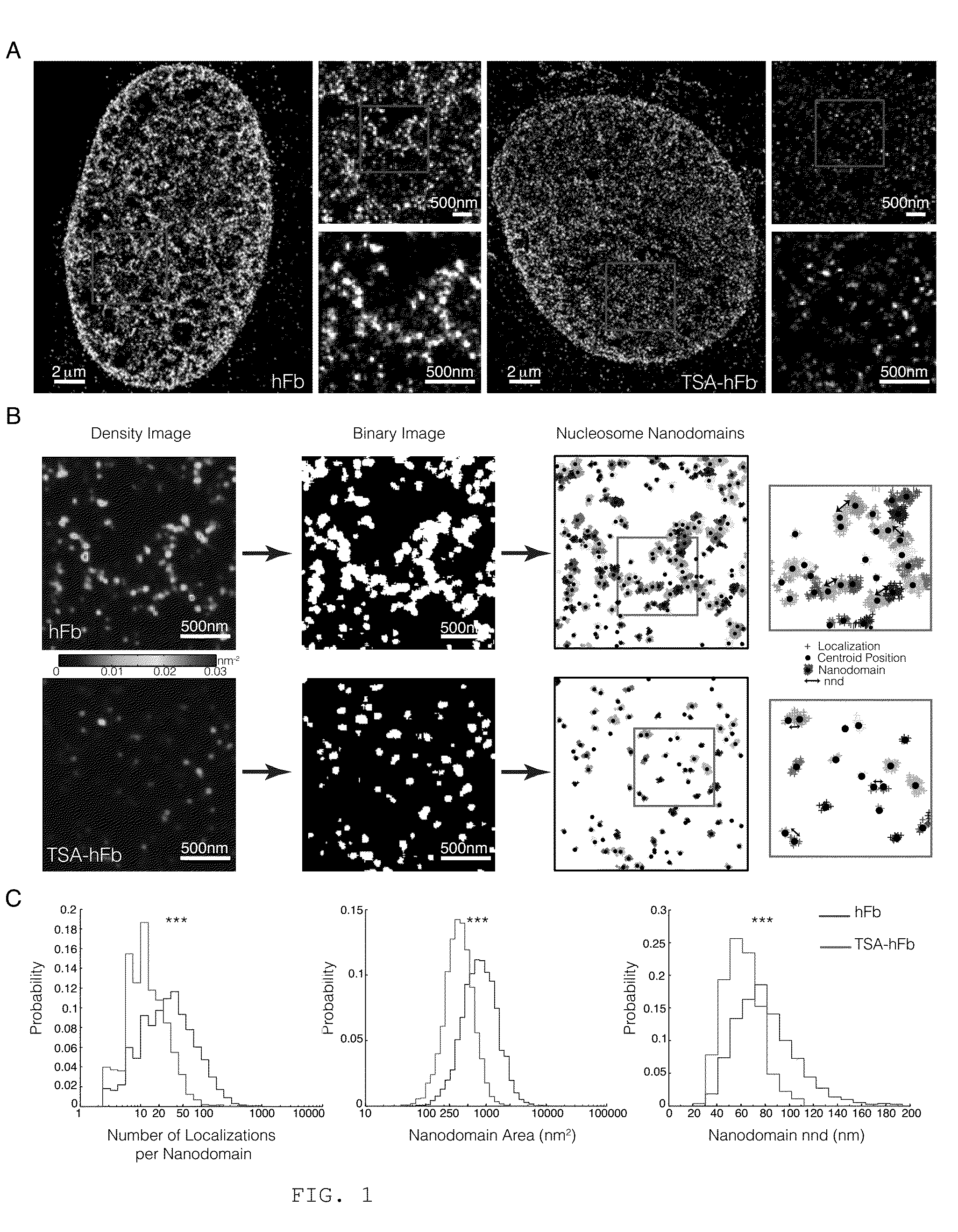
[0215]To reveal the organization of chromatin at nanoscale resolution, the inventors recorded STORM images of the core histone protein H2B in interphase human fibroblast nuclei (hFb). An antibody that recognizes native H2B was used. STORM images revealed a striking organization of H2B inside the nucleus (FIG. 1A, left), which was not evident with conventional fluorescence microscopy (FIG. 7A). H2B appeared clustered in discrete and spatially separated nanodomains (FIG. 1A, right zooms). The H2B nanodomain density (number of nanodomains per unit area) was ˜25% higher in the nuclear periphery, where the heterochromatin is thought to be located, compared to the nuclear interior. Since H2B is a core histone of the nucleosome octamer, its localization should reflect the arrangement of nucleosomes within the chromatin fiber. In accordance with this idea, another core histone protein of the nucleos...

example 2

Nucleosomes in Interphase Nuclei of Mouse Embryonic Stem Cells Form Discrete Nanodomains, Whose Organization Correlates with Ground State Pluripotency

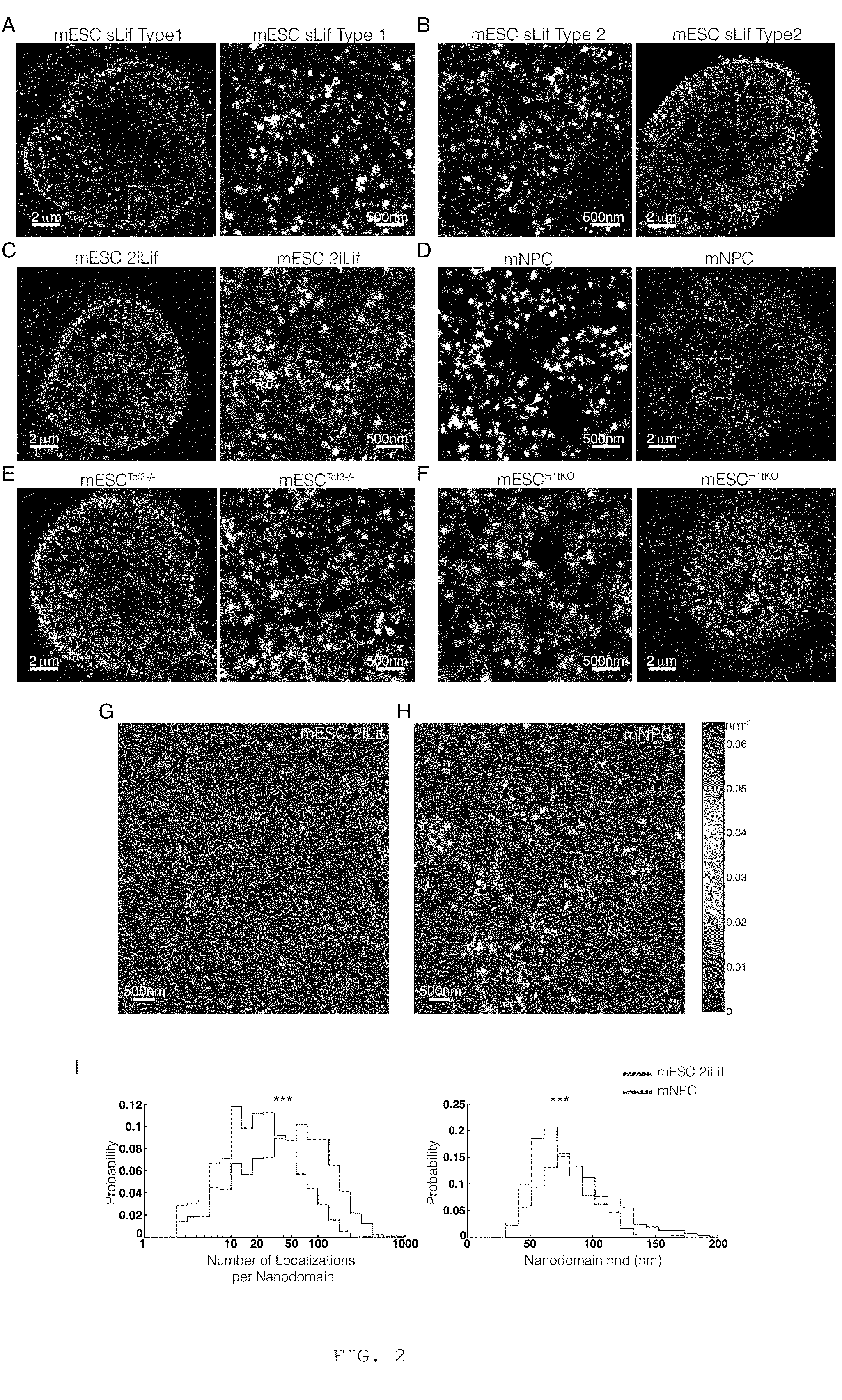
[0219]To assess the H2B organization of pluripotent stem cells, the inventors next imaged mouse embryonic stem cells (mESC) with STORM. mESCs were initially cultured in a medium containing serum and the Leukemia inhibitory factor (sLif) and H2B was labeled by immunofluorescence and imaged with STORM as before. STORM images of these mESCs showed two different categories of nuclei. The first category, Type 1, displayed nanodomains that appeared bright (i.e. contained a large number of localizations) similar to hFbs (FIG. 2A, yellow arrowheads). The second category, Type 2, on the other hand, displayed an increased amount of dimmer nanodomains (i.e. containing a small number of localizations) and the nanodomains were more dispersed within the nucleus, similar to TSA-hFbs (FIG. 2B, cyan arrowheads). It has been reported that mESCs culture...

example 3

Nanodomains Contain a Discrete Number of Nucleosomes and the Nucleosome Number Correlates with Cell Pluripotency

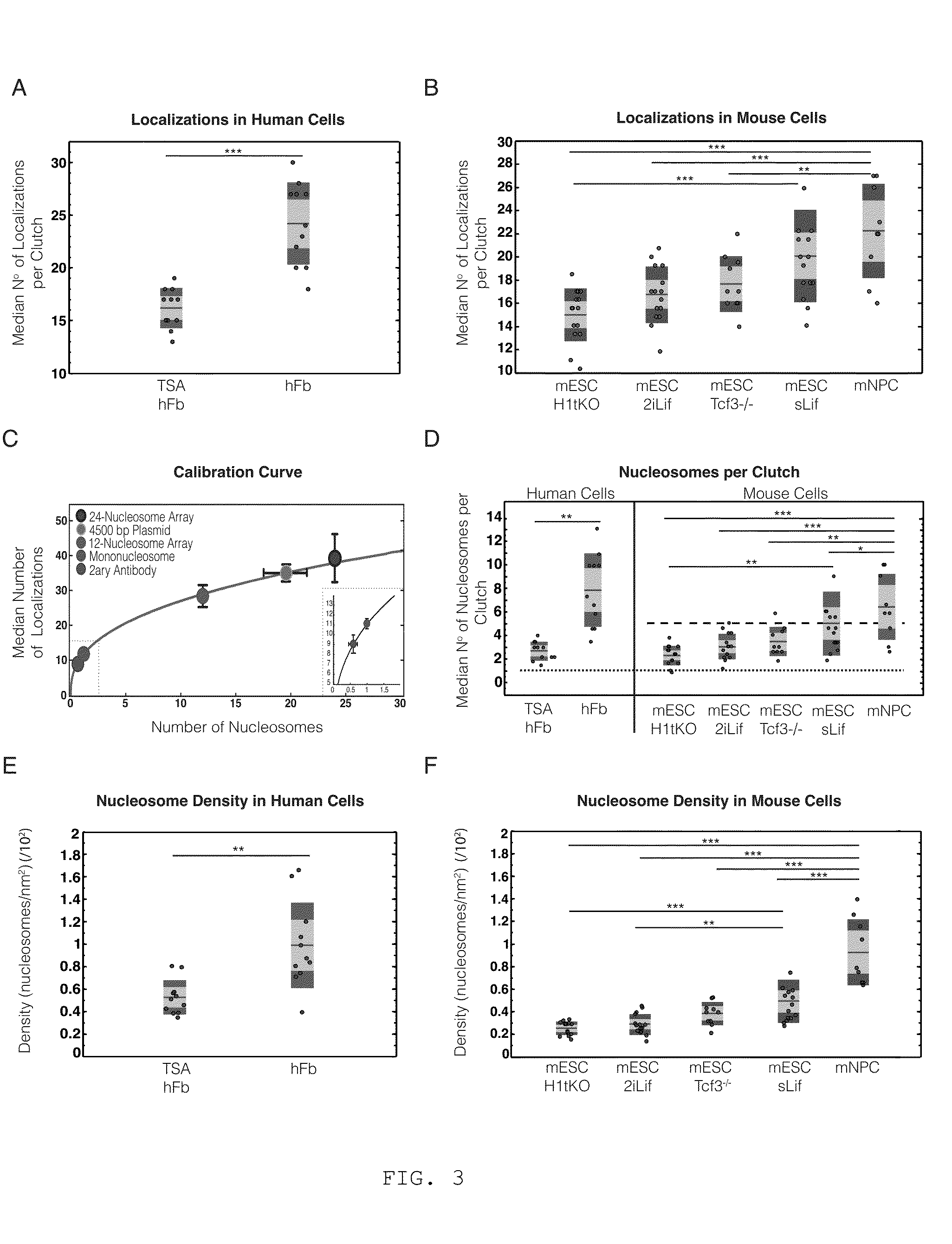
[0223]The inventors next aimed to further quantify the changes they observed in the number of localizations (and hence brightness) of nanodomains in terms of the number of nucleosomes. There is not a one-to-one relationship between the number of localizations in STORM images and the number of nucleosomes mainly for two reasons: i) the antibody epitope labeling efficiency may not be 100%, ii) each fluorophore can undergo multiple photoswitching events, resulting in multiple localizations arising from a single fluorophore. However, the epitope labeling efficiency of the H2B antibody should be comparable across the human cells (hFbs, TSA-hFbs) and likewise across the different mESCs analyzed. In addition, the antibodies used were always labeled with a similar dye composition (Extended Experimental Procedures) and each cell was imaged in the same way (Table I) to obtain compar...


PUM


Abstract

The present invention relates to methods for detecting the chromatin state of a cell based on recording a super resolution image of nucleosome organization and correlating said imaged with size of nucleosomal clutches, nucleosomal density and / or number of nucleosomes per nucleosomal clutches. Additionally, the invention relates to a kit comprising a first antibody capable of specifically binding to a histone protein and a photo switchable fluorophore linked-secondary antibody and the use of the kit of the invention for detecting the chromatin state of a cell and isolating a cell in an open chromatin state or in a close chromatin state. The invention also relates to a device adapted to detect the chromatin state of a cell.

Description

TECHNICAL FIELD OF INVENTION[0001]The present invention belongs to the field of methods for cell identification.BACKGROUND OF INVENTION[0002]Pluripotent stem cells have potential to differentiate into any of the three germ layers: endoderm, mesoderm, or ectoderm and provide a chance to obtain a renewable source of healthy cells and tissues to treat a wide array of diseases.[0003]Methods currently used to detect / isolate pluripotent cells have inherent experimental variability and low efficiency, and are (1) mechanical isolation based on morphology that requires experience, and is laborious and not efficient; (2) quantification of the endogenous expression of stem cell transcription factors (OCT4, SOX2, etc.) in live cells, which requires genome modification; (3) fluorescence-activated cell sorting (FACS)-based analysis using cell surface markers (SSEA-4, TRA-1-60, etc.), which requires use of antibody based staining that is inherently variable; and (4) more recently, a pluripotent st...

Claims


Application Information

Patent Timeline
IPC IPC(8): G01N33/68G01N21/64
CPCG01N33/6875G01N2458/00G01N21/6486G01N21/6458G01N2021/6439C07K16/18G01N21/6428G01N33/56966G01N2201/12G01N2333/47G06T7/0012
Inventor LAKADAMYALI, MELIKEMANZO, CARLORICCI, MARIA AURELIACOSMA, MARIA PIA
Owner FUNDACIO CENT DE REGULACIO GENOMICA
Learn More