Engineering Of A Novel Breast Tumor Microenvironment On A Microfluidic Chip

a microfluidic chip and microenvironment technology, applied in the field of engineering of a novel breast tumor microenvironment on a microfluidic chip, can solve the problems of cancer cell metastasis, difficult independent study of inability to test the effects of various microenvironmental cues using in vivo models, etc., to achieve excellent scaffolding materials, excellent diffusion properties, and excellent mechanical properties

Active Publication Date: 2016-04-07
ARIZONA STATE UNIVERSITY +1
View PDF6 Cites 8 Cited by
  • Summary
  • Abstract
  • Description
  • Claims
  • Application Information

AI Technical Summary

Benefits of technology

[0011]To address these challenges, we developed an innovative approach using microscale technologies and hydrogel based biomaterials to develop a three dimensional (3D) in vivo like breast tumor microenvironment on a microfluidic chip. Specifically, we used microengineering technology, hydrogel-based biomaterials, and co-culture of key cell types within the tumor stroma to develop an in vivo like 3D vascularized human breast tumor model microenvironment. Hydrogels are excellent scaffolding materials because of their 3D architecture resembling the native in vivo microenvironment, homogenous cellular distribution, excellent diffusion properties and tunable mechanical characteristics. To date, hydrogels have been extensively used to study vascularization / angiogenesis, cell invasion and tissue morphogenesis. Integration of microscale (i.e. microfluidics) technology and biology not only offers high throughput systems but also enables tight control of the cellular microenvironment by varying cell-cell, cell-substrate, and cell-soluble factors interactions. Using microfabrication techniques, it is possible to create precisely defined features and organized cellular constructs to address the limitations of the conventional 2D assays and 3D macroscale hydrogel constructs.
[0012]Surrounding stroma embedded with inflammatory and immune cell (i.e. FBs, macrophages) plays a crucial role in cancer progression. In particular, FBs have been shown to exhibit activated phenotype with significantly higher proliferation and ECM deposition (collagen I) within the tumor stroma. Moreover, there have been studies demonstrating the FBs and inflammatory cells are principal source of host-derived VEGF secretion, which result in increased vascular permeability and ultimately enhanced angiogenesis within the tumor microenvironment. Therefore, co-culture of FBs, macrophages and ECs along with metastatic breast cancer cells provides a promising path to assess the influence of other cells types on cancer migration through the surrounding stroma. The unique features of the disclosed devices and methods bridge the gap between conventional 2D / 3D assays and animal models to recapitulate the complexities associated within the native tumor stroma.

Problems solved by technology

However, using these models, there are certain difficulties to independently study the effects of various microenvironmental cues (i.e., cell-cell communication, mechanical properties of the matrix) on cancer cells metastasis.
Furthermore, extensive testing of therapeutic compounds using in vivo models is a costly process.
Although these assays have facilitated high throughput and economically efficient experimental analysis, they do not fully recapitulate the complexities of the native 3D tumor microenvironment.
The limiting aspects of 3D macroscale hydrogel models are the lack of precise control over cellular distribution, lack of vascularity and difficulties in establishing stable chemical gradients throughout the 3D matrix for potential drug screening applications.
While previous research has demonstrated that the crosstalk between the cancer cells and their surrounding microenvironment significantly influences their metastatic potential, the success of therapeutic approaches to suppress breast tumor progression has been insufficient.
A critical challenge in efficient translation of therapeutic strategies to clinical practice is the lack of physiologically relevant tumor models to study metastatic dispersion of cancer cells in response to microenvironmental stimuli within well-controlled experimental conditions.

Method used


Image

  • Engineering Of A Novel Breast Tumor Microenvironment On A Microfluidic Chip
  • Engineering Of A Novel Breast Tumor Microenvironment On A Microfluidic Chip
  • Engineering Of A Novel Breast Tumor Microenvironment On A Microfluidic Chip

Examples


example 1

An Exemplary Microfluidic Device

[0064]In this example, we describe a non-limiting exemplary embodiment of a microfluidic device according to the disclosed invention.

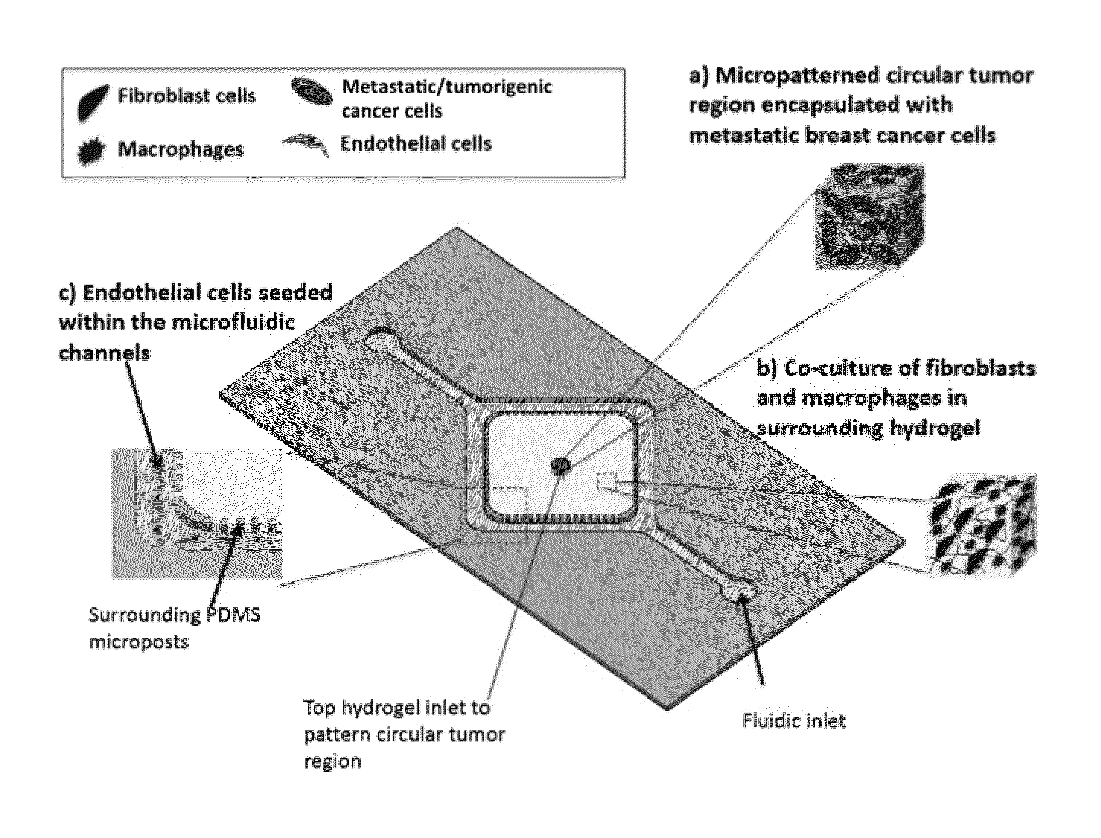
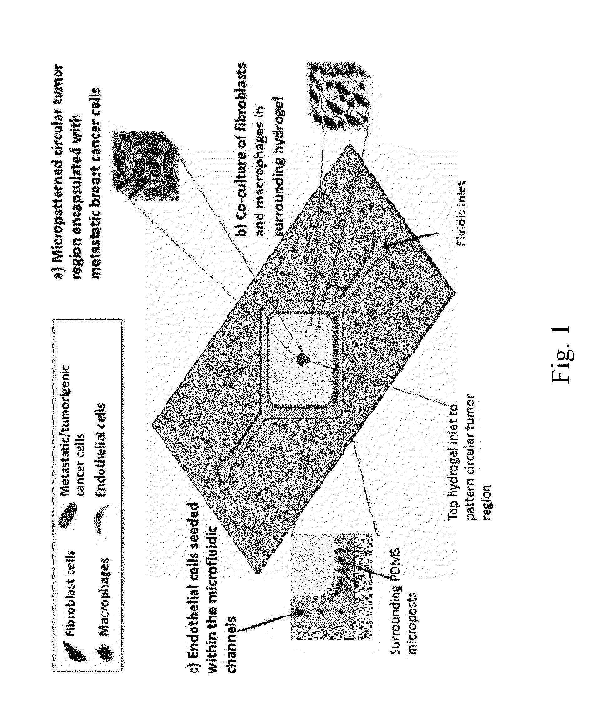
[0065]FIG. 1 shows the features of the exemplary microfluidic device, As shown in FIG. 1, the microfluidic device includes two separate regions of hydrogel constructs, one for the circular tumor construct (neoplastic lesion) encapsulated with metastatic breast cancer cells ((a); center circle), and the second one resembling the surrounding 3D ECM stroma for encapsulation of FBs and macrophages ((b); rectangle surrounding center circle). Two parallel microfluidic channels seeded with ECs are interconnected with the surrounding ECM hydrogel ((c), adjacent to the two long edges of rectangle of (b)). In particular, the microfluidic channels will be used for seeding of ECs resembling the vascular network ((c)). In addition, the microfluidic channels will be used to introduce anticancer drugs for testing.

[0066]Microfluidic Dev...

example 2

Production and Use of an Exemplary Microfluidic Device

[0081]In this proof of principal example, we demonstrate that a microfluidic device according to the disclosed invention is capable producing a well-defined tissue architecture, and that it enables high-contrast and real-time imaging for detailed invasion and intravasion studies.

[0082]Introduction.

[0083]The development of our breast cancer invasion platform has given our laboratory the opportunity to investigate and provide insight to key aspects of the metastatic cascade and tumorigenesis. This study validates our model as breast cancer invasion and demonstrates that our platform is capable of producing a well-defined tissue architecture and enabling high-contrast real-time imaging for detailed invasion and intravasation studies.

[0084]Creating a Spatially Organized Cancer Invasion Platform.

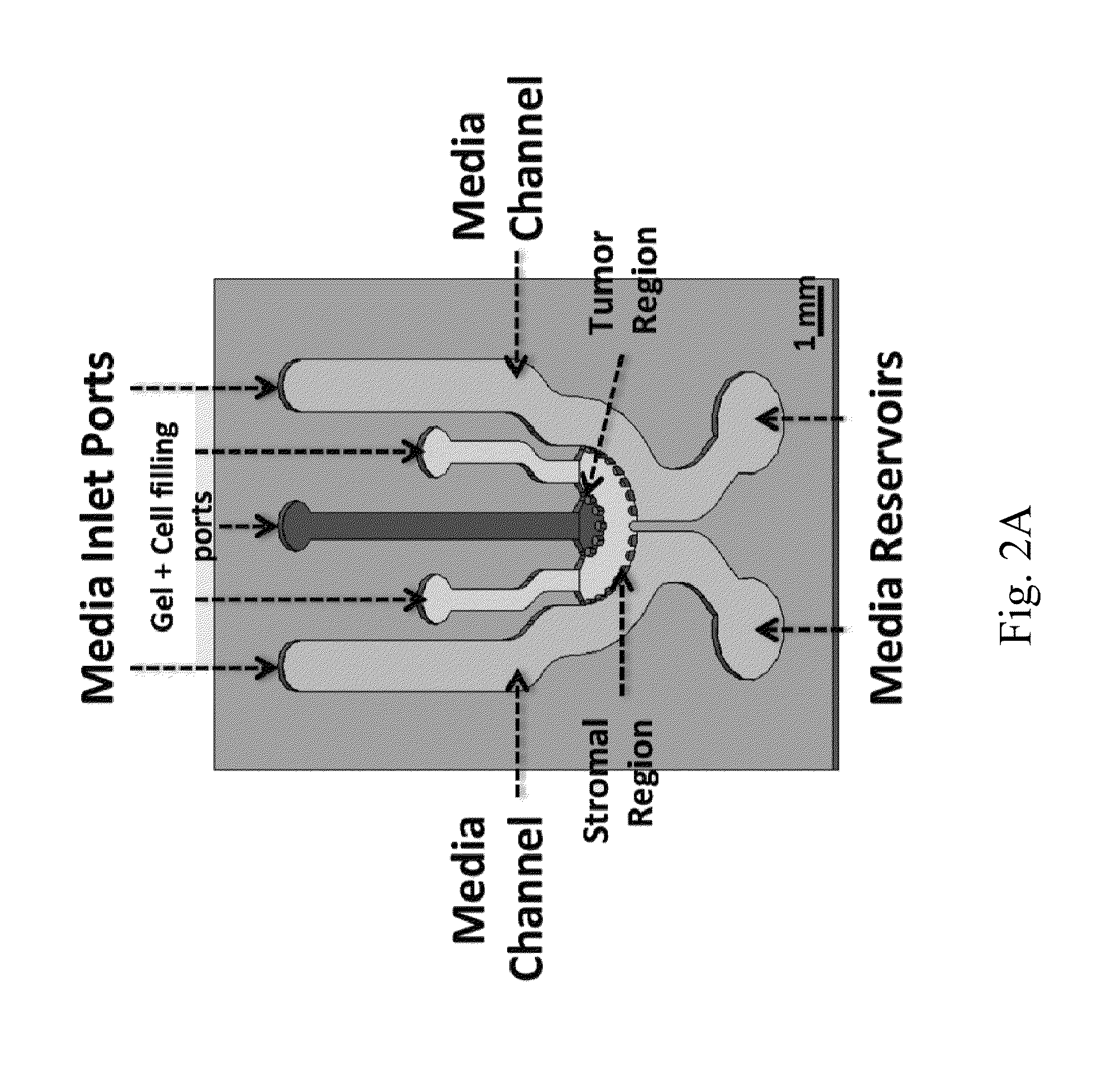
[0085]First, we developed a CAD design that caters to the native breast tumor architecture (FIG. 2A). It consisted of two distinct region mad...


PUM

PropertyMeasurementUnit
widthaaaaaaaaaa
optically transparentaaaaaaaaaa

Abstract

A microfluidic device for more accurately modeling the in vitro environment in which cancer occurs is disclosed. The device comprises a surface defining one or more microfluidic channels that may further comprise one or more endothelial cells, a first three dimensional scaffold comprising one or more cancer cells that is spatially separated from the one or more microfluidic channels, and a second three dimensional scaffold comprising one or more stromal cells, at least a portion of which is interposed between the one or more microfluidic channels and the first three dimensional scaffold. The second three dimensional scaffold is in fluid communication with both the first three dimensional scaffold and the one or more microfluidic channels. The device can be used to assay anti-cancer agents, or as a system for modeling the growth, behavior, or metastasis and tumor formation of cancer cells.

Description

CROSS-REFERENCE TO RELATED APPLICATIONS[0001]This application claims the benefit of U.S. provisional Application No. 62 / 058,186 filed on Oct. 1, 2014, which is incorporated by reference herein in its entirety.STATEMENT REGARDING FEDERALLY SPONSORED RESEARCH OR DEVELOPMENT[0002]Not applicable.BACKGROUND[0003]Metastasis, a process where cancer cells migrate to distant organs from the primary tumor site, is the key biological process responsible for 90% of all cancer related deaths. Metastatic dissemination of cancer cells is a multi-step biological process initiated by tumor angiogenesis and invasion of the cancer cells through their surrounding stroma toward the blood vessels.[0004]Tumor stroma is a structurally complex microenvironment hosting several cell types including endothelial cells (ECs), fibroblasts (FBs) and macrophages, packed in extracellular matrix (ECM), along with a dense network of capillaries. In this respect, the reciprocal interactions of the cancer cells with the...

Claims


Application Information

Patent Timeline
Patent Type & Authority Applications(United States)
IPC IPC(8): C12M3/06C12M1/00C12M1/12
CPCC12M23/16C12M29/00C12M25/14B01L3/502707B01L3/502715B01L2300/0816B01L2300/12G01N33/5011
Inventor NIKKHAH, MEHDIKAMM, ROGER D.TRUONG, DANH
Owner ARIZONA STATE UNIVERSITY
Why Eureka
Try Eureka