
Detection of hydroxymethylcytosine bases

a technology of hydroxymethyl cytosine and cytosine, which is applied in the field of molecular biology, can solve the problems of difficult detection of variation by current technologies, and achieve the effect of low resolution and high sensitivity

Active Publication Date: 2016-04-28
RAMOT AT TEL AVIV UNIV LTD
View PDF3 Cites 22 Cited by
  • Summary
  • Abstract
  • Description
  • Claims
  • Application Information

AI Technical Summary

Benefits of technology

The invention relates to methods of labeling DNA and imaging the labeled DNA molecule at the single molecule level with high sensitivity. These methods can specifically label epigenetic information and detect long-range genomic variations. The use of these methods can bridge the gap between sequencing methods and cytogenetic techniques, providing valuable information about the genetic identity of the observed molecule. The method is simple and quick and uses a reagent that is easy to prepare. Overall, the invention offers a new way to analyze DNA and its modifications with high precision.

Problems solved by technology

These dynamic, chemical modifications are a major source of genomic variation, yet this variation is difficult to detect by current technologies since it is masked by ensemble averaging.
However, due to the short sequence reads, sequencing reports on the population averaged distribution of 5hmC cannot resolve small sub-populations or characterize variation in the 5hmC patterns, which are required for identifying epigenetic modifications which may display high cell to cell variation due to their dynamic nature.

Method used


Image

  • Detection of hydroxymethylcytosine bases
  • Detection of hydroxymethylcytosine bases
  • Detection of hydroxymethylcytosine bases

Examples


example 1

5hmC Labeling

Material and Experimental Methods

Synthesis of UDP-6-Azide Glucose

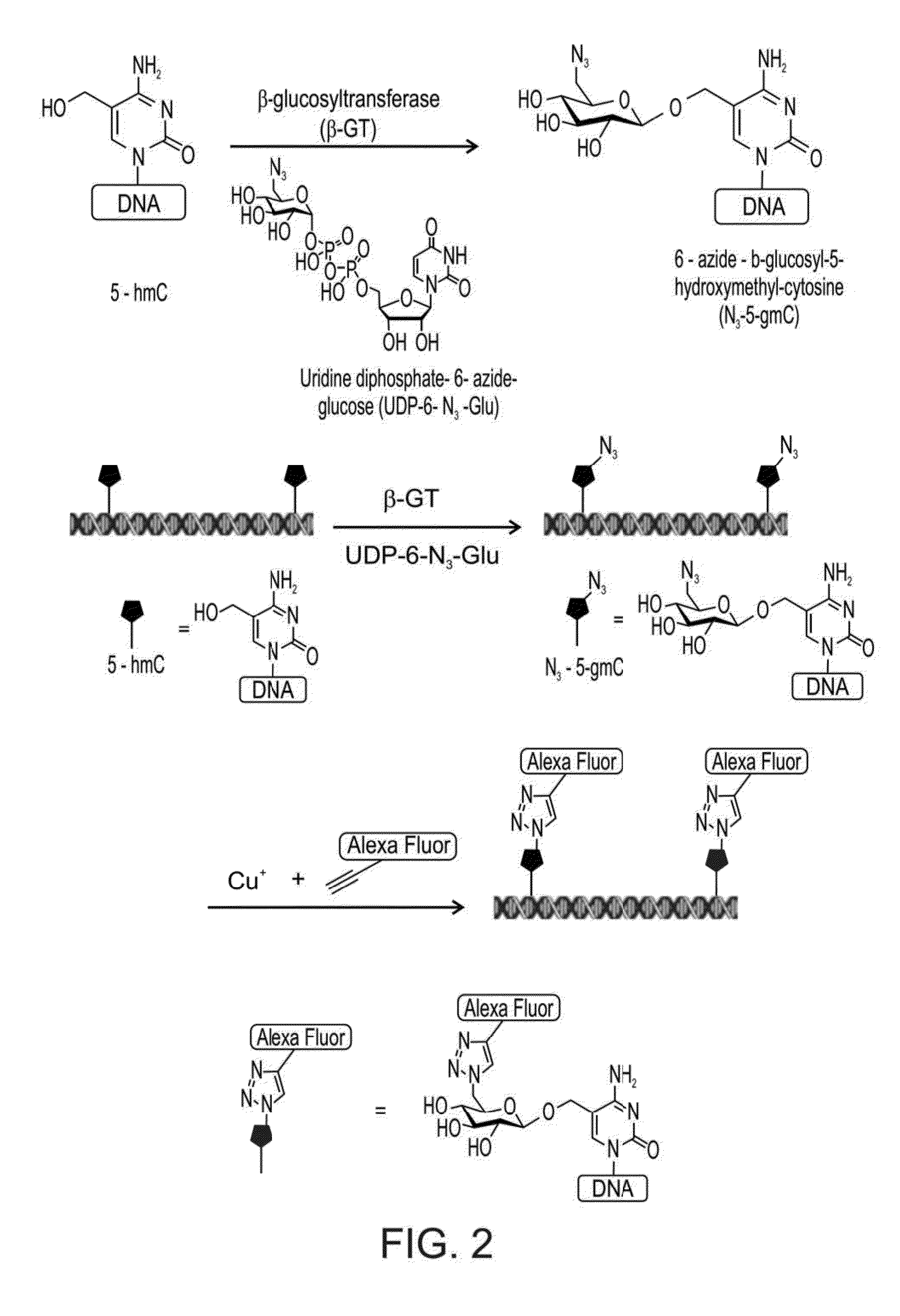
[0295]The T4 bacteriophage uses the enzyme β-glucosyltransferase (β-GT) to glucosilate hydroxylated bases in its DNA. Recently, a selective chemical method for the labeling of 5hmC was developed. In this method β-GT was exploited in order to transfer an azide-substituted glucose (UDP-6-N3-Glu) onto the hydroxyl group of 5hmC to form β-6-azide-glucosyl-5-hydroxymethyl-cytosine (5-N3-gmC). The presence of an azido moiety paves the way for 5hmC labeling using a click chemistry reaction by the addition of an alkyne substituted dye.

[0296]The main bottleneck of this labeling process is the cofactor UDP-6-N3-Glu, which can be synthesized or purchased but with very high costs.

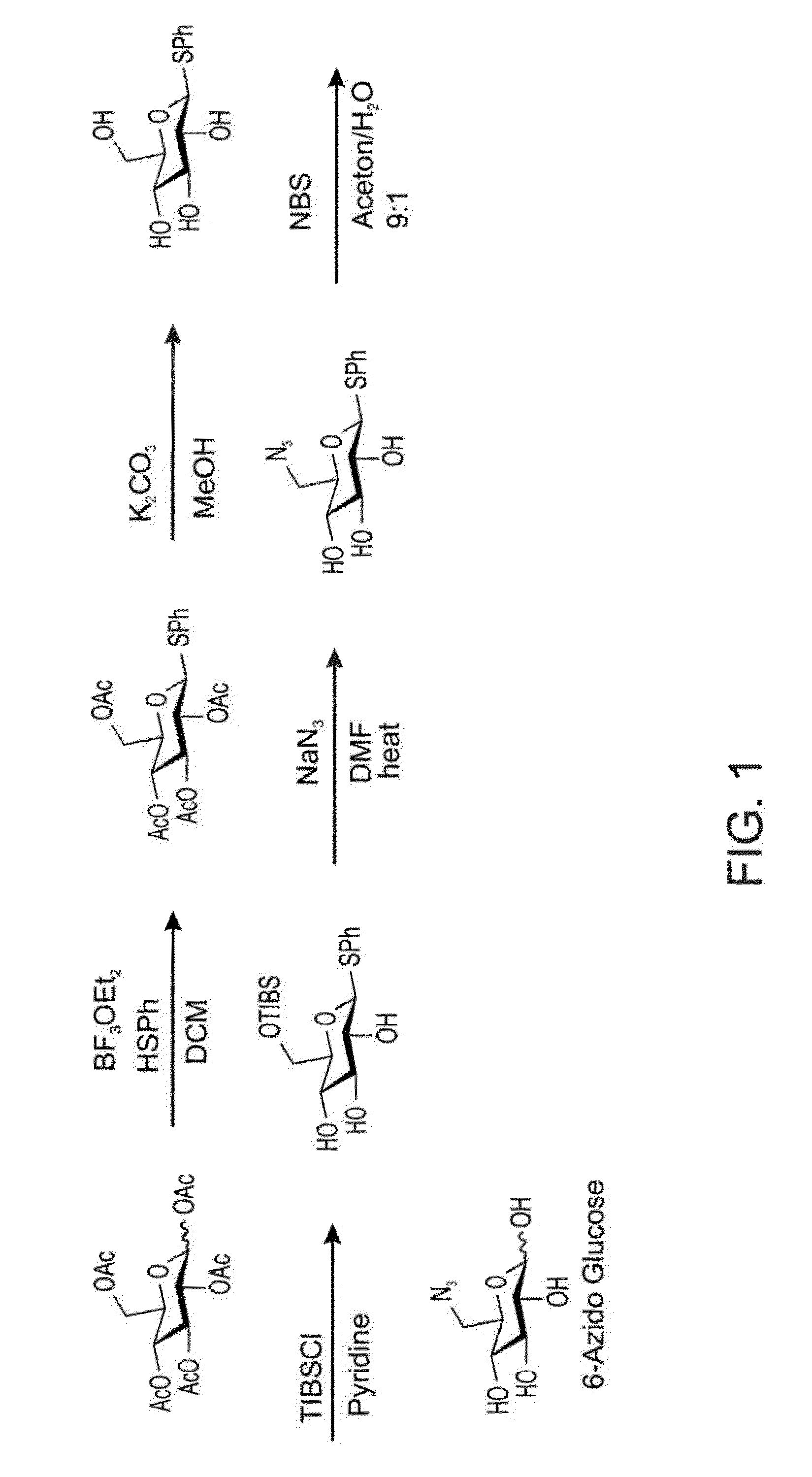
[0297]Herein, an enzymatic approach is used in order to have high yields of UDP-N3-Glu. A glucose azide (see FIG. 1) is used as a substrate for a sequential enzymatic cascade.

[0298]The glucose azide can be synthesized as described in FIG. 1, ...

example 2

5hmC Labeling is Sensitive and can be Done in High Throughput Settings

[0330]Example 1 above establishes the use of some embodiments of the present methodology for the quantification of global % hmC in a given sample. The technique is based on covalent labeling of hmC moieties by an enzymatic reaction, which is followed by the Huisgen cycloaddition of an alkyne to an azide moiety. Fluorescently labeled alkynes are used to fluorescently label hmC so that a ratiometric measurement of the adsorption intensities of the fluorophor relative to the DNA, at 260 nm, can be obtained. These measurements were conducted on a nanodrop spectrophotometer. A drop of 1 μl containing 1650 ng / μl of DNA was required in order to detect 0.02% hmC in a given sample. This is a highly concentrated sample which could sometimes be challenging to achieve. Each sample is measured separately since only one drop could be measured at a time.

[0331]Following is an improved ratiometric detection method which is based o...


PUM

PropertyMeasurementUnit
concentrationaaaaaaaaaa
thresholdaaaaaaaaaa

Abstract

Methodologies for labeling the epigenetic modification 5-hydroxymethyl-cytosine (5hmC) along a DNA molecule, and for imaging this epigenetic modification along a DNA molecule are disclosed. Related compositions and reagents, and methods of preparing same are also disclosed.

Description

FIELD AND BACKGROUND OF THE INVENTION[0001]The present invention, in some embodiments thereof, relates to molecular biology and, more particularly, but not exclusively, to detection and mapping of 5-hydroxymethyl cytosine bases within single or plurality of DNA molecules.[0002]Epigenetics refers to DNA and chromatic modifications that persist from one cell division to the next without change in the underlying DNA sequence. These dynamic, chemical modifications are a major source of genomic variation, yet this variation is difficult to detect by current technologies since it is masked by ensemble averaging.[0003]Epigenetic modifications include cytosine methylation (5mC) and the recently discovered cytosine hydroxymethylation (5hmC), which exhibits tissue and cell type specific distribution in mammalian genomes.[0004]In recent studies of genomic DNA from human and mouse brain tissue and mouse embryonic stem cells, it was found that a substantial fraction of 5-methyl-cytosine (5mC) in...

Claims


Application Information

Patent Timeline
Patent Type & Authority Applications(United States)
IPC IPC(8): C12Q1/68
CPCC12Q1/6806G01N21/6428G01N21/6458C12Q1/6827C12P19/305C12Q2537/164
Inventor EBENSTEIN, YUVALFRIDMAN, MICHA
Owner RAMOT AT TEL AVIV UNIV LTD
Learn More