Integrated fluorescence scanning system

a fluorescence scanning and integrated technology, applied in the field of fluorescence scanning systems, can solve the problems of short lifespan/high maintenance costs, high heat production, and high complexity of failure modes and maintenance/setup requirements, and achieve enhanced reproducibility, signal/noise and robustness, and short assay times.

Inactive Publication Date: 2016-05-05
LUMENCOR
View PDF9 Cites 7 Cited by
  • Summary
  • Abstract
  • Description
  • Claims
  • Application Information

AI Technical Summary

Benefits of technology

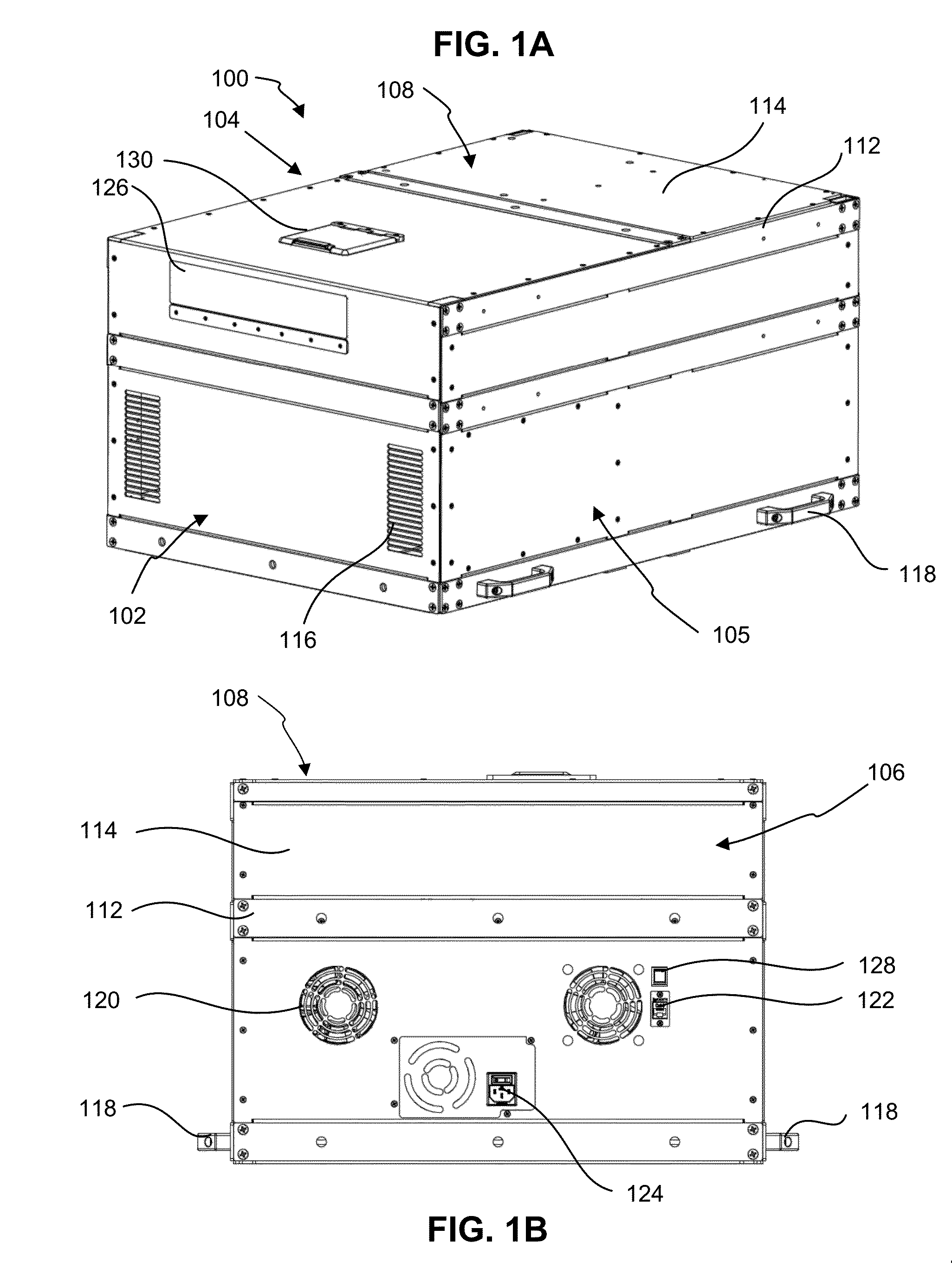
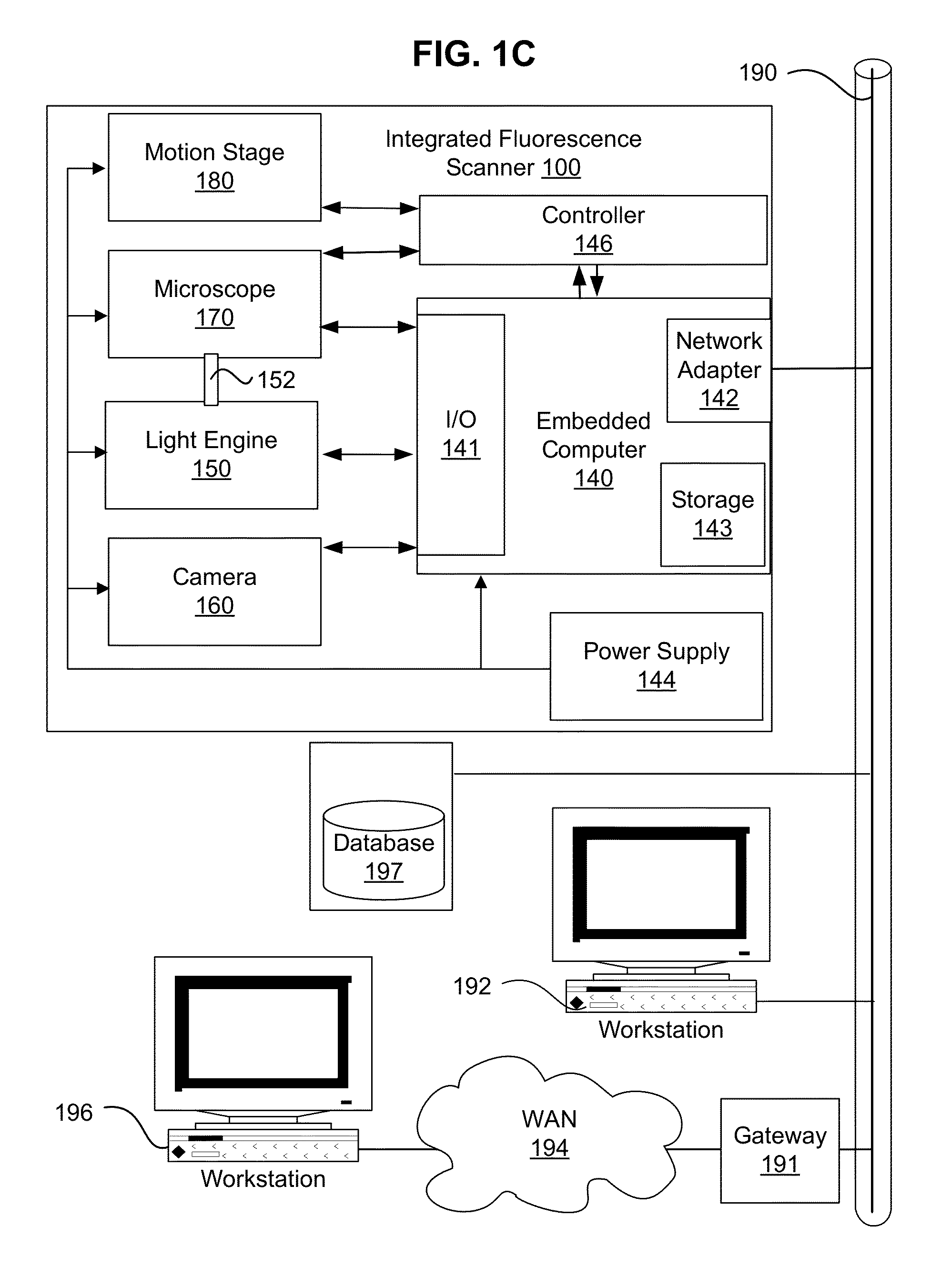
[0006]The present invention provides an integrated fluorescence scanner which overcomes the disadvantages of prior scanning systems. The present invention provides an integrated fluorescence scanner which incorporates a light engine, microscopes, imaging devices, and motion control into a network appliance that allows for automation of fluorescence microscopy.

Problems solved by technology

The instruments typically offer all the degrees of freedom for analysis that are expected of a microscope, yet are superfluous for a dedicated scanner or reader while adding complexity and unnecessary failure modes and mainte

Method used


Image

  • Integrated fluorescence scanning system
  • Integrated fluorescence scanning system
  • Integrated fluorescence scanning system

Examples


examples

[0087]FIGS. 6A and 6B show examples of scanning results obtained using a prototype of the integrated fluorescence scanner of FIGS. 1A-1C. FIG. 6A shows a fluorescence scanning image of an array of thin sections of mouse brain tissue laid out on a slide. The tissue is stained with DAPI fluorophore excited with UV light from the light engine and imaged in blue light at the camera. The resultant image is 35504×19639 pixels with, 16 bits per pixel intensity data comprising a 16×9 grid (162 tiles). FIG. 6B shows an array tomography image of mouse brain tissue on a slide. The image is 2048×2048 pixels with 16 bits per pixel. The camera used was A Hamamatsu™ Flash 4. A 60×1.4NA objective was used with oil immersion. The dimension of 1 pixel is 108 nm.

[0088]The foregoing description of the various embodiments of the present invention has been provided for the purposes of illustration and description. It is not intended to be exhaustive or to limit the invention to the precise forms disclose...


PUM


Abstract

An integrated fluorescence scanning system is provided. The integrated fluorescence scanner combines an embedded computer, light engine, microscope, and motion stage into a compact rack-mountable network appliance that allows for automation of fluorescence microscopy. In an embodiment, the integrated fluorescence scanner includes a solid-state light engine which can provide intense, pure, and stable light across the spectrum required for imaging of all fluorophores of interest. The embedded computer allows for autonomous operation, and network appliance features including synchronous multi-scanner operation and monitoring, multisite operation via a single control terminal, and calibration for inter and intra instrument consistency.

Description

CLAIM OF PRIORITY[0001]This application claims the benefit of priority to U.S. Provisional Application No. 62 / 072,195, filed Oct. 29, 2014 entitled “INTEGRATED FLUORESCENCE SCANNING SYSTEM” which application is incorporated herein in its entirety.FIELD OF THE INVENTION[0002]The present invention relates to a system and method for fluorescence scanning. In particular the present invention relates to an integrated fluorescence scanner featuring a solid state light source.BACKGROUND OF THE INVENTION[0003]Fluorescence microscopy utilizes an optical microscope configured to detect fluorescence and phosphorescence instead of or as well as reflection and absorption of light to study properties of specimens. In fluorescence microscopy, the specimen is illuminated with light of a specific wavelength an excitation filter or dichroic mirror is used to select the wavelength of light directed at the specimen. The excitation light excites fluorophores in the specimen causing the fluorophores to e...

Claims


Application Information

Patent Timeline
IPC IPC(8): G01N21/64
CPCG01N21/6458G01N2021/6471G01N21/6486G01N21/6452G01N21/64
Inventor JAFFE, CLAUDIA B.JAFFE, STEVEN M.
Owner LUMENCOR
Why Eureka
Try Eureka