X-ray phase-contrast imaging

a phase contrast and imaging technology, applied in the field of x-ray phase contrast imaging, can solve the problems of serious limitations in the field of view and the flux of photons, and the inability of conventional attenuation-based x-ray imaging to provide sufficient contrast for biological soft tissu

Active Publication Date: 2016-05-19
RENESSELAER POLYTECHNIC INST
View PDF4 Cites 20 Cited by
  • Summary
  • Abstract
  • Description
  • Claims
  • Application Information

AI Technical Summary

Benefits of technology

[0004]The subject invention provides novel and advantageous systems and methods for X-ray phase-contrast imaging (PCI), including the use of one or more period-varying or quasi-periodic gratings. An X-ray PCI system can include a phase grating that is period-varying or quasi-periodic and can be positioned between an object being imaged and a detector. A second grating, such as an absorption grating or an analyzer grating can also be present and disposed between

Problems solved by technology

Conventional attenuation-based X-ray imaging cannot provide sufficient contrast for biological soft tissue.
Conventional

Method used


Image

  • X-ray phase-contrast imaging
  • X-ray phase-contrast imaging
  • X-ray phase-contrast imaging

Examples


example 1

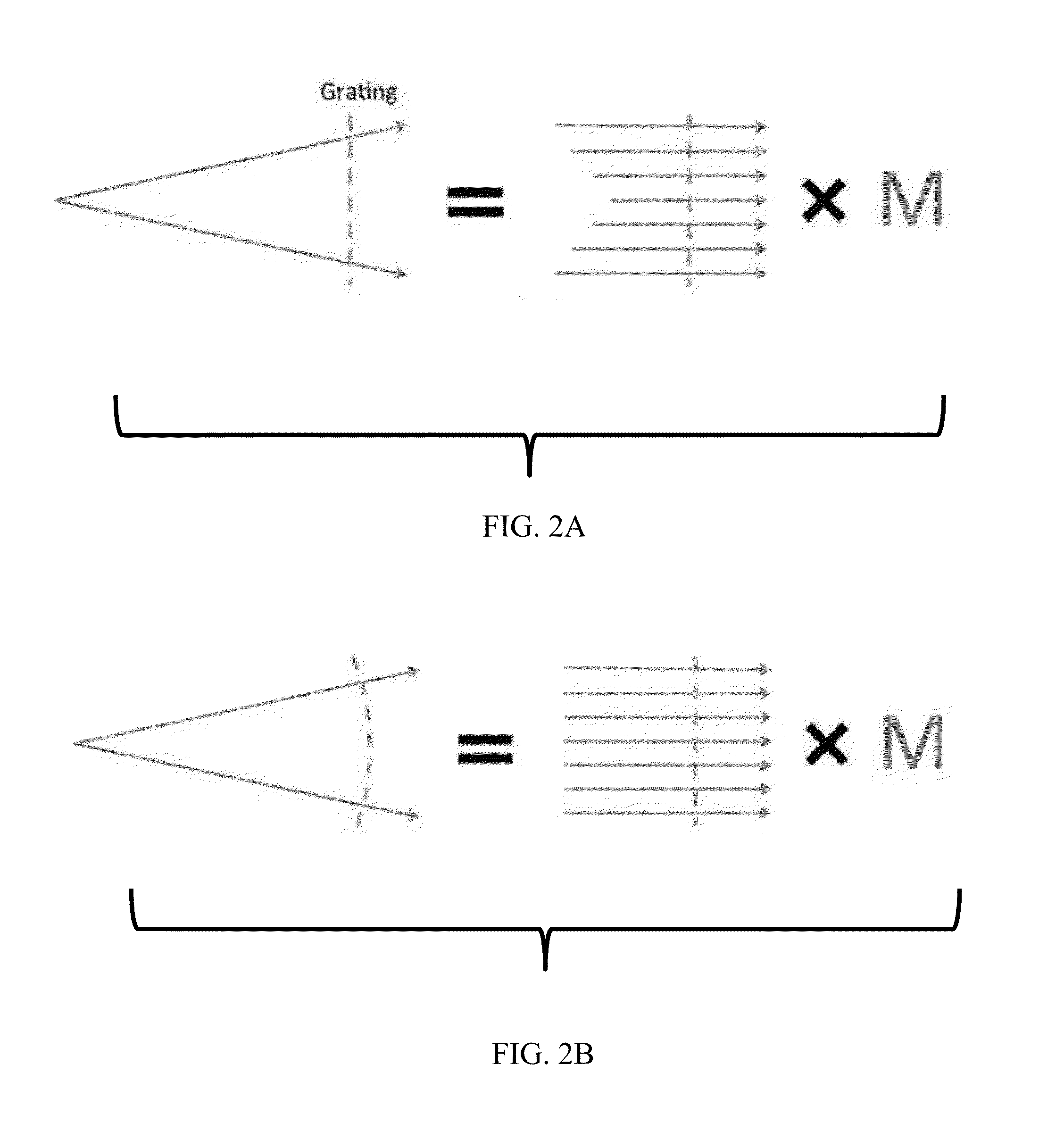
[0163]A numerical analysis was performed to show the modified interference pattern along the x-axis parallel to the grating plane for both a constant-period grating and a period-varying phase grating according to an embodiment of the subject invention. In this analysis, a pure phase grating with a λ phase shift was illuminated by a fully coherent parallel-beam of 28 keV.

[0164]FIG. 3A shows a plot of the period (in μm) along the x-axis (in μm) for the period-varying phase grating. FIG. 3B shows a plot of the period (in μm) along the x-axis (in μm) for the constant-period grating. FIG. 3C shows the interference pattern at different propagation distances from the grating for the period-varying phase grating. FIG. 3D shows the interference pattern at different propagation distances from the grating for the constant-period grating. FIG. 3E shows an enlarged view of the two boxes highlighted in FIG. 3D. The red arrows in FIG. 3E show the difference caused by the grating aperture widths (p...

example 2

[0165]To analyze the Talbot self-imaging performances with different combinations of gratings and detectors, as well as with periodic and quasi-periodic gratings designs, numerical experiments were performed.

[0166]The energy of incident X-rays was set to 28 keV. The distance between the X-ray source and the phase grating G1 grating was 1 m, the phase grating G1 and absorption grating G2 were separated by the first-order fractional Talbot distance, calculated according to Equation (10). The period of the phase grating G1 (pG1 was 4.8 μm, while the period of the absorption grating G2 (pG2) was determined according to imaging geometry. In the data collection step, the classic phase stepping method was employed, and the visibility of the stepping curve was used to quantify the imaging performance. The detector cell pitch was 50 μm.

[0167]First, for a grating-based phase-contrast imaging system, if the detector and the absorption grating have different curvatures as indicated by the red a...

example 3

[0171]To demonstrate the performance of the quasi-periodic grating, numerical simulations with Matlab (Mathwork) were conducted for both curved periodic phase grating G1 and quasiperiodic phase grating G1′ based on the general setup shown in FIG. 10B. In the simulation, the energy of the X-ray beam was 28 keV. The period of the phase grating G1 was set to 4.8 μm, with a radius, r0=1.0 m. The center of planar analyzer (absorption) grating G2 was at the first fractional Talbot image of the center of G1. 1 / rD−1 / r0=p2 / 8λr02. G1′ had the central period set to be the same as G1. The calculated period of G1′ as a function of 0 is shown in FIG. 11C, and the period of G2′ is calculated accordingly.

[0172]The period of the quasi-periodic grating is larger than 4.8 μm, and the same fabrication process for periodic grating can be adapted (see, e.g., David et al., “Fabrication of diffraction gratings for hard X-ray phase contrast imaging,” Microelectron. Eng. 84(5-8), 1172-1177 (2007), which is i...


PUM


Abstract

Systems and methods for X-ray phase-contrast imaging (PCI) are provided. A quasi-periodic phase grating can be positioned between an object being imaged and a detector. An analyzer grating can be disposed between the phase grating and the detector. Second-order approximation models for X-ray phase retrieval using paraxial Fresnel-Kirchhoff diffraction theory are also provided. An iterative method can be used to reconstruct a phase-contrast image or a dark-field image.

Description

CROSS-REFERENCE TO A RELATED APPLICATION[0001]The present application claims the benefit of U.S. Provisional Application Ser. No. 62 / 080,560, filed Nov. 17, 2014, which is incorporated by reference herein in its entirety, including any figures, tables, and drawings.BACKGROUND OF INVENTION[0002]Computed Tomography (CT) is a mainstay in diagnostic imaging. It plays key roles in disease detection and characterization, and also in patient follow-up during and after treatment. Biological soft tissue consists mainly of light elements, and its density is nearly uniform with little variation. Conventional attenuation-based X-ray imaging cannot provide sufficient contrast for biological soft tissue. X-ray phase imaging is sensitive to structural variation of soft tissue and offers superior contrast resolution for characterization of cancerous and other diseased tissues. The cross-section of an X-ray phase shift image is a thousand times greater than that of X-ray attenuation in soft tissue o...

Claims


Application Information

Patent Timeline
IPC IPC(8): A61B6/00G06T11/00G06T7/00
CPCA61B6/484A61B6/4035G06T2207/10116G06T7/0012G06T11/003A61B6/4291A61B6/032A61B6/4233A61B6/5205G06T11/005G21K1/025G21K2207/005G06T2211/461
Inventor WANG, GEXI, YANCONG, WENXIANG
Owner RENESSELAER POLYTECHNIC INST
Why Eureka
Try Eureka