
Method and apparatus for isolation, capture and molecular analysis of target particles

a target particle and molecular analysis technology, applied in the direction of fluid pressure measurement, liquid/fluent solid measurement, peptides, etc., can solve the problems of limited access, less effective treatment for patients, and outdated molecular information about tumors

Inactive Publication Date: 2016-07-21
PRECISION FOR MEDICINE TX INC
View PDF3 Cites 9 Cited by
  • Summary
  • Abstract
  • Description
  • Claims
  • Application Information

AI Technical Summary

Benefits of technology

The present invention is about an apparatus and method for processing samples of interest, particularly cancer cells, using dielectrophoresis in a continuous, semi-continuous, or intermittent system. The apparatus includes a microfluidic device with a chamber that can enrich and isolate target particles, such as cancer cells, from a suspension of particles. The apparatus can also have a reduced impact of steric hindrance, which can improve the efficiency of separating cancer cells from other cells in a sample. The method involves injecting the sample into the chamber and applying a dielectrophoretic force to separate the target particles from non-target particles. The apparatus and method can be used for diagnosis and monitoring of tumoral conditions in a sample.

Problems solved by technology

These methods have several limitations that include invasive, risky collection procedures for the patient, limited accessibility of the tumor tissue, dependence upon the cancer type and location, and the use of patient's archival tumor samples that may have been collected several years prior to metastatic relapse, making the molecular information about the patients' tumor outdated.
The combination of these factors results in less effective therapy for the patient and an unbeneficial economic impact to society.
Furthermore, because of the nature of metastatic cancer, oncologists often have to wait several months before they can determine if a specific treatment is beneficial to the patient.
Cancer cells display a broad spectrum of genetic alterations that include gene rearrangements, point mutations and gene amplifications, which lead to disturbances in molecular pathways regulating cell growth, survival and metastasis.
Common solid tumors, such as breast, lung, and colorectal cancer have been difficult to treat, perhaps in part because they are heterogeneous, with each subset of patients having different molecular abnormalities, and sometimes within the tumor itself.
Mutation analysis is usually performed on archived tumor tissue or on tissue from fresh biopsies, which can be a limiting factor for its use.
A sufficient amount of tissue for analysis is not available for about 25%-45% patients.
Historically, the detection and capture of such cells has been challenging (Gupta, et al., Biomicrofluidics 6, 024133 (2012)).
However, not all cancer cells express this marker.
Also, the CELLSEARCH® test does not provide the ability to generate any molecular information about the cancer.
Furthermore, other CTC technologies are limited in their ability to efficiently capture an adequate number of CTCs from whole blood and from various types of cancer.
Thus, currently available technologies do not solve the challenge of providing physicians and drug development companies conducting clinical trials in oncology with a broadly applicable, efficient, and economical method which allows fast analysis of predictive markers on CTCs for diagnosis, prognosis and therapy.

Method used


Image

  • Method and apparatus for isolation, capture and molecular analysis of target particles
  • Method and apparatus for isolation, capture and molecular analysis of target particles
  • Method and apparatus for isolation, capture and molecular analysis of target particles

Examples


example 1

[0102]In this Example, the recovery of SKOV3 ovarian cancer cells was compared between electrode 200 (illustrated in FIG. 2) and electrode 100 (illustrated in FIG. 1) using different elution buffer flow rates.

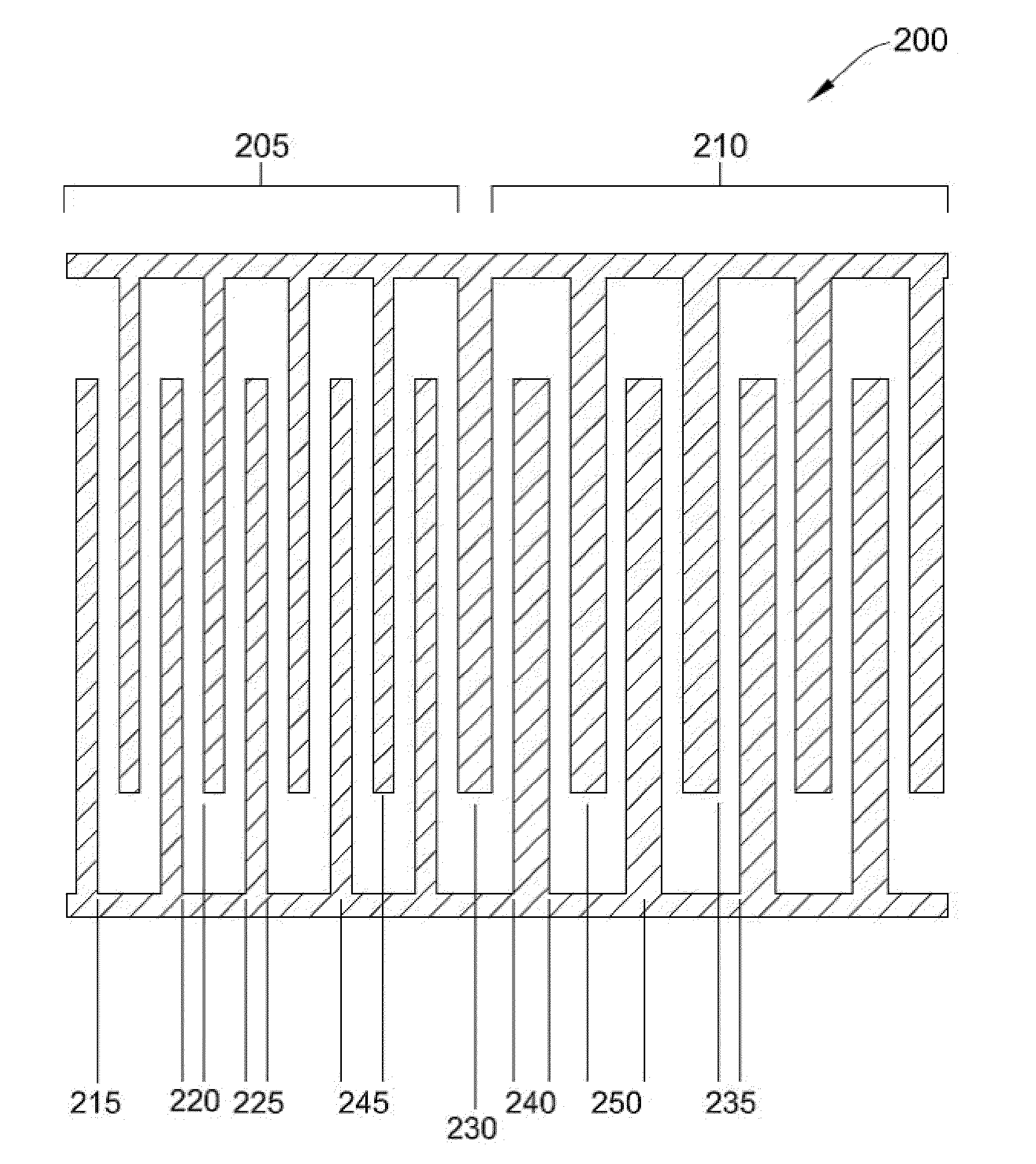
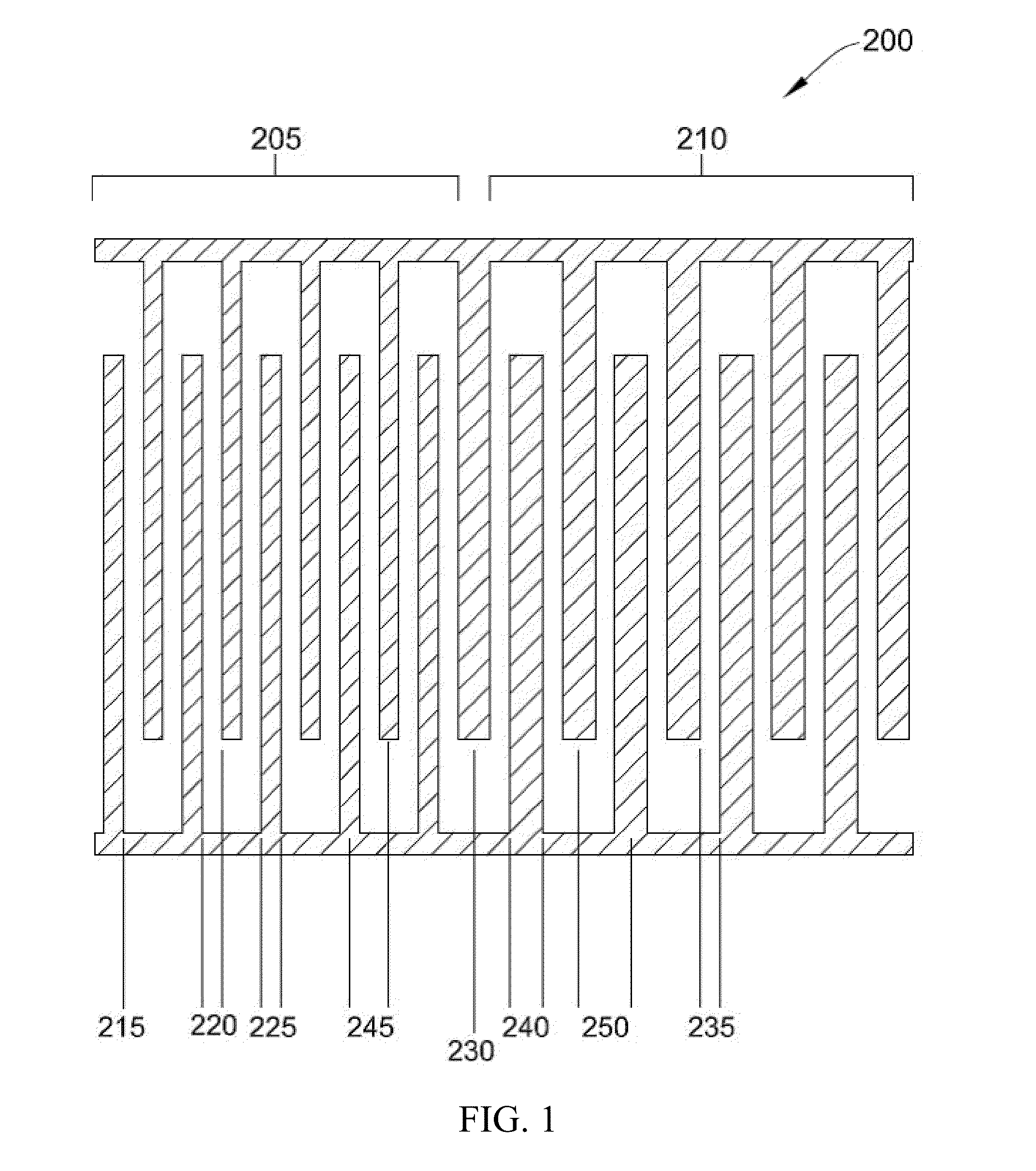
[0103]Specifically, PBMC were spiked with SKOV3 ovarian cancer cells. The data collected utilized a prior art electrode with an inter-electrode gap 110 of 50 microns and a conductive trace width 115 of 50 microns. In this particular Example, electrode 200 had two zones 205, 210 with a first inter-electrode gap 220 of 30 microns, a first conductive trace width 225 of 30 microns, a second inter-electrode gap 235 of 30 microns and a second conductive trace width 240 of 50 microns.

[0104]Table 1 summarizes a comparison between the recovery of SKOV3 cancer cells using electrode 200 compared with electrode 100 using different elution buffer flow rates.

TABLE 1Comparison of percent recovery with electrode configurations.% Recovery% Recoveryof SKOV3of SKOV3ovarian cancerovarian cancerElu...


PUM

PropertyMeasurementUnit
Electrical conductivityaaaaaaaaaa

Abstract

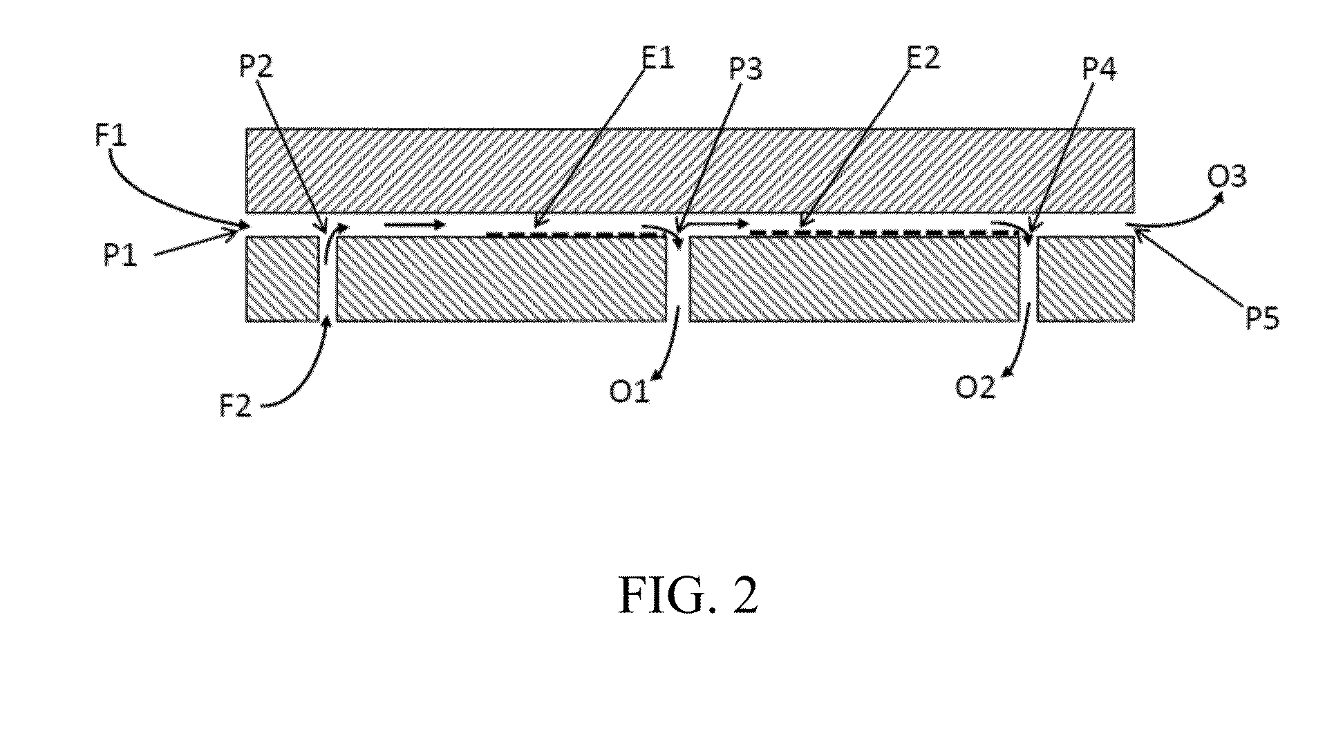
Methods and apparatuses for target particle separation and isolation are disclosed. The apparatuses are flow chambers that include an infusion port configured to introduce a sample, and an elution flow port configured to receive an elution buffer. The flow chamber also includes an electrode coupled to a floor of the flow chamber, wherein the electrode includes at least a first zone with a first inter-electrode pitch and a second zone with a second inter-electrode pitch. The first zone generates a first electric field and the second zone generates a second electric field of a different magnitude than the first electric field when an electric signal is applied. The first electric field and the second electric field separate the target particle from the fluidic suspension via dielectrophoresis. The flow chamber also includes a waste disposal port that discharges the waste material, and a collection port configured that collects a target particle enriched sample.

Description

CROSS-REFERENCE TO RELATED APPLICATIONS[0001]This international PCT Application claims the benefit of priority from U.S. Provisional Patent Application No. 61 / 871,624, filed Aug. 29, 2013, entitled, “APPARATUS AND METHOD FOR ISOLATION, CAPTURE AND MOLECULAR ANALYSIS OF TARGET PARTICLES DURING DIELECTROPHORESIS”, which is incorporated here by reference in its entirety.BACKGROUND OF THE INVENTION[0002]The present invention relates generally to methods and apparatuses for separation and isolation of target particles. More particularly, the present invention is directed to new device elements, apparatuses employing the device elements, and methods for providing improved efficient separation and isolation of one or more types of target particles. The methods employ integrated fluidic devices using dielectrophoresis that are designed to perform a continuous, semi-continuous, staged (interval or intermittent), or sporadic, processing of microfluids. The devices and apparatuses may employ a...

Claims


Application Information

Patent Timeline
IPC IPC(8): G01N1/10G01N33/483G01N27/447
CPCG01N1/10G01N27/44743G01N33/4833G01N27/44717G01N27/44769G01N30/0005G01N2030/0065B03C5/005B03C5/026B03C2201/26G01N2030/065
Inventor MENACHERY, ANOOPGUPTA, VISHALHASEGAWA, DAVID KPETHIG, RONALD
Owner PRECISION FOR MEDICINE TX INC
Learn More