Partial skull replacements capable of monitoring in real time and delivering substances into brain tissue, and uses thereof

Active Publication Date: 2016-09-15
RES & BUSINESS FOUND SUNGKYUNKWAN UNIV +1
View PDF0 Cites 9 Cited by
  • Summary
  • Abstract
  • Description
  • Claims
  • Application Information

AI Technical Summary

Benefits of technology

[0027]The partial skull replacement of the present invention includes PDMS as an active ingredient, and, besides visualizing brain tissues more clearly compared to a conventional cranial window technique using glass, has been recognized to be able to keep the brain healthy by having a minimal influence on intracranial pressure and CSF flow, allow the insertion of a pipette, etc. for the repetitive and simultaneous injection of a drug into brain tissues and for the measurement of brain signals, and enable ultrasonic penetration for the monitoring of brain tissues. Therefore, it is expected that the partial skull replacement of the present invention can be usefully employed in various methods of monitoring biological tissues within the skull.
[0028]In addition, in the case of the conventional wind

Problems solved by technology

Initially, such an artificial bone was developed using materials that were biologically inert, resulting in limited applications thereof due to the infection or inflammation of a surrounding tissue.
In particular, when the skull is replaced with a conventional glass

Method used


Image

  • Partial skull replacements capable of monitoring in real time and delivering substances into brain tissue, and uses thereof
  • Partial skull replacements capable of monitoring in real time and delivering substances into brain tissue, and uses thereof
  • Partial skull replacements capable of monitoring in real time and delivering substances into brain tissue, and uses thereof

Examples


example 1

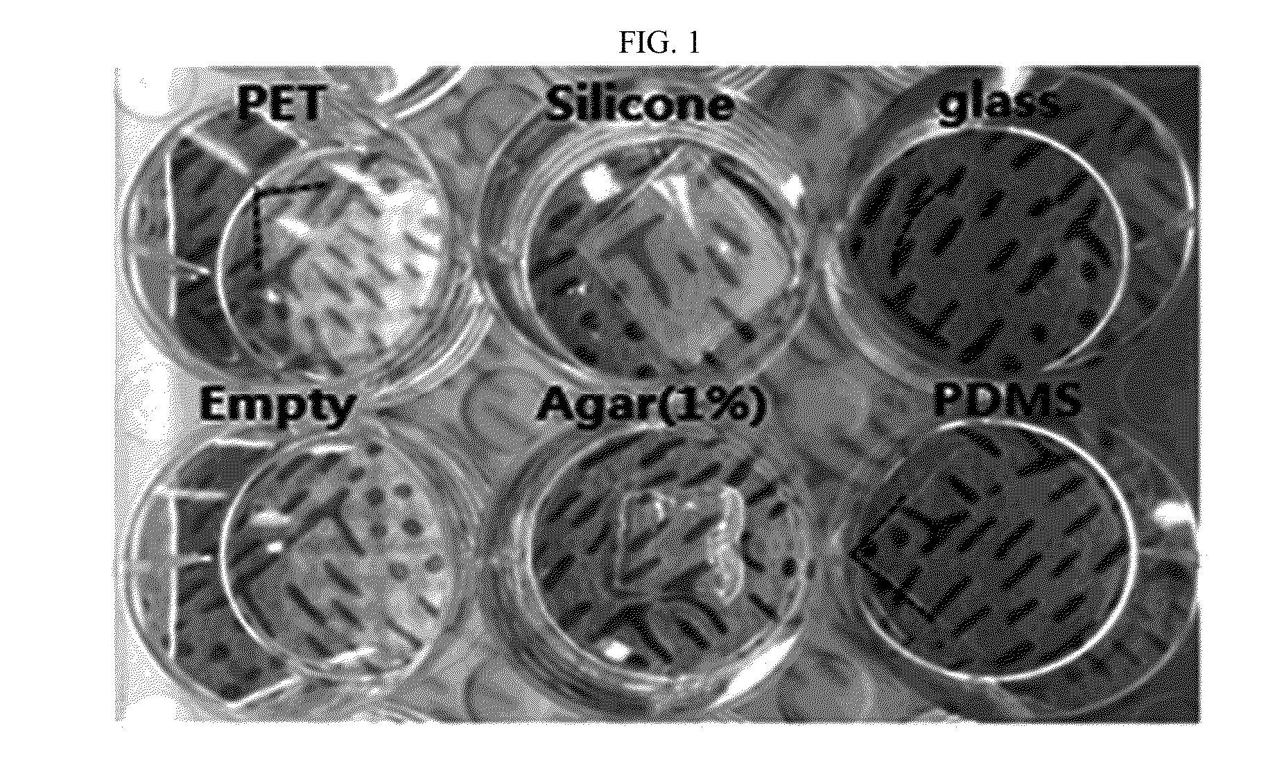
Search for Skull Substitute Material

[0062]1-1. Preparation of Substitute Materials

[0063]First, the present inventors searched for a material for a partial skull replacement for long-term monitoring of a surface of the brain within the skull. For this, glass, agarose (at 1% concentration), silicone, polyethylene terephthalate (PET), and PDMS were selected as candidate materials for a partial skull replacement and were each prepared in a cell culture dish (FIG. 1). Later, the cytotoxicity, hydrophilicity, and transparency of the materials were compared and evaluated.

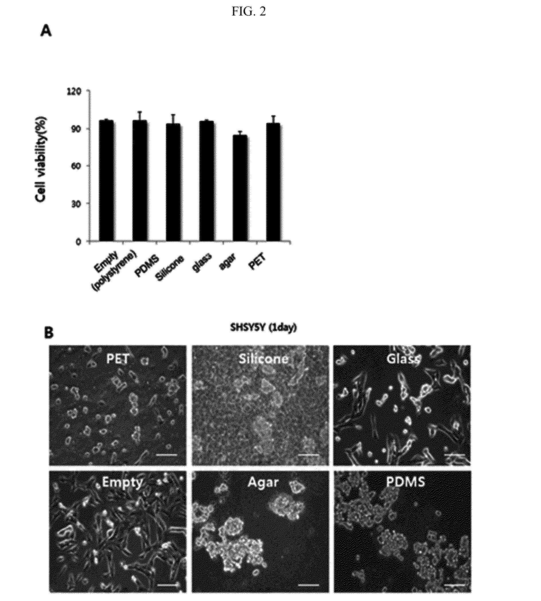
[0064]1-2. Evaluation of Cytotoxicity

[0065]In preparation for an application into biological tissue, the cytotoxicity of the candidate materials for a partial skull replacement prepared in Example 1-1 were evaluated. Specifically, SH-SY5Y cells, which are the human neuroblastoma cell lines, were cultured in cell culture dishes, each of which was treated with each candidate material for a partial skull replacement. 24 hours...

example 2

Preparation of Window by Attaching Artificial Skull Substitute

[0077]The present inventors intended to apply PDMS, which was selected to be most appropriate as the skull replacement material according to the above Example 1, to the skull of a rat.

[0078]As appears in FIG. 5, skin in the area corresponding to biological tissues to be observed by visualization was removed to cause the skull to be exposed (blue dot lines in FIG. 5A and FIG. 5B), and then, the skull was removed with a drill (FIG. 5C) to exposure brain under the skull. Then, meninges on the cortex was removed with a pair of fine forceps, and, before the brain expanded as a result of the removal of meninges, an artificial partial skull replacement consisting of PDMS with an appropriate thickness was applied on the cortical tissue, and attached, with an instant glue, to the skull in the affected area (FIG. 5D and FIG. 5E). Lastly, a dental resin was used to seal and cover all of the connection between the artificial partial ...

example 3

Confirmation of Excellence of Artificial Skull Substitute in Biological Tissue Monitoring

[0079]A cross-section of the skull to which the artificial partial skull replacement is attached according to Example 2 is provided the schematics in FIG. 6. As shown in FIG. 6, the artificial partial skull replacement cranial window of the present invention has a flexible form, which enables the partial skull replacement to be attached to the skull of a rat for the long-term monitoring of the brain and a biological structure within the skull in real time. The following experiments were conducted with the intention of confirming the above statement.

[0080]3-1. Evaluation of Stability of Brain Surface Tissues and Blood Vessels

[0081]Brain tissue and blood vessels within the skull were monitored for any change for a long period of 10 weeks from the day the artificial partial skull replacement was attached. As shown in FIG. 7, the results show that conditions enabling the observation of brain surface...


PUM

PropertyMeasurementUnit
Pressureaaaaaaaaaa
Areaaaaaaaaaaa
Biological propertiesaaaaaaaaaa

Abstract

The present invention relates to a partial skull replacement consisting of polydimethylsiloxane (PDMS) and a method of monitoring biological tissues within a skull using the partial skull replacement. The method of monitoring using the partial skull replacement of the present invention has a minimal influence on intracranial pressure and cerebrospinal fluid flow as compared to a conventional cranial window technique using glass, and, by way of visualizing biological tissues within a skull with enhanced clarity, is expected to be usefully employed as a method of monitoring biological tissues within the skull.

Description

CROSS-REFERENCE TO RELATED APPLICATION[0001]This application claims priority to and the benefit of Korean Patent Application No. 10-2015-0034628, filed on Mar. 12, 2015, the disclosure of which is incorporated herein by reference in its entirety.[0002]The present invention was made with the support of the Integrated Research of Basic Neuroscience and Biophysics # S-2014-1974-000-1 of the Institute for Basic Science (IBS).BACKGROUND[0003]1. Field of the Invention[0004]The present invention relates to a partial skull replacement consisting of polydimethylsiloxane (PDMS) and a method of using the same for monitoring biological tissue within a skull or injecting an agent.[0005]2. Discussion of Related Art[0006]A skull is a set of bones constituting the skeletal structure of a head and serves to hold a brain therein, giving protection for the same, and provides a space for sense organs such as eyes, ears, a nose, and a mouth. Being part of the skeleton in closest proximity to the brain, ...

Claims


Application Information

Patent Timeline
IPC IPC(8): A61L27/18A61M5/00A61K49/22A61B5/04A61L27/50A61K49/00A61M25/00A61B5/00
CPCA61L27/18A61M25/00A61M5/007A61B5/0071A61B5/04001A61M2210/0687A61B5/4094A61L27/50A61K49/0017A61K49/0073A61K49/226A61B5/0095A61B5/0042A61B5/0068A61B5/0084A61B5/0261A61B5/6848A61B5/6864A61B5/6868A61B8/00A61K49/0045A61B5/24C08L83/04A61B5/02A61B5/40A61F2/2875A61M39/0247A61M2039/025A61M2039/0276
Inventor KIM, SEONG-GISUH, MINAHHEO, CHAEJEONGBAAC, HYOUNG WON
Owner RES & BUSINESS FOUND SUNGKYUNKWAN UNIV
Why Eureka
Try Eureka