Functional imaging of photoreceptors in the retina by rhodopsin mapping

a technology of rhodopsin and photoreceptors, applied in the field of functional imaging of photoreceptors in the retina by rhodopsin mapping, can solve the problems of vision loss, personal tragedy, burden on society, and little progress in quantitative assessmen

Active Publication Date: 2017-02-09
UNIV OF MIAMI
View PDF3 Cites 7 Cited by
  • Summary
  • Abstract
  • Description
  • Claims
  • Application Information

AI Technical Summary

Benefits of technology

[0029]In some embodiments, the time delay between adjacent laser pulses is controlled by the difference in lengths between the optical fibers from which the pulses originate. Exemplary embodiments provide that the time delay between adjacent pulses is less than about 1 μs, and preferably between about 1 ns and about 10 ns. The time delay among pulses allows the detection of the reflection of these pulses from the retina with a single detector, e.g., a PMT.

Problems solved by technology

Loss of photoreceptors can lead to vision loss.
Loss of vision is not only a personal tragedy but also a burden to the society.
To date, however, little progress has been made in assessing quantitatively, via means of optical imaging, the function and structure of photoreceptors in vivo, which could provide crucial information for the diagnosis and monitoring of retinal degenerative disorders.

Method used


Image

  • Functional imaging of photoreceptors in the retina by rhodopsin mapping
  • Functional imaging of photoreceptors in the retina by rhodopsin mapping
  • Functional imaging of photoreceptors in the retina by rhodopsin mapping

Examples


example 1

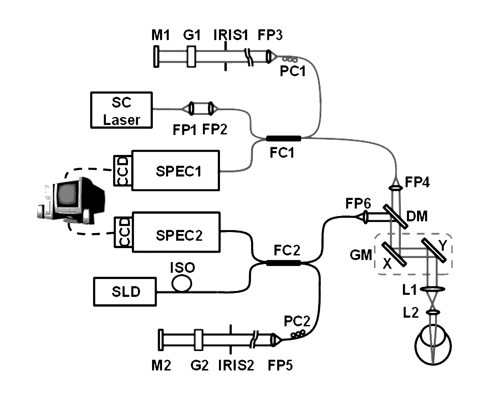
[0101]A schematic of the NIR / VIS-OCT imaging system is shown in FIG. 1. The system comprises two independent spectral domain-OCT subsystems, one of which is equipped with a near-infrared (NIR) light source for guiding the image alignment process in a dark environment and the other is equipped with a visible (VIS) light source for rhodopsin imaging.

[0102]The VIS-OCT employs a supercontinuum laser source (EXB-6, SuperK EXTREME, NKT Photonics, Denmark) equipped with a variable band-pass filter (SuperK Varia, NKT Photonics). The filtered light output (center wavelength: 520 nm, bandwidth: 9.3 nm, 80 MHz pulse rate) is delivered though a fiber delivery module. The VIS light is coupled into the source arm of a single-mode optical fiber-based Michelson interferometer by using a pair of identical fiber ports (PAF-X-11-A, Thorlabs). The NIR-OCT employs a superluminescent diode (SLD-37-HP, center wavelength: 840 nm, bandwidth: 50 nm, Superlum, Russia). The NIR- and VIS-OCT probe light beams a...

example 2

[0106]Two animal models were used for imaging rhodopsin with the NIR / VIS-OCT. The first model comprised albino rats with normal retina (Sprague Dawley, 200-400 g, Harlan Laboratories), and the second model comprised wild-type pigmented rats (Long Evans, 200-400 g, Harlan Laboratories).

[0107]The rats were dark-adapted for about 4 hours prior to each experiment (Behn, D. et al. Dark adaptation was faster in pigmented than albino rats. Doc Ophthalmol. 106, 153-159 (2003)). A cocktail containing ketamine (54 mg / kg body weight) and xylazine (6 mg / kg of body weight) was intraperitoneally injected for anesthesia. The rat's pupil was dilated with 0.5% tropicamide ophthalmic solution and 0.5% proparacaine hydrochloride ophthalmic solution. After the rats were sedated, a powerless contact lens was put on the eye to prevent cornea dehydration and cataract formation (Liu, X. et al. Effect of contact lens on optical coherence tomography imaging of rodent retina. Curr Eye Res. 38, 1235-1240 (2013...

example 3

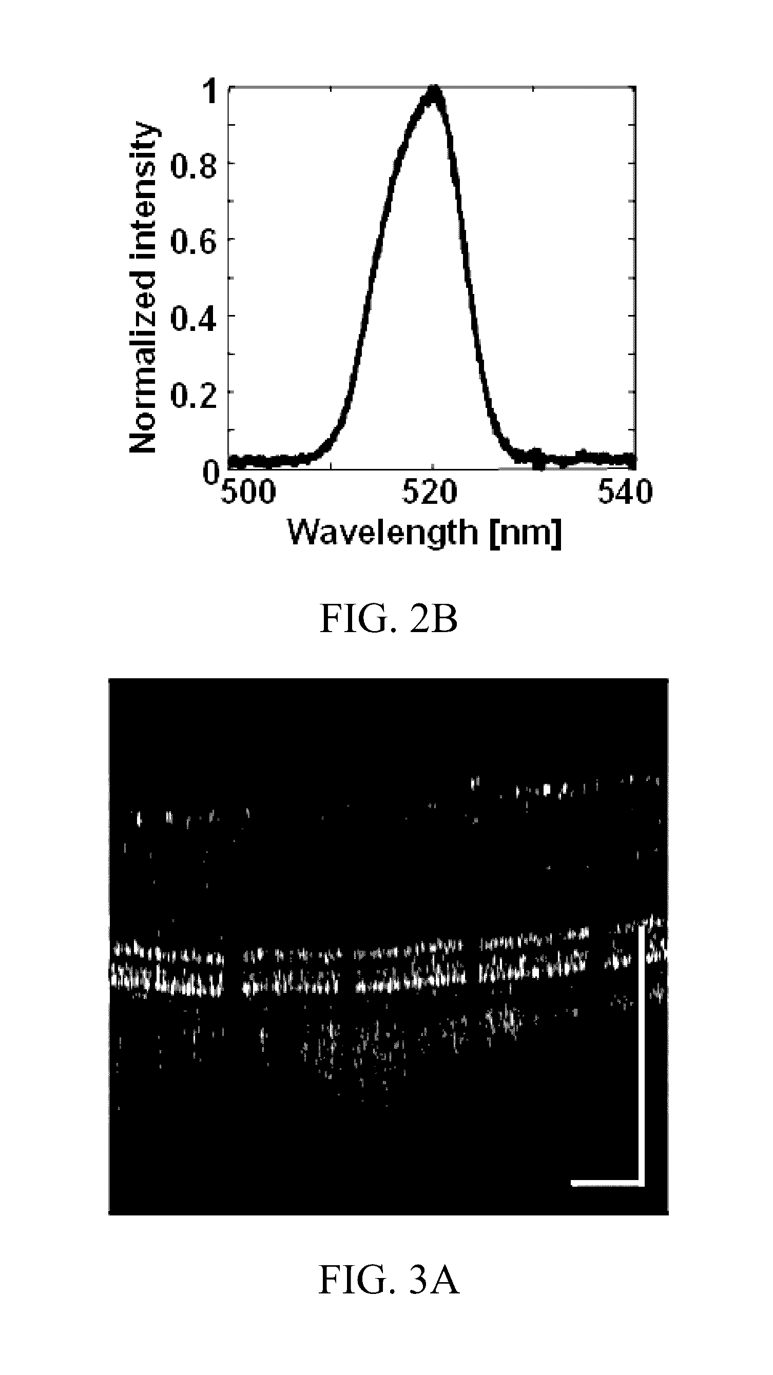
[0109]The following example pertains to imaging rhodopsin using an albino rat. The VIS source had a center wavelength of about 530 nm and a bandwidth of about 9.3, enabling a depth resolution of about 13 μm in air. The NIR source operated at a center wavelength of about 840 nm. Following dark-adaptation for about 4 hours, the retinal area of interest was located and the retinal image was optimized using the NIR-OCT, which was then turned off and the VIS-OCT was turned on to acquire a full 3D OCT dataset. The acquisition time was 1 second for an imaging volume containing 512×128 depth scans (A-scans).

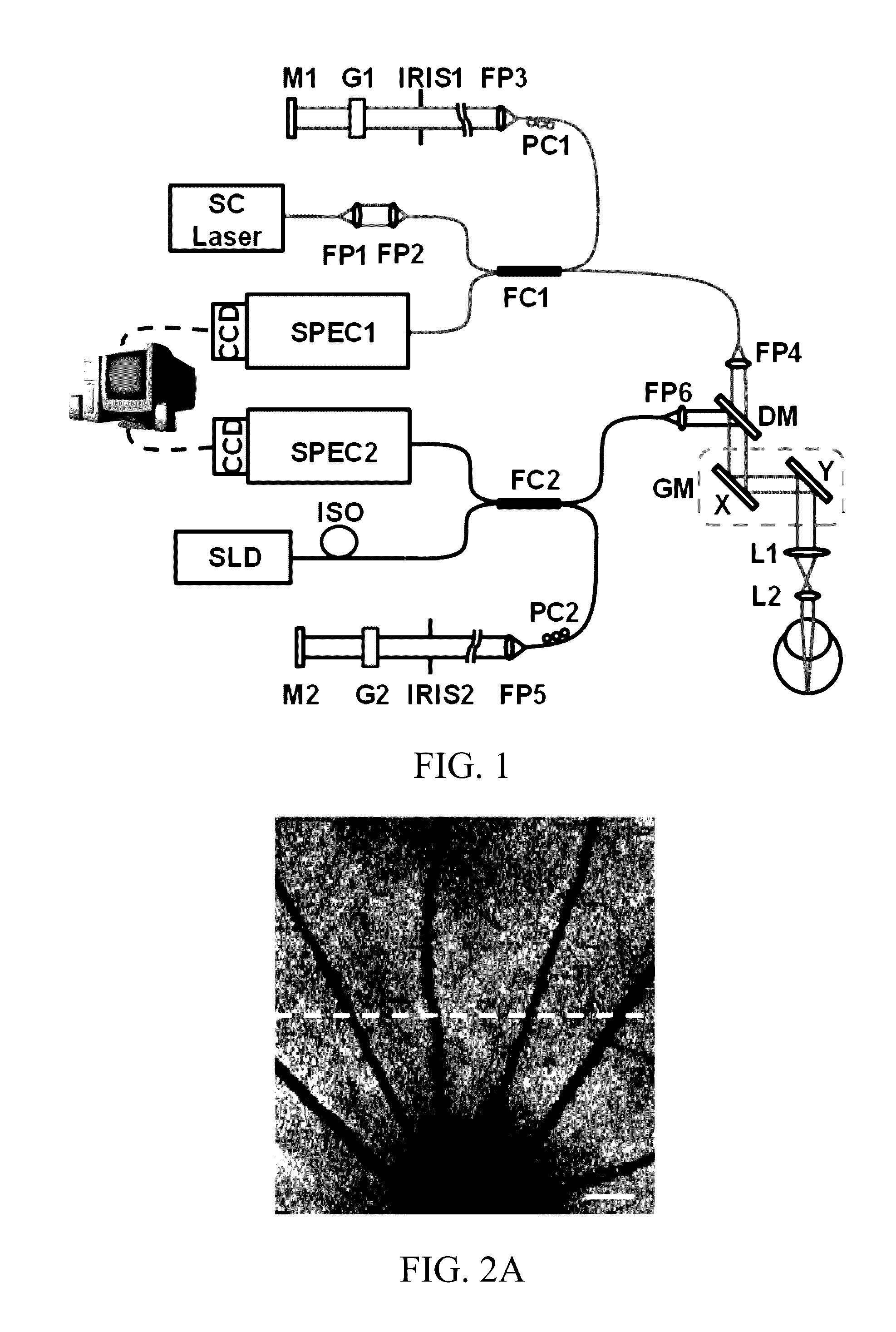
[0110]Rhodopsin was then bleached by bright room light (˜800 lx) for about 15 seconds, followed by acquisition of two light-adapted 3D images of the same retinal area. Each OCT cross-sectional image (B-scan) was manually segmented along the junction between the inner and outer segments (IS / OS) of the photoreceptors. An en-face view of the segmented OCT data was generated by summing the s...


PUM


Abstract

The subject invention provides novel apparatuses and methods for assessing the structure and function of photoreceptors in retina. In a specific embodiment, the assessment is accomplished by imaging rhodopsin, pigmented protein responsible for initiating vision acquisition of the eye, in living subjects. Advantageously, the technologies provided herein can be used for clinical evaluation of retinal photoreceptors for diagnosis, disease staging and follow-up, and outcome measurement for clinical trials of retinal degenerative disorders, including, for example, hereditary retinal degeneration and age-related macular degeneration.

Description

BACKGROUND[0001]Photoreceptor cells in the retina are sensory neurons that convert light signals into neuronal signals via phototransduction, a process that initiates visual senses. Phototransduction occurs in photoreceptors in specialized organelles, the outer segments. Loss of photoreceptors can lead to vision loss. This is especially prevalent in patients with hereditary retinal degenerations such as retinitis pigmentosa (RP), which affects over 100,000 Americans. Mutations in close to 60 genes are responsible for RP. Loss of vision is not only a personal tragedy but also a burden to the society. It is estimated that an RP patient has an average health care cost of $7,000 annually.[0002]Rhodopsin, the photosensitive molecule that absorbs photons and initiates phototransduction, is densely packed in the outer segments of rod photoreceptors. Rhodopsin has intrinsic absorbance in the visible spectrum in the dark adapted state. When exposed to light, the absorption spectrum of rhodop...

Claims


Application Information

Patent Timeline
Patent Type & Authority Applications(United States)
IPC IPC(8): A61B3/10A61B3/00A61B3/14A61B3/12
CPCA61B3/102A61B3/14A61B3/0025A61B3/12A61B3/1025
Inventor JIAO, SHULIANGWEN, RONGLAM, BYRON L.LIU, TAN
Owner UNIV OF MIAMI
Why Eureka
Try Eureka