

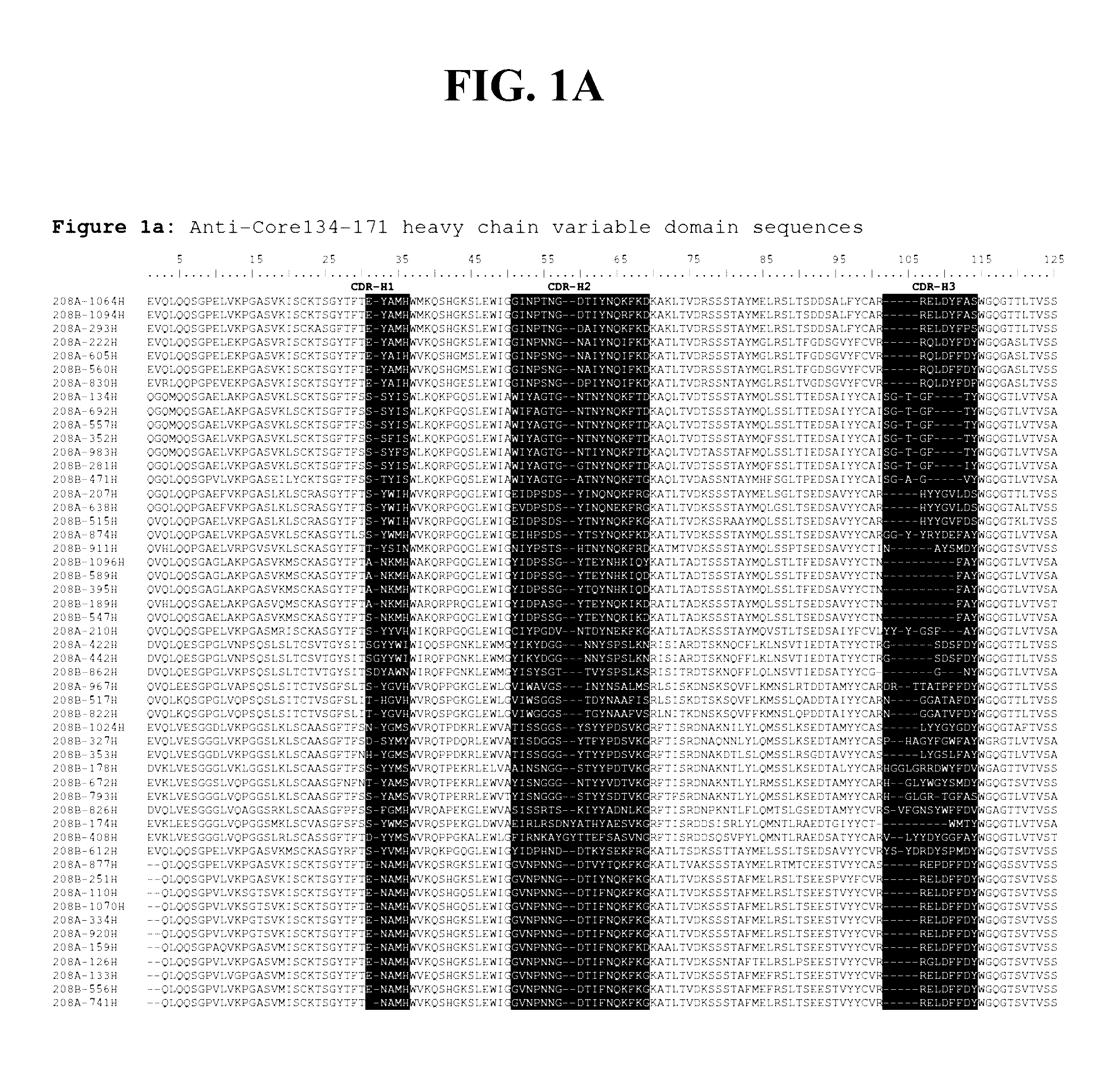
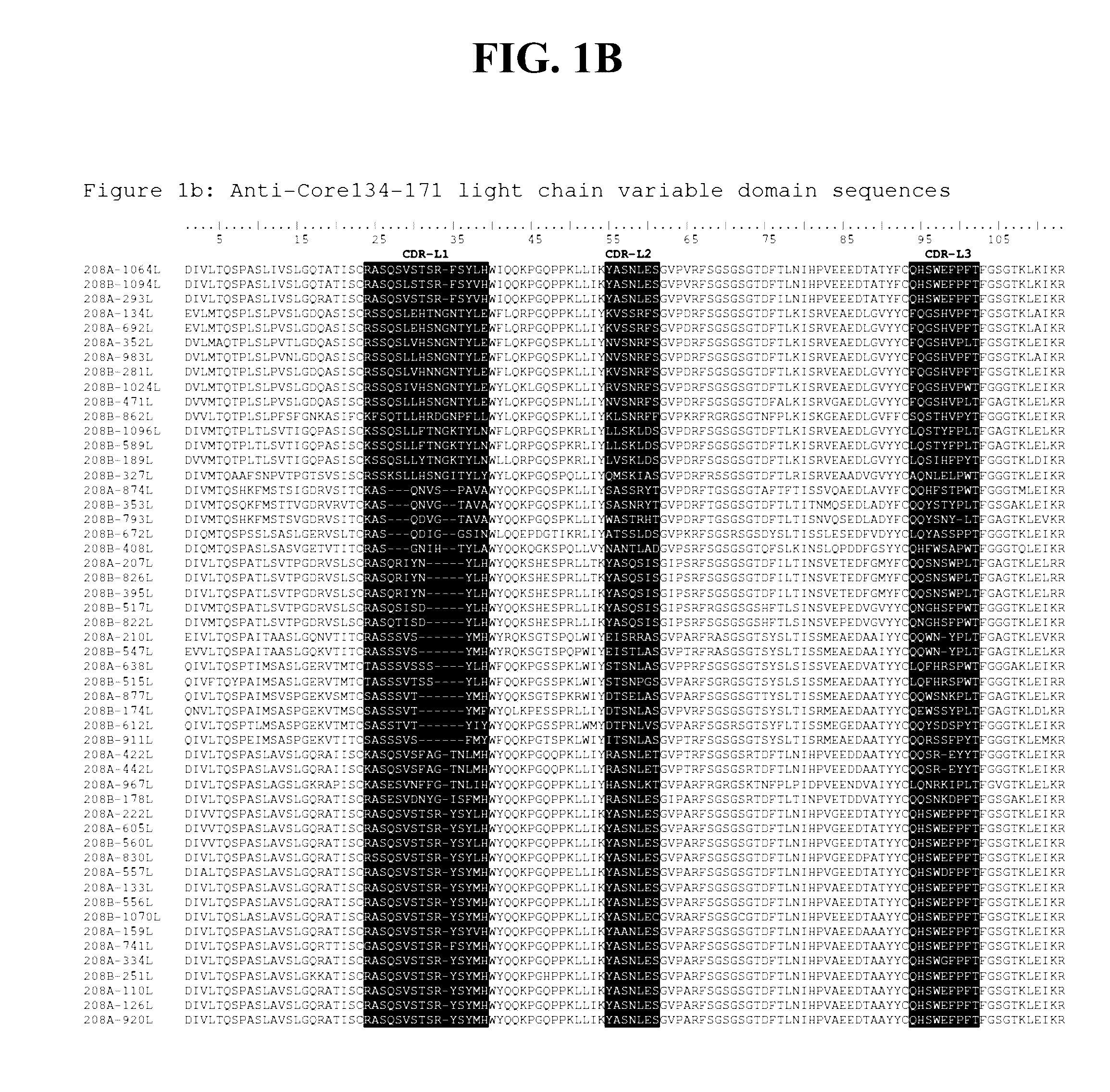
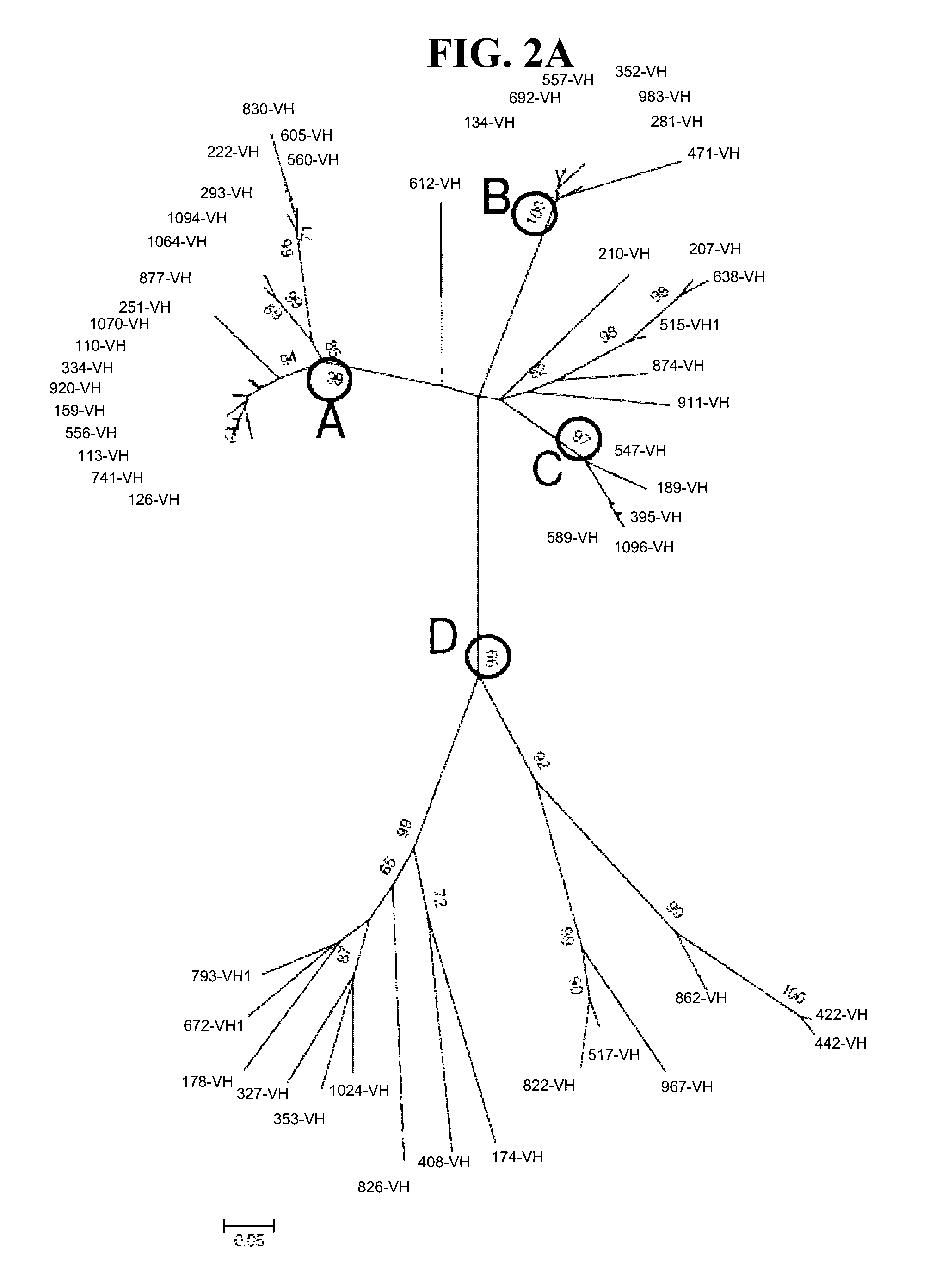
Detection methods employing hcv core lipid and DNA binding domain monoclonal antibodies
a technology of dna binding domain and core lipid, which is applied in the field of detection methods employing hcv core lipid binding domain and dna binding domain monoclonal antibodies or antibody fragments, can solve the problems of ineffective treatment in all cases, lack of vaccines, and antibody testing alone failing to detect hcv infected individuals, etc., and achieve enhanced signal intensity for core and 91-minicor
- Summary
- Abstract
- Description
- Claims
- Application Information
AI Technical Summary
Benefits of technology
Problems solved by technology
Method used
Image

Examples
example 1
Animal Immunizations
[0219]Female CAF1 / J and RBF / DnJ mice (both from The Jackson Laboratory, Bar Harbor, Me.) were immunized on weeks 0, 4 and 10 with 50 μg of an HCV core peptide corresponding to amino acids (all numbering per HCV-1) 134-171 covalently linked to BSA (ALRZ-8 immunogen).
[0220]HCV core peptide-BSA was prepared by AnaSpec, Inc. (Fremont, Calif.). The immunogen peptide was emulsified in Complete or Incomplete Adjulite Freund's Adjuvant (Pacific Immunology, Ramona, Calif.). Complete Freund's adjuvant was used for the primary immunization and Incomplete Freund's adjuvant for the second and third immunizations. Each inoculum was prepared by first diluting the HCV peptide-BSA to the appropriate concentration in sterile saline (0.9% sodium chloride), adding an equal volume of adjuvant and then mixing by passing back and forth between two syringes via a 3-way stopcock until a thick, stable emulsion was formed. Sera samples were taken 10-14 days following the 3rd immunization. ...
example 2
Screening of Mouse Sera for Antigen Immunoreactivity
[0221]Mouse sera samples collected 7-10 days after their final immunization were first tested in a 96-well micro titer enzyme immunoassay (EIA) for reactivity to each of three synthetic (Anaspec, Inc.) carboxy-terminal biotinylated HCV core peptides. The peptides used for screening were derived from the immunogen sequence described in Example 1 and had the following designations and sequences: Peptide 1 (all numbering per HCV-1), amino acids 134-151: MGYIPLVGAPLGGAARALAHG (SEQ ID NO:573); Peptide 2, amino acids 141-161: GAPLGGAARALAHGVRVLEDG (SEQ ID NO:574), Peptide 3, amino acids 151-171, LAHGVRVLEDGVNYATGNLPG (SEQ ID NO:575). Assay plates (NUNC Corporation, Naperville, Ill.) were coated with 100 μL / well of sheep anti-mouse IgG Fc specific antibody (Jackson ImmunoResearch, West Grove, Pa.) diluted to 2 μg / mL in phosphate-buffered saline (PBS). Plates were incubated at 37 deg C. for about 2 hours and then at about 21 deg C. for abo...
example 3
Screening of Mouse Sera for Relative Affinity
[0222]Relative affinity testing was completed for each sera sample—peptide combination for which a strong signal was seen in the previous assay. To determine the relative affinity of each serum sample for the individual core peptides, samples were tested for reactivity to limiting concentrations of each biotin labeled peptide. The assay format was identical to that described above, except that instead of preparing serial dilutions of mouse sera test samples, each sample was prepared at a single dilution, in blocking solution. Additionally, the individual peptides were tested at varying concentrations, beginning with a 500 nM solution in blocking solution followed by ten log 2 dilutions, also in blocking solution. Binding curves were generate and used to determine relative affinity for each sera sample. Based on these results, RBF / DnJ mice #306 and 315 were chosen for B cell fusion.
PUM
| Property | Measurement | Unit |
|---|---|---|
| length | aaaaa | aaaaa |
| full-length | aaaaa | aaaaa |
| full-length | aaaaa | aaaaa |
Abstract
Description
Claims
Application Information