System for interpretation of image patterns in terms of anatomical or curated patterns

a technology of image patterns and anatomical patterns, applied in image enhancement, instruments, image data processing, etc., can solve the problems of human bias in the analysis, difficult interpretation and analysis, etc., and achieve the effect of reducing the chance of human bias skewing the analysis, reducing the cost and time of interpretation, and increasing the reproducibility of the analysis

Active Publication Date: 2017-08-03
KATHOLIEKE UNIV LEUVEN +1
View PDF0 Cites 8 Cited by
  • Summary
  • Abstract
  • Description
  • Claims
  • Application Information

AI Technical Summary

Benefits of technology

[0034]An aspect of the disclosure is a system that can deliver anatomical interpretation of image patterns in an automated way, which holds strong potential for both unsupervised and supervised applications in the medical and biotechnology industry. In an unsupervised setting, the system can be used to obtain anatomical interpretations of an experimentally acquired image pattern without human intervention, thus reducing the chance of human bias skewing the analysis, increasing the reproducibility of the analysis, and reducing the cost and time of interpretation compared to manual interpretation by domain experts. The unsupervised aspect makes the body of anatomical information, currently locked into anatomical atlases, readily available to non-anatomy experts that need to acquire and analyze image patterns in an empirical modality. Additionally, due to the architecture of the system, it is possible to interpret millions of image patterns concurrently in a single calculation. This aspect not only increases the speed of interpretation of large sets of images, but also makes interpretation an option for images that hereto would not have merited the economical or time cost involved with manual interpretation. It is also important to note that the system is not inherently tied to anatomy information, and can essentially be used to give its best possible explanation for an image pattern using any user-provided database of patterns. This means that the same speed, reliability, and cost advantages over exhaustive manual interpretation can be brought to other forms of prior information or other data sources as well.
[0035]In a supervised setting, the system can form the backbone to a computer-assisted interpretation system that augments human interpretation by, for example, suggesting anatomy-supported labels for tissue sub-regions,

Problems solved by technology

However, this exploratory advantage is hampered by the large amount of data that a single IMS experiment can deliver, making interpretation and analysis difficult.
Proteomics 2011, 8:591-604; S. N. Whitehead, K. H. N. Chan, S. Gangaraju, J. Slinn, J. Li, and S. T. Hou, PLoS One 2011, 6, e20808; and T. Alexandrov, and J. H. Kobarg, Bioinformatics 2011, 27:i230-8), which poses a practical challeng

Method used


Image

  • System for interpretation of image patterns in terms of anatomical or curated patterns
  • System for interpretation of image patterns in terms of anatomical or curated patterns
  • System for interpretation of image patterns in terms of anatomical or curated patterns

Examples


examples

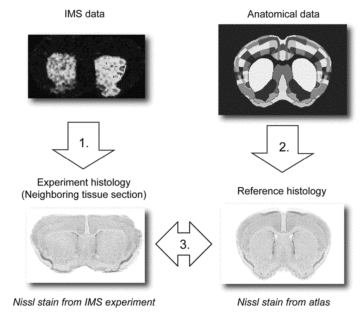
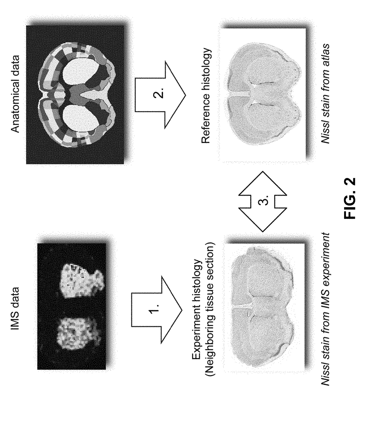
[0132]Since a substantial amount of IMS research focuses on rodent brain (K. Sköld, M. Svensson, A. Nilsson, X. Zhang, K. Nydahl, R. M. Caprioli, P. Svenningsson, and P. E. J. Andrén, Proteome Res. 2006, 5:262-269), the case studies herein use MALDI-TOF IMS data from mouse brain tissue as the empirical data source and the Allen Mouse Brain Atlas (E. S. Lein, M. J. Hawrylycz, N. Ao, M. Ayres, A. Bensinger, A. Bernard, A. F. Boe, M. S. Boguski, K. S. Brockway, E. J. Byrnes, and L. Chen, et al., Nature 2007, 445:168-176) as the curated anatomical data source. Both data types have been used in studies of neurodegenerative diseases such as Alzheimer's, Parkinson's, and healthy mouse brain (A. R. Jones, C. C. Overly, and S. M. Sunkin, Nat. Rev. Neurosci. 2009, 10:821-828; and J. Hanrieder, N. T. N. Phan, M. E. Kurczy, and A. G. Ewing, ACS Chem. Neurosci. 2013, 4:666-79). However, the methods developed herein are not specific to these case studies, a particular species, disease model, or a...

case study examples

Lipid Case Study Examples

[0190]FIG. 13 shows the correlation table for the lipid data (negative mode) with the atlas data. It shows areas of high positive, high negative, and low correlation. FIG. 14 displays several examples of anatomical structure / ion abundance combinations with high positive correlation.

Additional Automated Anatomical Interpretation Examples

[0191]Examples of automated anatomical interpretation applied to the protein-focused IMS case study are demonstrated in FIGS. 15A and 15B. When an ion image is given as input (top-left), the interpretation method provides an optimal anatomical explanation for the observed ion pattern (bottom), using the library of provided anatomical structures. Specifically, the ion intensity pattern is decomposed without user intervention into an optimal combination of contributing anatomical structures from the atlas. The interpretation method provides (i) the closest approximation of the measured ion image using atlas structures (top-middl...


PUM


Abstract

Imaging mass spectrometry (IMS) has become a prime tool for studying the distribution of biomolecules in tissue. Although IMS data sets can become very large, computational methods have made it practically feasible to search these experiments for relevant findings. However, these methods lack access to an important source of information that many human interpretations rely upon: anatomical insight. In this work, this need is addressed by (1) integrating a curated anatomical data source with an empirically acquired IMS data source, establishing an algorithm-accessible link between them; and (2) demonstrating the potential of such an IMS-anatomical atlas link by applying it toward automated anatomical interpretation of ion distributions in tissue.

Description

CROSS-REFERENCE TO RELATED APPLICATIONS[0001]This application is a national phase entry under 35 U.S.C. §371 of International Patent Application PCT / BE2015 / 000034, filed Aug. 3, 2015, designating the United States of America and published in English as International Patent Publication WO 2016 / 015108 A1 on Feb. 4, 2016, which claims the benefit under Article 8 of the Patent Cooperation Treaty to U.S. Provisional Patent Application Ser. No. 62 / 031,965, filed Aug. 1, 2014.TECHNICAL FIELD[0002]This application relates generally to a system or a method for automated interpretation of images in a computer-readable environment and 1) generates an image to interpret by a technique that images the spatial distribution of a particular property of a sample of a molecular or chemical composition, 2) links a body of curated patterns or a set of reference image patterns to interpret the image, and 3) spatially maps the body of curated patterns or the reference image patterns to the image to inter...

Claims


Application Information

Patent Timeline
IPC IPC(8): G06K9/00H01J49/00G06T7/00G06K9/20G06K9/62
CPCG06K9/00147G06K9/2054G06K9/6218G06T7/0012G06T2207/30048G06T2207/10081G06T2207/10116G06T2207/10132H01J49/0004G06T2207/10072G06T2207/10104G06T2207/10128G06T7/11G06T7/143G06V20/698G06F18/23
Inventor CAPRIOLI, RICHARD M.DE MOOR, BARTVAN DE PLAS, RAFVERBEECK, NICOWAELKENS, ETIENNE
Owner KATHOLIEKE UNIV LEUVEN
Why Eureka
Try Eureka