
Measurement of blood volume using velocity-selective pulse trains on MRI

a pulse train and velocity-selective technology, applied in the field of medical imaging, can solve the problems of methodological problems, gd-enhanced mri techniques, and the difficulty in reducing the accuracy of mri quantification, so as to reduce the effect of eddy curren

Active Publication Date: 2017-10-12
THE JOHN HOPKINS UNIV SCHOOL OF MEDICINE
View PDF0 Cites 3 Cited by
  • Summary
  • Abstract
  • Description
  • Claims
  • Application Information

AI Technical Summary

Benefits of technology

This patent describes a method for improving the accuracy of magnetic resonance imaging (MRI) to measure blood volume. The method uses a special labeling module and velocity-sensitized pulses to reduce eddy currents and improve the image quality. The method also includes a subtraction-based method to cancel out signals from static tissue and measures blood volume based on its relaxation factors. Overall, this method can provide a more accurate and reliable way to measure blood volume in MRI scans.

Problems solved by technology

For patients with occluded carotid arteries, increased oxygenation extraction fraction (OEF) and CBV yet at normal CBF (autoregulatory vasodilation) were associated with higher risk of stroke.
For absolute quantification of CBV, this technique faces several challenges, e.g., identification of global arterial input function (AIF) from a pure blood voxel, consideration of the delay and dispersion of the local AIF, and non-linear response of intravascular Gd concentration.
However, the risks of developing nephrogenic systemic fibrosis (NSF) limit Gd-enhanced MRI techniques on patient population with impaired renal function and the finding of gadolinium deposition in neuro tissues even for people without kidney diseases warrant caution of repeat use of certain Gd-based contrast agents.
Despite gaining popularity in clinical studies for different organs, the quantification precision and accuracy of this fitting-based method still suffers methodological issues such as requirement of high signal-to-noise ratio (SNR) and the validity of the bi-exponential model, especially in the brain where CBV is smaller than 5% and pulsatile flow of cerebrospinal fluid (CSF) is present.
A further overlooked drawback of the IVIM signal model is its neglecting of the T1 and T2 relaxation factors in each compartment, thus the results are dependent on the employed repetition time (TR) and echo time (TE) respectively.

Method used


Image

  • Measurement of blood volume using velocity-selective pulse trains on MRI
  • Measurement of blood volume using velocity-selective pulse trains on MRI
  • Measurement of blood volume using velocity-selective pulse trains on MRI

Examples


Embodiment Construction

[0020]The presently disclosed subject matter now will be described more fully hereinafter with reference to the accompanying Drawings, in which some, but not all embodiments of the inventions are shown. Like numbers refer to like elements throughout. The presently disclosed subject matter may be embodied in many different forms and should not be construed as limited to the embodiments set forth herein; rather, these embodiments are provided so that this disclosure will satisfy applicable legal requirements. Indeed, many modifications and other embodiments of the presently disclosed subject matter set forth herein will come to mind to one skilled in the art to which the presently disclosed subject matter pertains, having the benefit of the teachings presented in the foregoing descriptions and the associated Drawings. Therefore, it is to be understood that the presently disclosed subject matter is not to be limited to the specific embodiments disclosed and that modifications and other...


PUM


Abstract

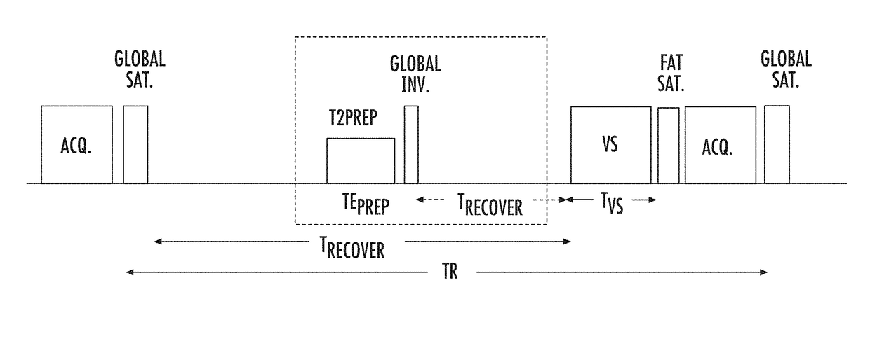
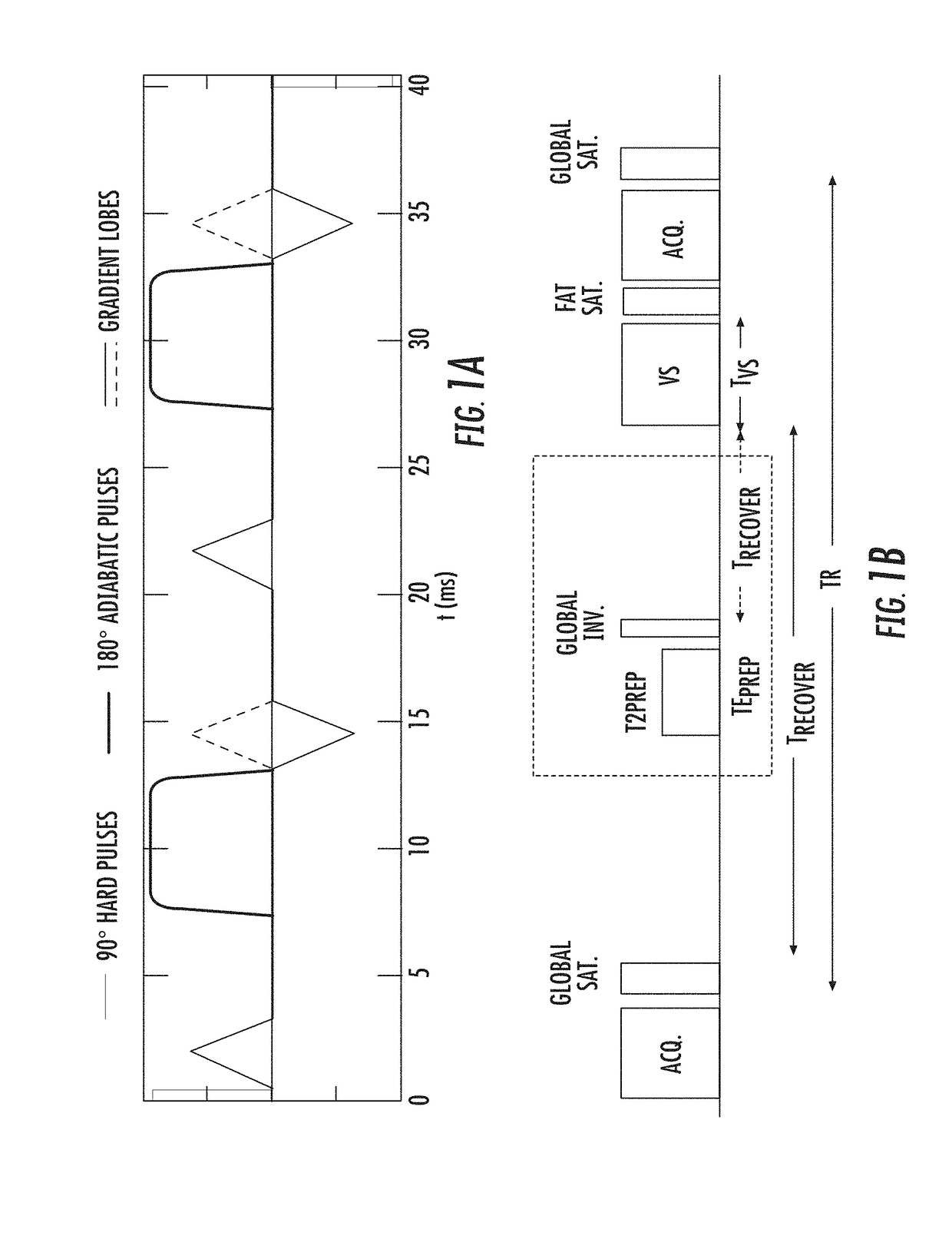
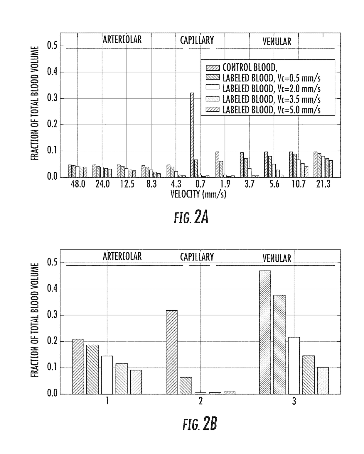
The present invention is directed to a system and method for measuring blood volume using non-contrast-enhanced magnetic resonance imaging. The method of the present invention includes a subtraction-based method using a pair of acquisitions immediately following velocity-sensitized pulse trains for the label module and its corresponding control module, respectively. The signal of static tissue is canceled out and the difference signal comes from the flowing blood compartment above a cutoff velocity. After normalizing to a proton density-weighted image acquired separately and scaled with the blood T1 and T2 relaxation factors, quantitative measurement of blood volume is then obtained.

Description

CROSS REFERENCE TO RELATED APPLICATION[0001]This application claims the benefit of U.S. Provisional Patent Application No. 62 / 321,429 filed on Apr. 12, 2016, which is incorporated by reference, herein, in its entirety.GOVERNMENT SUPPORT[0002]This invention was made with government support under grant number NIH K25 HL121192 and P41 EB015909 awarded by the National Institutes of Health. The government has certain rights in the invention.FIELD OF THE INVENTION[0003]The present invention relates generally to medical imaging. More particularly, the present invention relates to a system and method for measurement of blood volume using MRI.BACKGROUND OF THE INVENTION[0004]Velocity-selective (VS) pulse trains can provide unique functionalities when designing pulse sequences for various Magnetic Resonance Imaging (MRI) based hemodynamic evaluation: MR angiography (MRA); blood flow (perfusion), blood volume, or transit time; oxygen extraction fraction; metabolic rate of oxygen.[0005]Quantita...

Claims


Application Information

Patent Timeline
Patent Type & Authority Applications(United States)
IPC IPC(8): G01R33/50G01R33/56
CPCG01R33/5601G01R33/50G01R33/5607G01R33/56366
Inventor QIN, QIN
Owner THE JOHN HOPKINS UNIV SCHOOL OF MEDICINE
Learn More