Engineering of a Novel Breast Tumor Microenvironment on a Microfluidic Chip

Active Publication Date: 2018-02-22
ARIZONA STATE UNIVERSITY
View PDF0 Cites 8 Cited by
  • Summary
  • Abstract
  • Description
  • Claims
  • Application Information

AI Technical Summary

Benefits of technology

[0011]To address these challenges, we developed an innovative approach using microscale technologies and hydrogel based biomaterials to develop a three dimensional (3D) in vivo like breast tumor microenvironment on a microfluidic chip. Specifically, we used microengineering technology, hydrogel-based biomaterials, and co-culture of key cell types within the tumor stroma to develop an in vivo like 3D vascularized human breast tumor model microenvironment. Hydrogels are excellent scaffolding materials because of their 3D architecture resembling the native in vivo microenvironment, homogenous cellular distribution, excellent diffusion p

Problems solved by technology

However, using these models, there are certain difficulties to independently study the effects of various microenvironmental cues (i.e., cell-cell communication, mechanical properties of the matrix) on cancer cells metastasis.
Furthermore, extensive testing of therapeutic compounds using in vivo models is a costly process.
Although these assays have facilitated high throughput and economically efficient experimental analysis, they do not fully recapitulate the complexities of the native 3D tumor microenvironment.
The limiting aspects of 3D macroscale hydrogel models are the lack of precise control over cellular distribution, lack of vascularity and difficulties

Method used


Image

  • Engineering of a Novel Breast Tumor Microenvironment on a Microfluidic Chip
  • Engineering of a Novel Breast Tumor Microenvironment on a Microfluidic Chip
  • Engineering of a Novel Breast Tumor Microenvironment on a Microfluidic Chip

Examples


example 1

An Exemplary Microfluidic Device

[0090]In this example, we describe a non-limiting exemplary embodiment of a microfluidic device according to the disclosed invention.

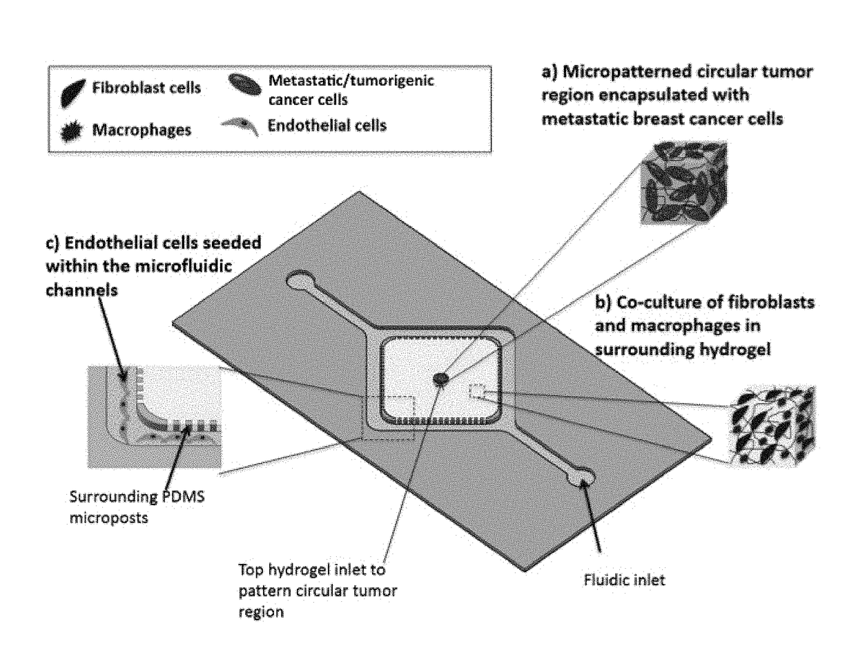
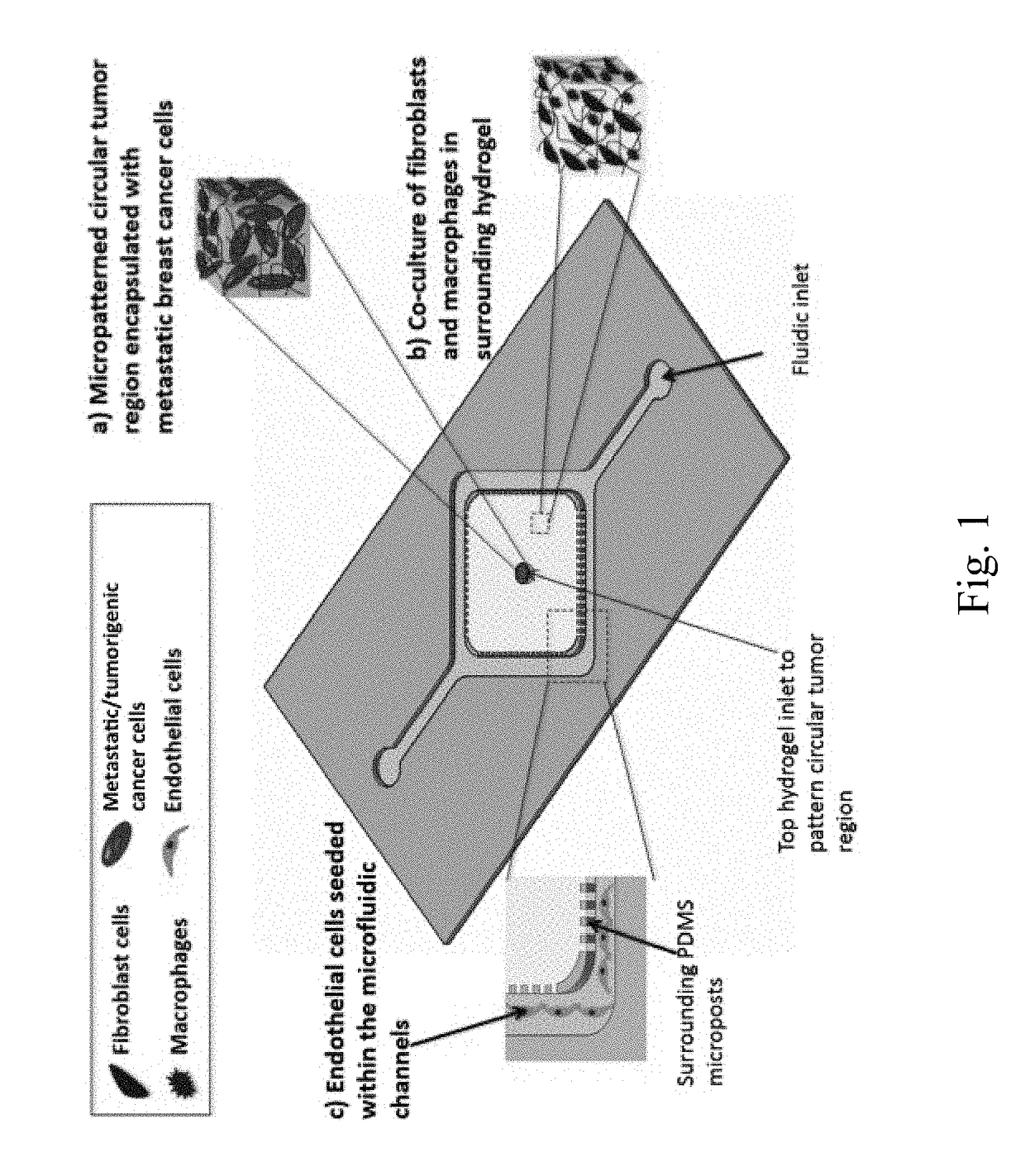
[0091]FIG. 1 shows the features of the exemplary microfluidic device. As shown in FIG. 1, the microfluidic device includes two separate regions of hydrogel constructs, one for the circular tumor construct (neoplastic lesion) encapsulated with metastatic breast cancer cells ((a); center circle), and the second one resembling the surrounding 3D ECM stroma for encapsulation of FBs and macrophages ((b); rectangle surrounding center circle). Two parallel microfluidic channels seeded with ECs are interconnected with the surrounding ECM hydrogel ((c), adjacent to the two long edges of rectangle of (b)) . In particular, the microfluidic channels will be used for seeding of ECs resembling the vascular network ((c)). In addition, the microfluidic channels will be used to introduce anticancer drugs for testing.

Microfluidic Device

[0...

example 2

Production and Use of an Exemplary Microfluidic Device

[0104]In this proof of principal example, we demonstrate that a microfluidic device according to the disclosed invention is capable producing a well-defined tissue architecture, and that it enables high-contrast and real-time imaging for detailed invasion and intravasion studies.

Introduction

[0105]The development of our breast cancer invasion platform has given our laboratory the opportunity to investigate and provide insight to key aspects of the metastatic cascade and tumorigenesis. This study validates our model as breast cancer invasion and demonstrates that our platform is capable of producing a well-defined tissue architecture and enabling high-contrast real-time imaging for detailed invasion and intravasation studies.

Creating a Spatially Organized Cancer Invasion Platform

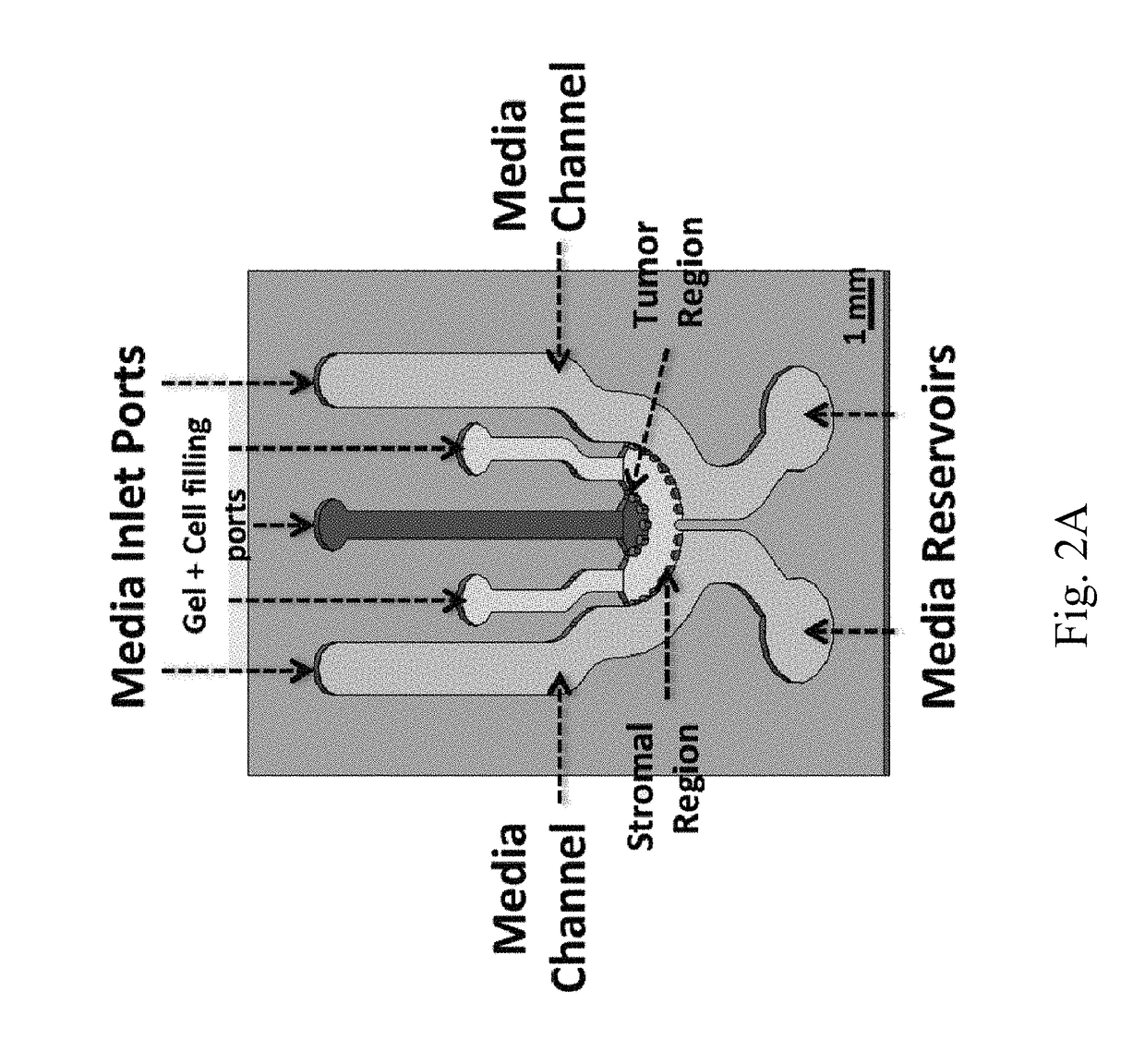
[0106]First, we developed a CAD design that caters to the native breast tumor architecture (FIG. 2A). It consisted of two distinct region made up of two co...

example 3

Another Exemplary Microfluidic Device

[0111]In this example, we describe additional non-limiting exemplary embodiments of a microfluidic device according to the disclosed invention.

Introduction

[0112]In this newly developed platform, we created a novel microfluidic platform comprised of concentric three-layer cell-laden hydrogels, which allowed for simultaneous observation of vascular network maturation as well as breast cancer cell invasion and intravasation. We demonstrated that highly metastatic MDA-MB-231 disseminated from the primary tumor region and invaded into the adjacent stroma layer, where the presence of a vascular endothelial network in the third layer of the platform drastically increased cancer cell motility and invasion. Most importantly, by day 6 of culture, cancer cells could be visualized intravasating into the vasculature. Additionally, the presence of the invading cancer cells significantly reduced vessel diameter and increased permeability, consistent with previo...


PUM


Abstract

A microfluidic device for more accurately modeling the in vitro environment in which cancer occurs is disclosed. The microfluidic device includes a surface defining one or more microfluidic channels, a first three dimensional scaffold comprising one or more cancer cells that is spatially separated from the one or more microfluidic channels, a second three dimensional scaffold, at least a portion of which is contacting and in fluid communication with the first three dimensional scaffold, and that is spatially separated from the one or more microfluidic channels, and a third three dimensional scaffold, at least a portion of which is contacting and in fluid communication with the one or more microfluidic channels and the second three dimensional scaffold. The device can be used to assay anti-cancer agents, or as a system for modeling the growth, behavior, or metastasis and tumor formation of cancer cells.

Description

CROSS-REFERENCE TO RELATED APPLICATIONS[0001]This application is a continuation-in-part of U.S. patent application Ser. No. 14 / 872,920 filed Oct. 1, 2015 which claims the benefit of U.S. provisional Application No. 62 / 058,186 filed on Oct. 1, 2014, which are incorporated by reference herein in their entirety.STATEMENT REGARDING FEDERALLY SPONSORED RESEARCH OR DEVELOPMENT[0002]Not applicable.BACKGROUND[0003]Metastasis, a process where cancer cells migrate to distant organs from the primary tumor site, is the key biological process responsible for 90% of all cancer related deaths. Metastatic dissemination of cancer cells is a multi-step biological process initiated by tumor angiogenesis and invasion of the cancer cells through their surrounding stroma toward the blood vessels.[0004]Tumor stroma is a structurally complex microenvironment hosting several cell types including endothelial cells (ECs), fibroblasts (FBs) and macrophages, packed in extracellular matrix (ECM), along with a de...

Claims


Application Information

Patent Timeline
IPC IPC(8): G01N33/50B01L3/00
CPCG01N33/5011B01L3/502707B01L3/502715B01L2300/0816B01L2300/12C12M23/16C12M25/14
Inventor NIKKHAH, MEHDITRUONG, DANH
Owner ARIZONA STATE UNIVERSITY
Why Eureka
Try Eureka